
NALP1 (NAcht, Leucine-rich repeat and PYD domain containing protein 1; also NAC, CARD7, DEFCAP and CLR17.1) is a 160-170 kDa member of the NLRP family of molecules. It is expressed in dendritic cells, neutrophils, T and B cells, alveolar macrophages, permatogonia, neurons, plus intestinal columnar epithelium, and is found in both cytoplasm and nucleus. NALP1 promotes apoptosis plus IL-1 beta and IL-18 maturation by activating caspase-1 and -5. It does so by forming a 700 kDa inflammasome comprised of NALP1, ASC plus caspase-1 and -5. Bacterial wall peptidoglycan binds to NALP1, promoting ATP binding, NALP1 oligomerization, and caspase activation. Human NALP1 is 1473 amino acids (aa) in length. It contains an N-terminal DAPIN domain (aa 1-92), a NACHT domain (aa 328-637), seven consecutive LRRs (aa 704-1236) and one CARD region (aa 1374-1463). Alternate splice forms exist that range in size from 70 kDa-150 kDa. Either individually, or in combination, there can be a 19 aa substitution for either aa 1353-1473 or aa 1368-1472, a deletion of aa 91-260, 958-987 or 1262-1305, and a 43 aa insert after Leu785. Over aa 1-323, human NALP1 shares less that 20% aa identity with mouse NALP1.

Scientific images may be resized, cropped, or adjusted for brightness or contrast for presentation purposes. These updates are limited to presentation and do not affect the underlying scientific interpretation of the data. Where available, additional source data may be provided upon request.
Key Product Details
Species Reactivity
Validated:
Cited:
Applications
Validated:
Cited:
Label
Antibody Source
Product Specifications
Immunogen
Met1-Asp323 (Leu155His)
Accession # Q9C000
Specificity
Clonality
Host
Isotype
Scientific Data Images for Human NLRP1/NALP1 Antibody
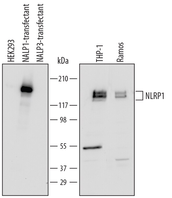
Detection of Human NLRP1/NALP1 by Western Blot.
Western blot shows lysates of HEK293 human embryonic kidney cell line either mock transfected, transfected with human NLRP1/NALP1 or transfected with NALP3, THP-1 human acute monocytic leukemia cell line, and Ramos human Burkitt's lymphoma cell line. PVDF Membrane was probed with 0.5 µg/mL of Sheep Anti-Human NLRP1/NALP1 Antigen Affinity-purified Polyclonal Antibody (Catalog # AF6788) followed by HRP-conjugated Anti-Sheep IgG Secondary Antibody (Catalog # HAF016). Specific bands were detected for NLRP1/NALP1 at approximately 160 kDa (as indicated). This experiment was conducted under reducing conditions and using Immunoblot Buffer Group 2.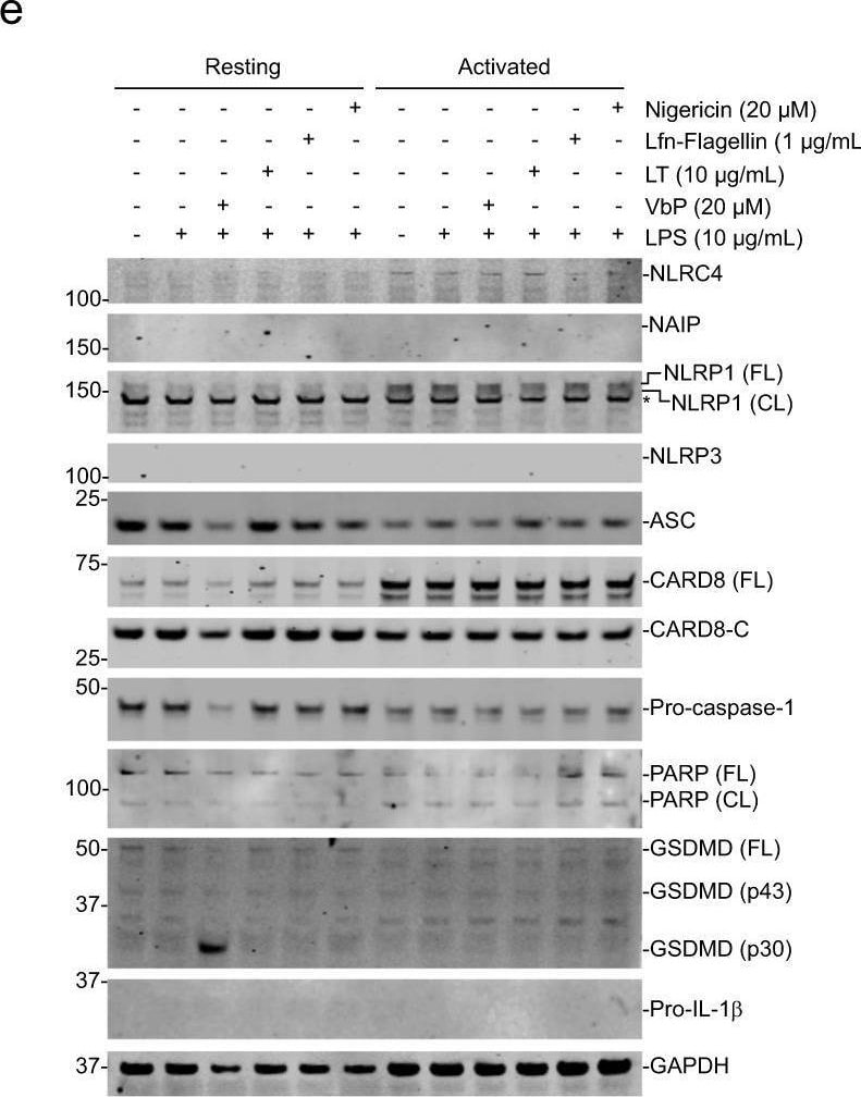
Detection of NLRP1/NALP1 by Western Blot
DPP8/9 inhibitors activate the CARD8 inflammasome in T cells. A–c Human primary CD3+ T cells were pre-treated with the indicated compounds for 30 min prior to the addition of VbP (2 μM, 6 h). Cell viability was assessed using Cell-TiterGlo. Data are means ± s.e.m. of four or five biological replicates. ***p < 0.001 by two-sided Student’s t test. NS not significant. d Immunoblots of CARD8 KO CD3+ T-cell lysates treated with VbP (20 μM, 24 h). e, f CD3+ T cells (Donor 008) were primed with LPS (10 μg/mL, 4 h) before treatment with the indicated concentrations of VbP (6 h), LT (6 h), Lfn-flagellin (3 h), or nigericin (1 h). Lysates were then evaluated by immunoblotting (e) and supernatants were analyzed for LDH release (f) and IL-1 beta secretion (g). Image collected and cropped by CiteAb from the following open publication (https://pubmed.ncbi.nlm.nih.gov/32796818), licensed under a CC-BY license. Not internally tested by R&D Systems.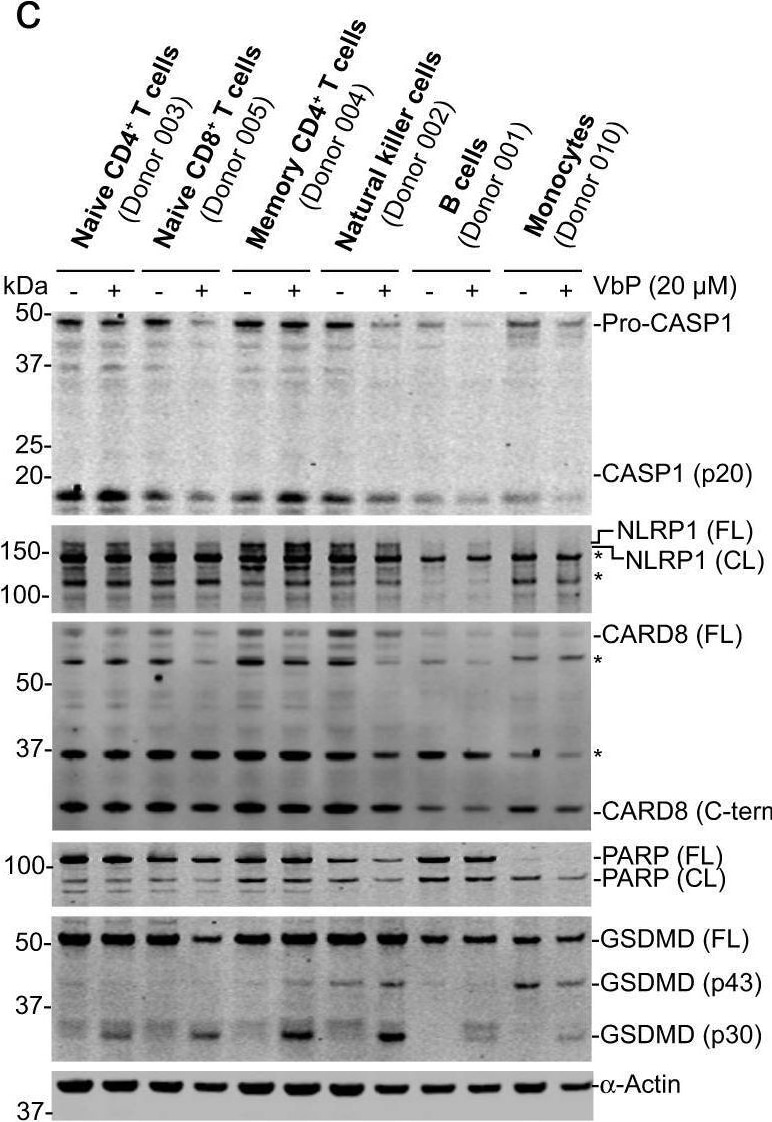
Detection of NLRP1/NALP1 by Western Blot
DPP8/9 inhibitors induce pyroptosis in primary lymphocytes.a Domain structures of human NLRP1 and CARD8. Note that the ZU5 and UPA sub-domains together are referred to as a FIIND. b, c The indicated primary cells were treated with VbP (24 h) before cell death was assessed by Cell-TiterGlo (b) and lysates were evaluated by immunoblotting (c). Data are means ± s.e.m. of four biological replicates. d CD3+ T cells were treated with VbP (20 μM, 24 h) or 8j (20 μM, 24 h) before lysates were evaluated by immunoblotting. Immunoblots are representative of three independent experiments. Image collected and cropped by CiteAb from the following open publication (https://pubmed.ncbi.nlm.nih.gov/32796818), licensed under a CC-BY license. Not internally tested by R&D Systems.Applications for Human NLRP1/NALP1 Antibody
Western Blot
Sample: HEK293 human embryonic kidney cell line either transfected with human NLRP1/NALP1 or transfected with NALP3, THP‑1 human acute monocytic leukemia cell line, and Ramos human Burkitt's lymphoma cell line
Reviewed Applications
Read 1 review rated 5 using AF6788 in the following applications:
Formulation, Preparation, and Storage
Purification
Reconstitution
Sterile PBS to a final concentration of 0.2 mg/mL. For liquid material, refer to CoA for concentration.
Formulation
Shipping
Stability & Storage
- 12 months from date of receipt, -20 to -70 °C as supplied.
- 1 month, 2 to 8 °C under sterile conditions after reconstitution.
- 6 months, -20 to -70 °C under sterile conditions after reconstitution.
Calculators
Background: NLRP1/NALP1
Long Name
Alternate Names
Gene Symbol
UniProt
Additional NLRP1/NALP1 Products
Product Documents for Human NLRP1/NALP1 Antibody
Certificate of Analysis
To download a Certificate of Analysis, please enter a lot or batch number in the search box below.
Note: Certificate of Analysis not available for kit components.
Product Specific Notices for Human NLRP1/NALP1 Antibody
For research use only
Related Research Areas
Citations for Human NLRP1/NALP1 Antibody
 Powered by Bioz
Customer Reviews for Human NLRP1/NALP1 Antibody (1)
Have you used Human NLRP1/NALP1 Antibody?
Submit a review and receive an Amazon gift card!
$25/€18/£15/$25CAN/¥2500 for a review with an image
$10/€7/£6/$10CAN/¥1110 for a review without an image
Submit a review
-
Application: ELISASample Tested: HeLa human cervical epithelial carcinoma cell lineSpecies: HumanVerified Customer | Posted 09/07/2017
There are no reviews that match your criteria.
Protocols
Find general support by application which include: protocols, troubleshooting, illustrated assays, videos and webinars.
- Cellular Response to Hypoxia Protocols
- R&D Systems Quality Control Western Blot Protocol
- Troubleshooting Guide: Western Blot Figures
- Western Blot Conditions
- Western Blot Protocol
- Western Blot Protocol for Cell Lysates
- Western Blot Troubleshooting
- Western Blot Troubleshooting Guide
- View all Protocols, Troubleshooting, Illustrated assays and Webinars
Associated Pathways
