
The neuregulin family of structurally related glycoproteins comprises products from four distinct but related genes, Nrg-1, Nrg-2, Nrg-3, and Nrg-4. Through alternative splicing or the use of alternative promoters, Nrg-1 has been shown to encode more than 14 soluble or transmembrane proteins. The extracellular domain of the transmembrane NRG1 isoforms can be proteolytically cleaved to release soluble growth factors. All NRG1 isoforms contain an EGF‑like domain ( alpha - or beta -splice variant that differ in their C-terminal region) that is required for their direct binding to the ErbB3 or ErbB4 receptor tyrosine kinases. The ErbB3 or ErbB4 subsequently recruits and heterodimerizes with ErbB2, resulting in tyrosine phosphorylation and NRG1 signaling. NRG1 isoforms can be classified into three major subtypes. Type I (Neu Differentiation Factor, NDF; Heregulin, HRG; Acetylcholine Receptor Inducing Activity, ARIA) and type II (Glial Growth Factor, GGF) NRG1s have an immunoglobulin (Ig)-like domain N-terminal to the EGF‑like domain. Type I NRG1s differ from type II NRG1s by having a glycosylation-rich domain between the Ig‑like and the EGF‑like domains. Type III NRG1s (Sensory and Motor neuron-Derived Factor) lacks the Ig‑like domain but has a cysteine rich domain (CRD) instead. NRG1 isoforms show distinct spatial and temporal expression patterns. These proteins play important roles during development of both the nervous system and the heart. They have been shown to regulate the selective expression of neurotransmitter receptors in neurons and at the neuromuscular junction, and promote the differentiation and development of Schwann cells from neural crest stem cells. NRG1s have also been shown to be involved in the establishment of the oligodendroglial lineage.
Human Neuregulin-1 beta 1/NRG1 beta 1 EGF Domain Antibody
R&D Systems | Catalog # AF-396-NA


Scientific images may be resized, cropped, or adjusted for brightness or contrast for presentation purposes. These updates are limited to presentation and do not affect the underlying scientific interpretation of the data. Where available, additional source data may be provided upon request.
Key Product Details
Species Reactivity
Validated:
Cited:
Applications
Validated:
Cited:
Label
Antibody Source
Product Specifications
Immunogen
Thr176-Lys246
Accession # NP_039250
Specificity
Clonality
Host
Isotype
Endotoxin Level
Scientific Data Images for Human Neuregulin-1 beta 1/NRG1 beta 1 EGF Domain Antibody
Cell Proliferation Induced by NRG1‑ beta 1/HRG1‑ beta 1 and Neutralization by Human NRG1‑ beta 1/HRG1‑ beta 1 Antibody.
Recombinant Human NRG1-beta 1/ HRG1-beta 1 EGF Domain (Catalog # 396-HB) stimulates proliferation in the MCF-7 human breast cancer cell line in a dose-dependent manner (orange line). Proliferation elicited by Recombinant Human NRG1-beta 1/ HRG1-beta 1 EGF Domain (0.75 ng/mL) is neutralized (green line) by increasing concentrations of Goat Anti-Human NRG1-beta 1/HRG1-beta 1 EGF Domain Antigen Affinity-purified Polyclonal Antibody (Catalog # AF-396-NA). The ND50 is typically 0.5-2.0 µg/mL.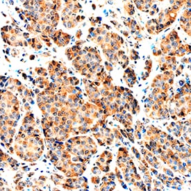
NRG1‑ beta 1/HRG1‑ beta 1 in Human Breast.
NRG1-beta 1/HRG1-beta 1 was detected in immersion fixed paraffin-embedded sections of human breast using Goat Anti-Human NRG1-beta 1/HRG1-beta 1 EGF Domain Antigen Affinity-purified Polyclonal Antibody (Catalog # AF-396-NA) at 1 µg/mL overnight at 4 °C. Tissue was stained using the Anti-Goat HRP-DAB Cell & Tissue Staining Kit (brown; Catalog # CTS008) and counterstained with hematoxylin (blue). Specific staining was localized to cytoplasm. View our protocol for Chromogenic IHC Staining of Paraffin-embedded Tissue Sections.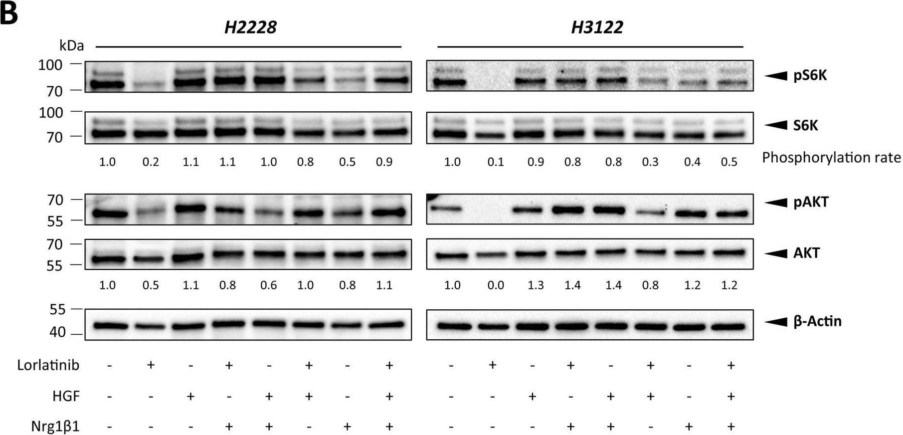
Detection of NRG1-beta 1/HRG1-beta 1 EGF by Western Blot
FB2-conditioned medium (CM) (A) and CAF-associated ligands HGF and Nrg1 beta 1 (B) influence the activation of AKT signaling upon ALK signaling perturbation via lorlatinib in H2228 and H3122 cells, as shown by representative western blots of AKT/pAKT and S6K/pS6K in H2228 and H3122 cells. Phosphorylation rates were normalized to those of beta -actin. AKT, protein kinase B; S6K, ribosomal S6 kinase Image collected and cropped by CiteAb from the following open publication (https://cancerandmetabolism.biomedcentral.com/articles/10.1186/s40170-0…), licensed under a CC-BY license. Not internally tested by R&D Systems.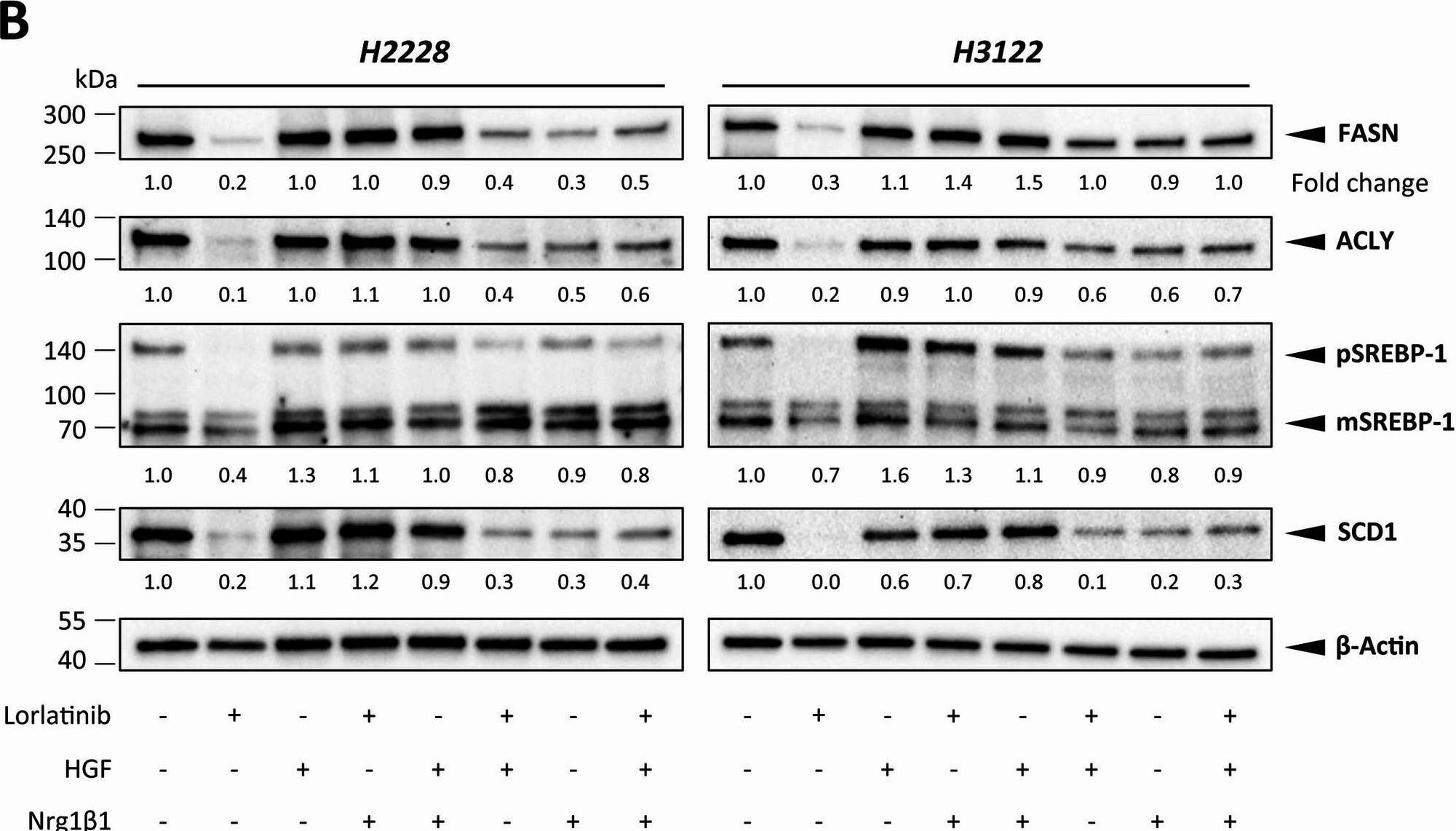
Detection of NRG1-beta 1/HRG1-beta 1 EGF by Western Blot
FB2-conditioned medium (CM) (A) and CAF-associated ligands HGF and Nrg1 beta 1 (B) influence the expression of fatty acid metabolism-regulating targets upon ALK signaling perturbation via lorlatinib in H2228 and H3122 cells, as shown by representative western blots of FASN, ACLY, and SREBP-1 in H2228 and H3122 cells. Expression fold changes were normalized to those of beta -actin. ACLY, ATP-citrate lyase; FASN, fatty acid synthase; pSREBP-1, precursor sterol-responsive element binding protein-1; mSREBP-1, mature SREBP-1 Image collected and cropped by CiteAb from the following open publication (https://cancerandmetabolism.biomedcentral.com/articles/10.1186/s40170-0…), licensed under a CC-BY license. Not internally tested by R&D Systems.Applications for Human Neuregulin-1 beta 1/NRG1 beta 1 EGF Domain Antibody
Immunohistochemistry
Sample: Immersion fixed paraffin-embedded sections of human breast
Western Blot
Sample: Recombinant Human NRG1-beta 1/HRG1-beta 1 EGF Domain (Catalog # 396-HB)
Neutralization
Formulation, Preparation, and Storage
Purification
Reconstitution
Reconstitute at 0.2 mg/mL in sterile PBS. For liquid material, refer to CoA for concentration.
Formulation
Shipping
Stability & Storage
- 12 months from date of receipt, -20 to -70 °C as supplied.
- 1 month, 2 to 8 °C under sterile conditions after reconstitution.
- 6 months, -20 to -70 °C under sterile conditions after reconstitution.
Calculators
Background: Neuregulin-1 beta 1/NRG1 beta 1
References
- Buonanno, A., and Fischbach, G.D. (2001) Curr. Opin. Neurobiol. 11:287.
- Adlkofer, K. and Lai, C. (2000) Glia 29:104.
- Garratt, A.N. et al. (2000) BioEssays 22:987.
Alternate Names
Gene Symbol
UniProt
Additional Neuregulin-1 beta 1/NRG1 beta 1 Products
Product Documents for Human Neuregulin-1 beta 1/NRG1 beta 1 EGF Domain Antibody
Certificate of Analysis
To download a Certificate of Analysis, please enter a lot or batch number in the search box below.
Note: Certificate of Analysis not available for kit components.
Product Specific Notices for Human Neuregulin-1 beta 1/NRG1 beta 1 EGF Domain Antibody
For research use only
Related Research Areas
Citations for Human Neuregulin-1 beta 1/NRG1 beta 1 EGF Domain Antibody
 Powered by Bioz
Customer Reviews for Human Neuregulin-1 beta 1/NRG1 beta 1 EGF Domain Antibody
There are currently no reviews for this product. Be the first to review Human Neuregulin-1 beta 1/NRG1 beta 1 EGF Domain Antibody and earn rewards!
Have you used Human Neuregulin-1 beta 1/NRG1 beta 1 EGF Domain Antibody?
Submit a review and receive an Amazon gift card!
$25/€18/£15/$25CAN/¥2500 for a review with an image
$10/€7/£6/$10CAN/¥1110 for a review without an image
Submit a review
Protocols
Find general support by application which include: protocols, troubleshooting, illustrated assays, videos and webinars.
- Antigen Retrieval Protocol (PIER)
- Antigen Retrieval for Frozen Sections Protocol
- Appropriate Fixation of IHC/ICC Samples
- Cellular Response to Hypoxia Protocols
- Chromogenic IHC Staining of Formalin-Fixed Paraffin-Embedded (FFPE) Tissue Protocol
- Chromogenic Immunohistochemistry Staining of Frozen Tissue
- ClariTSA™ Fluorophore Kits
- Detection & Visualization of Antibody Binding
- Fluorescent IHC Staining of Frozen Tissue Protocol
- Graphic Protocol for Heat-induced Epitope Retrieval
- Graphic Protocol for the Preparation and Fluorescent IHC Staining of Frozen Tissue Sections
- Graphic Protocol for the Preparation and Fluorescent IHC Staining of Paraffin-embedded Tissue Sections
- Graphic Protocol for the Preparation of Gelatin-coated Slides for Histological Tissue Sections
- IHC Sample Preparation (Frozen sections vs Paraffin)
- Immunofluorescent IHC Staining of Formalin-Fixed Paraffin-Embedded (FFPE) Tissue Protocol
- Immunohistochemistry (IHC) and Immunocytochemistry (ICC) Protocols
- Immunohistochemistry Frozen Troubleshooting
- Immunohistochemistry Paraffin Troubleshooting
- Preparing Samples for IHC/ICC Experiments
- Preventing Non-Specific Staining (Non-Specific Binding)
- Primary Antibody Selection & Optimization
- Protocol for Heat-Induced Epitope Retrieval (HIER)
- Protocol for Making a 4% Formaldehyde Solution in PBS
- Protocol for VisUCyte™ HRP Polymer Detection Reagent
- Protocol for the Preparation & Fixation of Cells on Coverslips
- Protocol for the Preparation and Chromogenic IHC Staining of Frozen Tissue Sections
- Protocol for the Preparation and Chromogenic IHC Staining of Frozen Tissue Sections - Graphic
- Protocol for the Preparation and Chromogenic IHC Staining of Paraffin-embedded Tissue Sections
- Protocol for the Preparation and Chromogenic IHC Staining of Paraffin-embedded Tissue Sections - Graphic
- Protocol for the Preparation and Fluorescent IHC Staining of Frozen Tissue Sections
- Protocol for the Preparation and Fluorescent IHC Staining of Paraffin-embedded Tissue Sections
- Protocol for the Preparation of Gelatin-coated Slides for Histological Tissue Sections
- R&D Systems Quality Control Western Blot Protocol
- TUNEL and Active Caspase-3 Detection by IHC/ICC Protocol
- The Importance of IHC/ICC Controls
- Troubleshooting Guide: Immunohistochemistry
- Troubleshooting Guide: Western Blot Figures
- Western Blot Conditions
- Western Blot Protocol
- Western Blot Protocol for Cell Lysates
- Western Blot Troubleshooting
- Western Blot Troubleshooting Guide
- View all Protocols, Troubleshooting, Illustrated assays and Webinars