Human PAK2 Antibody (677411)
R&D Systems | Catalog # MAB6849

Scientific images may be resized, cropped, or adjusted for brightness or contrast for presentation purposes. These updates are limited to presentation and do not affect the underlying scientific interpretation of the data. Where available, additional source data may be provided upon request.
Key Product Details
Species Reactivity
Validated:
Cited:
Applications
Validated:
Cited:
Label
Antibody Source
Product Specifications
Immunogen
Leu127-Gln221
Accession # Q13177
Specificity
Clonality
Host
Isotype
Scientific Data Images for Human PAK2 Antibody (677411)
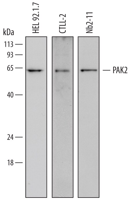
Detection of Human, Mouse, and Rat PAK2 by Western Blot.
Western blot shows lysates of HEL 92.1.7 human erythroleukemic cell line, CTLL-2 mouse cytotoxic T cell line, and Nb2-11 rat lymphoma cell line. PVDF membrane was probed with 2 µg/mL of Mouse Anti-Human PAK2 Monoclonal Antibody (Catalog # MAB6849) followed by HRP-conjugated Anti-Mouse IgG Secondary Antibody (Catalog # HAF007). A specific band was detected for PAK2 at approximately 63 kDa (as indicated). This experiment was conducted under reducing conditions and using Immunoblot Buffer Group 1.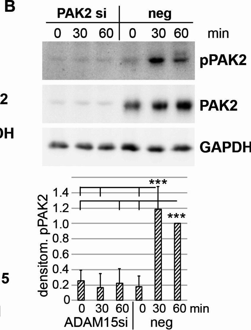
Detection of erw2itdfglz6sbdcfwsn.jpg by Western Blot
ADAM15- and N-cadherin (NCAD) dependent activation of PAK2 upon mechanical strain. (A–D) Immunoblots for pPAK2. (A) of RASFs seeded at different cell densities and mechanically strained for 30 and 60 min. (B) from strained RASFs with PAK2 silencing (si) and treated with a negative control siRNA (neg). (C, D) immunoblots for pPAK2, ADAM15 and NCAD from RASFs treated with ADAM15 and NCAD siRNA; right panels, showing the mean ± SD of densitometric analysis of 6 different RASF cell lines. ***p < 0.0005, Student’s t-test. GAPDH served as a loading control. Image collected and cropped by CiteAb from the following open publication (https://www.nature.com/articles/s41598-025-94012-2), licensed under a CC-BY license. Not internally tested by R&D Systems.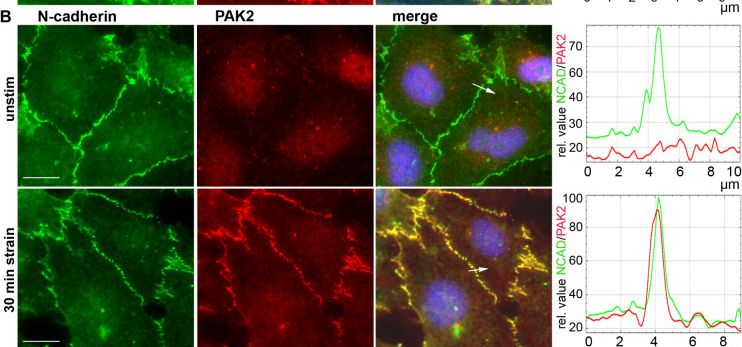
Detection of PAK2 by Immunocytochemistry/ Immunofluorescence
Mechano-induced recruitment of PAK2 together with Nck to the cell membrane. (A, B) RASFs grown on Bioflex plates and either unstimulated or strained for 30 min were fluorescently stained for (A) NCAD and Nck and (B) NCAD and PAK2. Adjacent to images: quantification of the pixel density of NCAD /Nck or NCAD /PAK2 along the white arrow. (C) double staining of NCAD and PAK2 in RASFs with prior silencing of Nck, right, pixel density along arrow. Cell nuclei were counterstained with DAPI. Objective 40x, scale bar = 10 μm. (D) immunoblots for PAK2, pPAK2, Nck and NCAD. RASFs pre-treated with Nck siRNA (I) and a nonsilencing siRNA (N) were mechanically strained for 15 and 30 min, the cell surface biotinylated with non-membrane-permeable biotin reagent and purified on streptavidin-conjugated magnetic beads. Contr: non biotinylated cell lysates enriched on streptavidin beads served as background control. TL = total lysates from 30 min time point. GAPDH was used to control the purity of the plasma membrane fractions. Image collected and cropped by CiteAb from the following open publication (https://www.nature.com/articles/s41598-025-94012-2), licensed under a CC-BY license. Not internally tested by R&D Systems.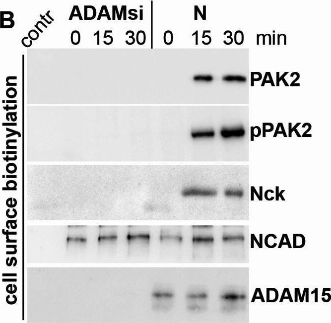
Detection of PAK2 by Western Blot
Binding of PAK2 to ADAM15/NCAD complex at cell membrane is crucially dependent on ADAM15. (A) IPs of strained RASFs using ADAM15 or NCAD antibodies and detection of PAK2 and Nck. TL = total lysate. IgG, mouse IgG served as background control. (B) cell surface biotinylation and enrichment of membrane fractions on streptavidin beads of strained cells with ADAM15-silenced or nonsilenced (N). Contr: non biotinylated cell lysates enriched on streptavidin beads served as background control. (C, D) IPs using ADAM15 antibodies in (C) RASFs with silenced and nonsilenced ADAM15 expression (N) and (D) in chondrocyte cell lines transfected with full length ADAM15 (full-A15) or ADAM15 lacking the cytoplasmic domain ( delta cyto). (E, F) analogous to (C, D) IPs using N-cadherin antibodies, showing precipitates of PAK2 and Nck in ADAM15-expressing cells only. (E) right, densitometric evaluation of PAK2 from experiments obtained from 4 donors. Image collected and cropped by CiteAb from the following open publication (https://www.nature.com/articles/s41598-025-94012-2), licensed under a CC-BY license. Not internally tested by R&D Systems.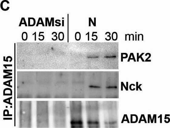
Detection of PAK2 by Western Blot
Binding of PAK2 to ADAM15/NCAD complex at cell membrane is crucially dependent on ADAM15. (A) IPs of strained RASFs using ADAM15 or NCAD antibodies and detection of PAK2 and Nck. TL = total lysate. IgG, mouse IgG served as background control. (B) cell surface biotinylation and enrichment of membrane fractions on streptavidin beads of strained cells with ADAM15-silenced or nonsilenced (N). Contr: non biotinylated cell lysates enriched on streptavidin beads served as background control. (C, D) IPs using ADAM15 antibodies in (C) RASFs with silenced and nonsilenced ADAM15 expression (N) and (D) in chondrocyte cell lines transfected with full length ADAM15 (full-A15) or ADAM15 lacking the cytoplasmic domain ( delta cyto). (E, F) analogous to (C, D) IPs using N-cadherin antibodies, showing precipitates of PAK2 and Nck in ADAM15-expressing cells only. (E) right, densitometric evaluation of PAK2 from experiments obtained from 4 donors. Image collected and cropped by CiteAb from the following open publication (https://www.nature.com/articles/s41598-025-94012-2), licensed under a CC-BY license. Not internally tested by R&D Systems.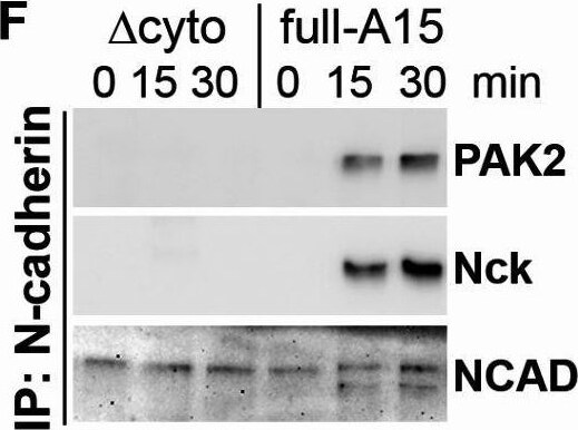
Detection of PAK2 by Western Blot
Binding of PAK2 to ADAM15/NCAD complex at cell membrane is crucially dependent on ADAM15. (A) IPs of strained RASFs using ADAM15 or NCAD antibodies and detection of PAK2 and Nck. TL = total lysate. IgG, mouse IgG served as background control. (B) cell surface biotinylation and enrichment of membrane fractions on streptavidin beads of strained cells with ADAM15-silenced or nonsilenced (N). Contr: non biotinylated cell lysates enriched on streptavidin beads served as background control. (C, D) IPs using ADAM15 antibodies in (C) RASFs with silenced and nonsilenced ADAM15 expression (N) and (D) in chondrocyte cell lines transfected with full length ADAM15 (full-A15) or ADAM15 lacking the cytoplasmic domain ( delta cyto). (E, F) analogous to (C, D) IPs using N-cadherin antibodies, showing precipitates of PAK2 and Nck in ADAM15-expressing cells only. (E) right, densitometric evaluation of PAK2 from experiments obtained from 4 donors. Image collected and cropped by CiteAb from the following open publication (https://www.nature.com/articles/s41598-025-94012-2), licensed under a CC-BY license. Not internally tested by R&D Systems.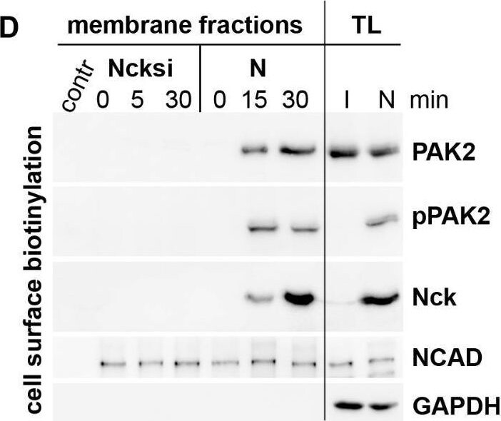
Detection of PAK2 by Western Blot
Mechano-induced recruitment of PAK2 together with Nck to the cell membrane. (A, B) RASFs grown on Bioflex plates and either unstimulated or strained for 30 min were fluorescently stained for (A) NCAD and Nck and (B) NCAD and PAK2. Adjacent to images: quantification of the pixel density of NCAD /Nck or NCAD /PAK2 along the white arrow. (C) double staining of NCAD and PAK2 in RASFs with prior silencing of Nck, right, pixel density along arrow. Cell nuclei were counterstained with DAPI. Objective 40x, scale bar = 10 μm. (D) immunoblots for PAK2, pPAK2, Nck and NCAD. RASFs pre-treated with Nck siRNA (I) and a nonsilencing siRNA (N) were mechanically strained for 15 and 30 min, the cell surface biotinylated with non-membrane-permeable biotin reagent and purified on streptavidin-conjugated magnetic beads. Contr: non biotinylated cell lysates enriched on streptavidin beads served as background control. TL = total lysates from 30 min time point. GAPDH was used to control the purity of the plasma membrane fractions. Image collected and cropped by CiteAb from the following open publication (https://www.nature.com/articles/s41598-025-94012-2), licensed under a CC-BY license. Not internally tested by R&D Systems.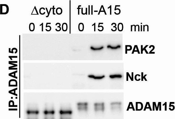
Detection of PAK2 by Western Blot
Binding of PAK2 to ADAM15/NCAD complex at cell membrane is crucially dependent on ADAM15. (A) IPs of strained RASFs using ADAM15 or NCAD antibodies and detection of PAK2 and Nck. TL = total lysate. IgG, mouse IgG served as background control. (B) cell surface biotinylation and enrichment of membrane fractions on streptavidin beads of strained cells with ADAM15-silenced or nonsilenced (N). Contr: non biotinylated cell lysates enriched on streptavidin beads served as background control. (C, D) IPs using ADAM15 antibodies in (C) RASFs with silenced and nonsilenced ADAM15 expression (N) and (D) in chondrocyte cell lines transfected with full length ADAM15 (full-A15) or ADAM15 lacking the cytoplasmic domain ( delta cyto). (E, F) analogous to (C, D) IPs using N-cadherin antibodies, showing precipitates of PAK2 and Nck in ADAM15-expressing cells only. (E) right, densitometric evaluation of PAK2 from experiments obtained from 4 donors. Image collected and cropped by CiteAb from the following open publication (https://www.nature.com/articles/s41598-025-94012-2), licensed under a CC-BY license. Not internally tested by R&D Systems.Applications for Human PAK2 Antibody (677411)
Western Blot
Sample: HEL 92.1.7 human erythroleukemic cell line, CTLL‑2 mouse cytotoxic T cell line, and Nb2‑11 rat lymphoma cell line
Formulation, Preparation, and Storage
Purification
Reconstitution
Sterile PBS to a final concentration of 0.5 mg/mL. For liquid material, refer to CoA for concentration.
Formulation
Shipping
Stability & Storage
- 12 months from date of receipt, -20 to -70 °C as supplied.
- 1 month, 2 to 8 °C under sterile conditions after reconstitution.
- 6 months, -20 to -70 °C under sterile conditions after reconstitution.
Calculators
Background: PAK2
Long Name
Alternate Names
Gene Symbol
UniProt
Additional PAK2 Products
Product Documents for Human PAK2 Antibody (677411)
Certificate of Analysis
To download a Certificate of Analysis, please enter a lot or batch number in the search box below.
Note: Certificate of Analysis not available for kit components.
Product Specific Notices for Human PAK2 Antibody (677411)
For research use only
Related Research Areas
Citations for Human PAK2 Antibody (677411)
 Powered by Bioz
Customer Reviews for Human PAK2 Antibody (677411)
There are currently no reviews for this product. Be the first to review Human PAK2 Antibody (677411) and earn rewards!
Have you used Human PAK2 Antibody (677411)?
Submit a review and receive an Amazon gift card!
$25/€18/£15/$25CAN/¥2500 for a review with an image
$10/€7/£6/$10CAN/¥1110 for a review without an image
Submit a review
Protocols
Find general support by application which include: protocols, troubleshooting, illustrated assays, videos and webinars.
- Cellular Response to Hypoxia Protocols
- R&D Systems Quality Control Western Blot Protocol
- Troubleshooting Guide: Western Blot Figures
- Western Blot Conditions
- Western Blot Protocol
- Western Blot Protocol for Cell Lysates
- Western Blot Troubleshooting
- Western Blot Troubleshooting Guide
- View all Protocols, Troubleshooting, Illustrated assays and Webinars