
Macrophage stimulating protein receptor (MSP R), also known as Ron, is one of a small family of receptor tyrosine kinases (RTKs) that also includes human Met and chicken Sea. Each family member is synthesized as a single-chain precursor that is cleaved into a mature disulfide-linked heterodimer composed of an extracellular alpha chain and a membrane-spanning beta chain with intrinsic tyrosine kinase activity. MSP R/Ron is overexpressed in a number of primary breast carcinomas and non-small cell lung carcinomas, suggesting a role for this RTK in mammary and lung tumor progression. Phosphorylation of MSP R/Ron at Y1238/Y1239 in the activation loop of the kinase domain is responsible for kinase activity.
Human phospho-MSPR/Ron (Y1238/Y1239) Antibody
R&D Systems | Catalog # AF1947


Scientific images may be resized, cropped, or adjusted for brightness or contrast for presentation purposes. These updates are limited to presentation and do not affect the underlying scientific interpretation of the data. Where available, additional source data may be provided upon request.
Key Product Details
Validated by
Biological Validation
Species Reactivity
Validated:
Human
Cited:
Human, Mouse
Applications
Validated:
Western Blot
Cited:
Immunohistochemistry, Western Blot, Immunocytochemistry, Immunoprecipitation
Label
Unconjugated
Antibody Source
Polyclonal Rabbit IgG
Loading...
Product Specifications
Immunogen
Phosphopeptide containing human MSP R/Ron Y1238/Y1239 sites
Specificity
The antibody detects endogenous human MSP R/Ron phosphorylated at Y1238/Y1239 using Western blots.
Clonality
Polyclonal
Host
Rabbit
Isotype
IgG
Scientific Data Images for Human phospho-MSPR/Ron (Y1238/Y1239) Antibody
Detection of Human Phospho-MSP R/Ron (Y1238/Y1239) by Western Blot.
Western blot shows lysates of MDA-MB-453 human breast cancer cell line untreated (-) or treated (+) with 100 µM pervanadate (PV) for 10 minutes. The membranes were untreated or treated with calf intestinal phosphatase (CIP). PVDF membrane was probed with 1 µg/mL of Human Phospho-MSP R/Ron (Y1238/Y1239) Antigen Affinity-purified Polyclonal Antibody, followed by HRP-conjugated Anti-Rabbit IgG Secondary Antibody (Catalog # HAF008). A specific band was detected for Phospho-MSP R/Ron (Y1238/Y1239) at approximately 170 kDa (as indicated). This experiment was conducted under reducing conditions and using Immunoblot Buffer Group 1.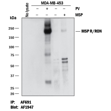
Detection of Human Phospho-MSP R/Ron (Y1238/Y1239) by Western blot.
Human Phospho-MSP R/Ron (Y1238/Y1239) was immunoprecipitated from 1 mg of lysates of MDA-MB-453 human breast cancer cell line untreated (-) or treated (+) with 100 uM Pervanadate (PV) for 10 minutes or 100 ng/mL Recombinant Human MSP/MST1 Protein (Catalog # 352-MS) for 10 minutes. MSP R/Ron (Y1238/Y1239) was immunoprecipiated with 1 ug Goat Anti-Human MSP R/Ron Antigen Affinity-purified Polyclonal Antibody(Catalog # AF691). The MSP R/Ron (Y1238/Y1239)-antibody complexes were absorbed using Protein G Sepharose. Immunoprecipitated MSP R/Ron (Y1238/Y1239) was detected by Western blot using 0.1 µg/mL of Rabbit Anti-Human Phospho-MSP R/Ron (Y1238/Y1239) Antigen Affinity-purified Polyclonal Antibody (Catalog # AF1947).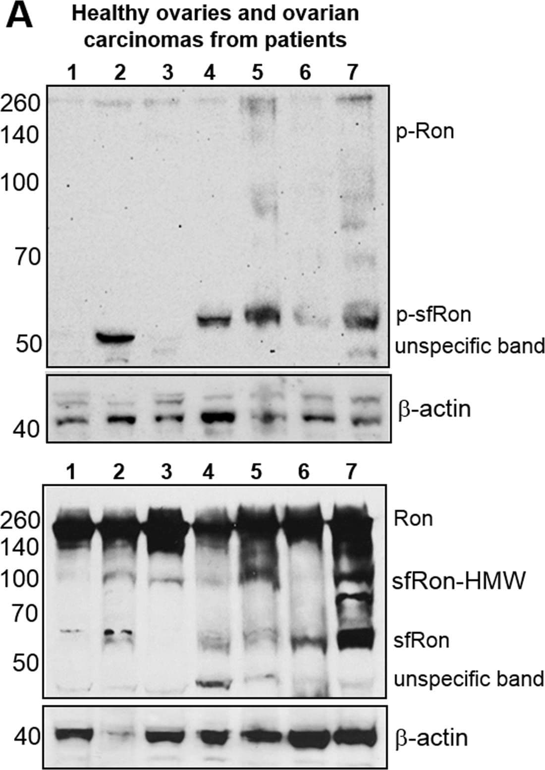
Detection of Human Human Phospho-MSPR/Ron (Y1238/Y1239) Antibody by Western Blot
The expression and activity of Ron receptor isoforms in primary tumors and high-grade serous ovarian (HG-SOC) PDXsA. Upper panel displays western blot analysis of tumor lysates from patients assayed for phosphorylated (active) sfRon and Ron. Lower panel shows western blot analysis of total levels of Ron isoforms. The higher molecular weight sfRon bands (sfRon-HMW), which are putative, posttranslationally modified sfRon forms were also noted. The blots were stripped and re-probed for beta -actin. Lanes represent: healthy ovary (1, 2, 3); ovarian adenocarcinoma (4); carcinosarcoma (5); endometrioid adenocarcinoma (6) and HG-SOC (7). B. Upper panel displays western blot analysis of tumor lysates from HG-SOC PDXs assayed for phosphorylated Ron isoforms. Lower panel shows western blot analysis of total levels of Ron isoforms. The blots were stripped and re-probed for beta -actin or GAPDH. C. The expression of Ron isoforms was assessed by WES capillary electrophoresis-based protein assay in OVCAR3-sfRon cell line engineered to express sfRon vs. parental OVACR3 cells and compared with sfRon positive or negative HG-SOC PDXs. GAPDH was used as loading control. Image collected and cropped by CiteAb from the following publication (https://pubmed.ncbi.nlm.nih.gov/27551332), licensed under a CC-BY license. Not internally tested by R&D Systems.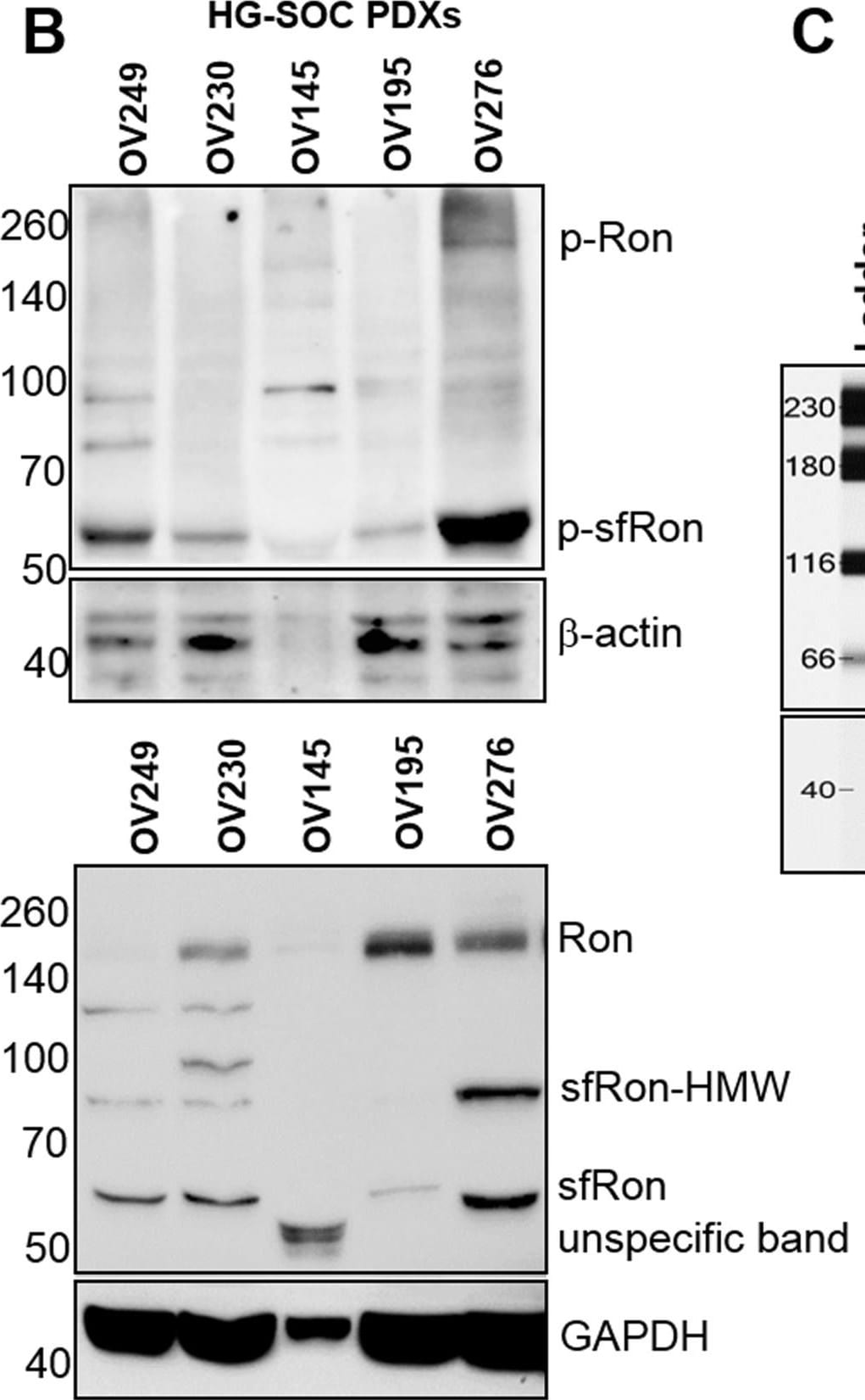
Detection of Human MSPR/Ron by Western Blot
The expression and activity of Ron receptor isoforms in primary tumors and high-grade serous ovarian (HG-SOC) PDXsA. Upper panel displays western blot analysis of tumor lysates from patients assayed for phosphorylated (active) sfRon and Ron. Lower panel shows western blot analysis of total levels of Ron isoforms. The higher molecular weight sfRon bands (sfRon-HMW), which are putative, posttranslationally modified sfRon forms were also noted. The blots were stripped and re-probed for beta -actin. Lanes represent: healthy ovary (1, 2, 3); ovarian adenocarcinoma (4); carcinosarcoma (5); endometrioid adenocarcinoma (6) and HG-SOC (7). B. Upper panel displays western blot analysis of tumor lysates from HG-SOC PDXs assayed for phosphorylated Ron isoforms. Lower panel shows western blot analysis of total levels of Ron isoforms. The blots were stripped and re-probed for beta -actin or GAPDH. C. The expression of Ron isoforms was assessed by WES capillary electrophoresis-based protein assay in OVCAR3-sfRon cell line engineered to express sfRon vs. parental OVACR3 cells and compared with sfRon positive or negative HG-SOC PDXs. GAPDH was used as loading control. Image collected and cropped by CiteAb from the following open publication (https://pubmed.ncbi.nlm.nih.gov/27551332), licensed under a CC-BY license. Not internally tested by R&D Systems.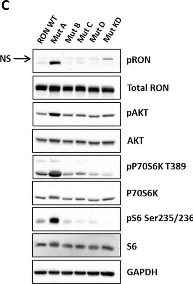
Detection of MSPR/Ron by Western Blot
Inducible expression of different RON mutants in T47D cells and analysis of downstream PI3K/mTORC1 signaling. a Schematic presentation of RON kinase structure is shown in the left panel. Mutations that were introduced to RON are shown in the right panel. Mutated residues are highlighted in pink. b Inducible expression of RON mutants in T47D cells at the protein (upper panel) and mRNA level (lower panel). T47D cells transduced with different RON mutants in a Tet-inducible lentiviral plasmid were treated with 500 ng/ml doxycycline for 48 h, and analyzed by Western blot. Equal level of RON expression is apparent among the mutants, which is comparable to RON wild type (WT). Graph shows qRT-PCR analysis of T47D cells expressing RON mutants in the presence and absence of doxycycline (500 ng/ml) for 48 h with primers specific to RON. GAPDH was used as the internal control. c Signaling analysis of the PI3K/mTORC1 pathway in T47D cells expressing RON WT and mutants. T47D-RON WT and mutants were treated with 100 ng/ml doxycycline for 48 h, followed by serum starvation for 24 h. Western blot shows robust activation of AKT, P70S6K, and rpS6, as readouts for activation of PI3K and mTORC1 kinase pathways, in Mutant A compared to the other mutants. GAPDH was used as the internal control. See also Supplementary Figure S8 for signaling analysis of mutants, and Supplementary Figure S9 for the detailed signaling analysis of RON-Mut A in the context of MSP-dependent and -independent RON activation. Line indicates the separation between lanes on the same Western blots. PSI plexin–semaphorin–integrin, IPT immunoglobulin–plexin–transcription, TM transmembrane. Image collected and cropped by CiteAb from the following open publication (https://pubmed.ncbi.nlm.nih.gov/30456298), licensed under a CC-BY license. Not internally tested by R&D Systems.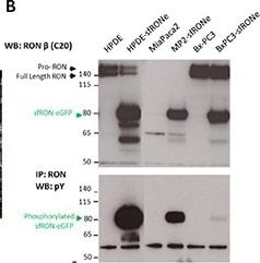
Detection of MSPR/Ron by Western Blot
Short form RON induces oncogenic phenotypes and has transforming capacityA. HPDE cell line expressing the sfRON-eGFP fusion protein showed no change in cell morphology but expressed high levels of the protein. B. The sfRONe protein is expressed in 3 different cell lines at similar levels. The protein is constitutively active in each cell line but with different levels of phosphorylation. C. MSP ligand activates full length RON but not short form RON. D. Signaling downstream of sfRON activates the MAPK and AKT pathway. E. Short form RON expression in the HPDE cell line increases cell motility more than full length RON activation with MSP alone. Image collected and cropped by CiteAb from the following open publication (https://pubmed.ncbi.nlm.nih.gov/27323855), licensed under a CC-BY license. Not internally tested by R&D Systems.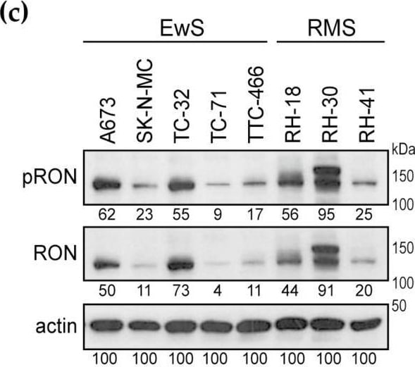
Detection of MSPR/Ron by Western Blot
RON is expressed in Ewing sarcomas and cell lines. (a) Relative RON transcript expression in Ewing sarcoma primary tumors from patients with localized (non-met) or metastatic (met) disease in comparison to MSC cultures, as determined by qPCR. (b) Respective RON expression in Ewing sarcoma cell lines (EwS) compared to MSC cultures. (c) RON protein is expressed and phosphorylated in Ewing sarcoma and rhabdomyosarcoma (RMS) cell lines. Cells were grown in standard tissue culture conditions. Following analysis of phospho-RON, blots were stripped and re-probed for total RON expression; 10% gel; numbers indicate densitometry readings relative to respective actin loading control. Image collected and cropped by CiteAb from the following open publication (https://pubmed.ncbi.nlm.nih.gov/32272784), licensed under a CC-BY license. Not internally tested by R&D Systems.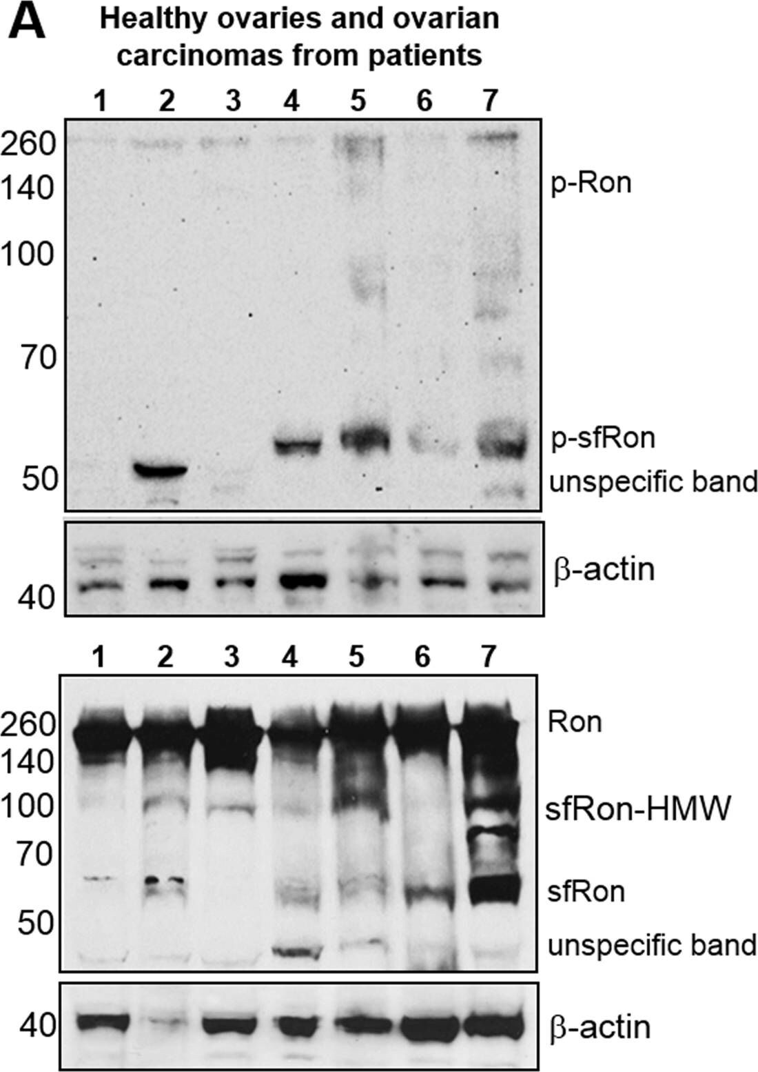
Detection of MSPR/Ron by Western Blot
The expression and activity of Ron receptor isoforms in primary tumors and high-grade serous ovarian (HG-SOC) PDXsA. Upper panel displays western blot analysis of tumor lysates from patients assayed for phosphorylated (active) sfRon and Ron. Lower panel shows western blot analysis of total levels of Ron isoforms. The higher molecular weight sfRon bands (sfRon-HMW), which are putative, posttranslationally modified sfRon forms were also noted. The blots were stripped and re-probed for beta -actin. Lanes represent: healthy ovary (1, 2, 3); ovarian adenocarcinoma (4); carcinosarcoma (5); endometrioid adenocarcinoma (6) and HG-SOC (7). B. Upper panel displays western blot analysis of tumor lysates from HG-SOC PDXs assayed for phosphorylated Ron isoforms. Lower panel shows western blot analysis of total levels of Ron isoforms. The blots were stripped and re-probed for beta -actin or GAPDH. C. The expression of Ron isoforms was assessed by WES capillary electrophoresis-based protein assay in OVCAR3-sfRon cell line engineered to express sfRon vs. parental OVACR3 cells and compared with sfRon positive or negative HG-SOC PDXs. GAPDH was used as loading control. Image collected and cropped by CiteAb from the following open publication (https://www.genesandcancer.com/lookup/doi/10.18632/genesandcancer.109), licensed under a CC-BY license. Not internally tested by R&D Systems.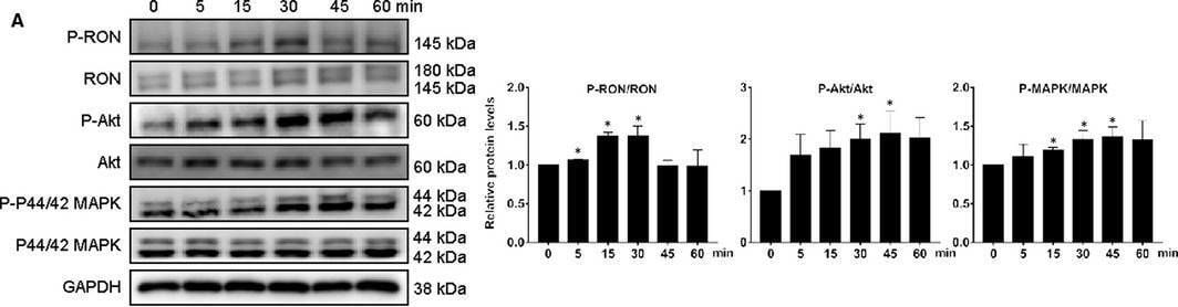
Detection of MSPR/Ron by Western Blot
RON promoted EMT through Akt and MAPK signalling pathways in human endometrial epithelial cells and inflammatory cytokines participate in this process. A, MSP treatment increased the phosphorylation of RON, P44/42 MAPK and Akt in control endometrial epithelial cells. Three times experiments were repeated and the values were expressed as mean ± SD (*P < .05, **P < .01, compared with control group). B, the effects of inhibitors on MSP‐induced EMT (BMS, BMS777607; PD, PD98059; TCN, triciribine), three times experiments were repeated and the values were expressed as mean ± SD (*P < .05, **P < .01, ***P < .001 compared with control group, #P < .05, ##P < .01, ###P < .001 compared with MSP group). Three times experiments were repeated and the values were expressed as mean ± SD (*P < .05, **P < .01, ***P < .001 compared with control group). Image collected and cropped by CiteAb from the following open publication (https://pubmed.ncbi.nlm.nih.gov/33410267), licensed under a CC-BY license. Not internally tested by R&D Systems.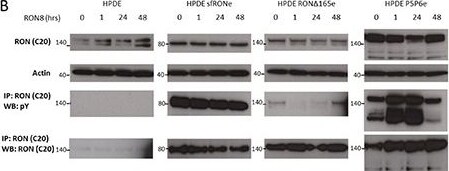
Detection of MSPR/Ron by Western Blot
A RON specific small molecule inhibitor blocks isoform constitutive activation while monoclonal antibody therapy does notA. RON8 is a monoclonal antibody directed at the ligand binding extracellular domain of RON which blocks MSP induced phosphorylation in the HPDE cells line. B. Isoform expressing cell lines were not inhibited by RON8. RON delta 165 phosphorylation was decreased up to 24 hours of treatment but then returned to baseline levels. C. LDC047896 treatment for 24 hours at 1 μM prior to stimulation with RON ligand MSP blocked full length RON phosphorylation. Constitutive activation of sfRON and P5P6 was removed at a dose of 1 μM. Similarly, constitutive activation of full length RON in the 34E cell line was removed at a 1 μM dose. D. Activation of flRON was blocked at a 1 μM concentration after stimulation with MSP, but it did not block MAPK phosphorylation. MAPK signaling in the isoform expressing and 34E cell lines was decreased at a dose of 1 μM but did not decrease AKT signaling. D, Viability of HPDE sfRON and P5P6 was decreased significantly (p 0.001 and 0f.007) but not in other lines. The western blots in this image were derived from different gels. Image collected and cropped by CiteAb from the following open publication (https://pubmed.ncbi.nlm.nih.gov/27323855), licensed under a CC-BY license. Not internally tested by R&D Systems.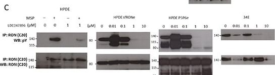
Detection of MSPR/Ron by Western Blot
A RON specific small molecule inhibitor blocks isoform constitutive activation while monoclonal antibody therapy does notA. RON8 is a monoclonal antibody directed at the ligand binding extracellular domain of RON which blocks MSP induced phosphorylation in the HPDE cells line. B. Isoform expressing cell lines were not inhibited by RON8. RON delta 165 phosphorylation was decreased up to 24 hours of treatment but then returned to baseline levels. C. LDC047896 treatment for 24 hours at 1 μM prior to stimulation with RON ligand MSP blocked full length RON phosphorylation. Constitutive activation of sfRON and P5P6 was removed at a dose of 1 μM. Similarly, constitutive activation of full length RON in the 34E cell line was removed at a 1 μM dose. D. Activation of flRON was blocked at a 1 μM concentration after stimulation with MSP, but it did not block MAPK phosphorylation. MAPK signaling in the isoform expressing and 34E cell lines was decreased at a dose of 1 μM but did not decrease AKT signaling. D, Viability of HPDE sfRON and P5P6 was decreased significantly (p 0.001 and 0f.007) but not in other lines. The western blots in this image were derived from different gels. Image collected and cropped by CiteAb from the following open publication (https://pubmed.ncbi.nlm.nih.gov/27323855), licensed under a CC-BY license. Not internally tested by R&D Systems.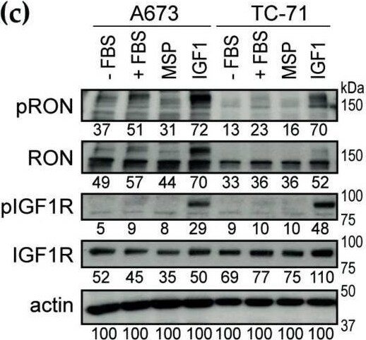
Detection of MSPR/Ron by Western Blot
IMC-RON8 antibody inhibits migration but not proliferation of Ewing sarcoma cell lines in vitro. (a) IMC-RON8 does not significantly reduce monolayer cell viability of EwS and RMS cell lines. Cells were grown in standard conditions and treated as indicated. After 72 h, relative cell viability was measured by MTT assay. (b) IMC-RON8 impairs migration of Ewing sarcoma cells. Cells were cultured in serum-free medium, seeded into trans-well chambers containing 100 ng/mL IMC-RON8 where indicated and allowed to migrate towards 10% serum. Graphs represent mean ± SD of triplicate experiments. (c) RON is cross-activated by IGF1–IGF1R signaling but not vice versa. Cells were starved in serum-free medium for 24 h before stimulation with fetal bovine serum (FBS) (10%), MSP (400 ng/mL) or IGF1 (100 ng/mL) for 30 min. Following analysis of phospho-RON, blots were stripped and re-probed for total RON expression; numbers indicate densitometry readings relative to respective actin loading control. (d) Dose-response of the EwS and RMS cell lines to the anti-IGF1R antibody IMC-A12. Assays were performed as in (a); significances refer to maximum dose compared to non-treated cells. (e) Combined treatment with IMC-RON8 plus IMC-A12 does not result in synergistic effects on cell viability. MTT assay was performed as in (a). Graphs (a), (d) and (e) represent the mean ± SD of at least three independent experiments. (f) Combined treatment with IMC-RON8 plus IMC-A12 does not reveal synergistic effects on migration. Assays were performed as in (b) with 100 ng/mL IMC-A12 and 1000 ng/mL IMC-RON8 as indicated. Significance is indicated as p < 0.05 (*), p < 0.01 (**) and p < 0.001 (***), while ns indicates a non-significant p-value. Image collected and cropped by CiteAb from the following open publication (https://pubmed.ncbi.nlm.nih.gov/32272784), licensed under a CC-BY license. Not internally tested by R&D Systems.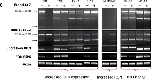
Detection of MSPR/Ron by Western Blot
DNA demethylating agents modulate RON isoform expressionA. Pancreatic cancer cell lines were treated with Decitabine (DAC, 4μM) for 96 hours and assessed for percent genomic DNA methylation. B. The treatment was effective at decreasing DNA methylation and depleting its target – DNA methyltransferase 1 (DNMT1) in all cell lines. C. PCR showed that in a subset of cell lines which normally express RON (34E, 79E, Panc1, HPDE) DAC treatment decreased flRON expression and isoform expression. In cell lines which do not express RON (MiaPaca2) DAC treatment induces its expression. D. The PCR data is confirmed using western blot with the same trends of up and downregulation seen in concordant cell lines. The western blots in this image derive from different gels. Image collected and cropped by CiteAb from the following open publication (https://pubmed.ncbi.nlm.nih.gov/27323855), licensed under a CC-BY license. Not internally tested by R&D Systems.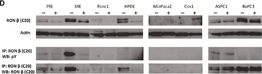
Detection of MSPR/Ron by Western Blot
DNA demethylating agents modulate RON isoform expressionA. Pancreatic cancer cell lines were treated with Decitabine (DAC, 4μM) for 96 hours and assessed for percent genomic DNA methylation. B. The treatment was effective at decreasing DNA methylation and depleting its target – DNA methyltransferase 1 (DNMT1) in all cell lines. C. PCR showed that in a subset of cell lines which normally express RON (34E, 79E, Panc1, HPDE) DAC treatment decreased flRON expression and isoform expression. In cell lines which do not express RON (MiaPaca2) DAC treatment induces its expression. D. The PCR data is confirmed using western blot with the same trends of up and downregulation seen in concordant cell lines. The western blots in this image derive from different gels. Image collected and cropped by CiteAb from the following open publication (https://pubmed.ncbi.nlm.nih.gov/27323855), licensed under a CC-BY license. Not internally tested by R&D Systems.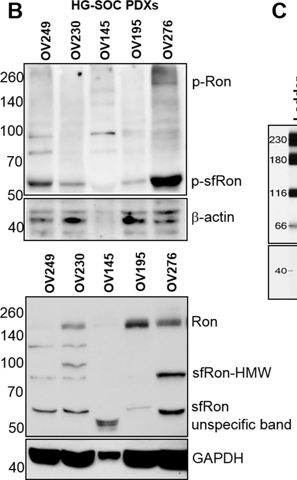
Detection of MSPR/Ron by Western Blot
The expression and activity of Ron receptor isoforms in primary tumors and high-grade serous ovarian (HG-SOC) PDXsA. Upper panel displays western blot analysis of tumor lysates from patients assayed for phosphorylated (active) sfRon and Ron. Lower panel shows western blot analysis of total levels of Ron isoforms. The higher molecular weight sfRon bands (sfRon-HMW), which are putative, posttranslationally modified sfRon forms were also noted. The blots were stripped and re-probed for beta -actin. Lanes represent: healthy ovary (1, 2, 3); ovarian adenocarcinoma (4); carcinosarcoma (5); endometrioid adenocarcinoma (6) and HG-SOC (7). B. Upper panel displays western blot analysis of tumor lysates from HG-SOC PDXs assayed for phosphorylated Ron isoforms. Lower panel shows western blot analysis of total levels of Ron isoforms. The blots were stripped and re-probed for beta -actin or GAPDH. C. The expression of Ron isoforms was assessed by WES capillary electrophoresis-based protein assay in OVCAR3-sfRon cell line engineered to express sfRon vs. parental OVACR3 cells and compared with sfRon positive or negative HG-SOC PDXs. GAPDH was used as loading control. Image collected and cropped by CiteAb from the following open publication (https://www.genesandcancer.com/lookup/doi/10.18632/genesandcancer.109), licensed under a CC-BY license. Not internally tested by R&D Systems.Applications for Human phospho-MSPR/Ron (Y1238/Y1239) Antibody
Application
Recommended Usage
Western Blot
1 µg/mL
Sample: MDA‑MB‑453 human breast cancer cell line treated with Pervanadate or Recombinant Human MSP/MST1 Protein (Catalog # 352-MS)
Sample: MDA‑MB‑453 human breast cancer cell line treated with Pervanadate or Recombinant Human MSP/MST1 Protein (Catalog # 352-MS)
Formulation, Preparation, and Storage
Purification
Antigen and protein A Affinity-purified
Reconstitution
Reconstitute at 0.2 mg/mL in sterile PBS. For liquid material, refer to CoA for concentration.
Loading...
Formulation
Lyophilized from a 0.2 μm filtered solution in PBS with Trehalose. *Small pack size (SP) is supplied either lyophilized or as a 0.2 µm filtered solution in PBS.
Shipping
Lyophilized product is shipped at ambient temperature. Liquid small pack size (-SP) is shipped with polar packs. Upon receipt, store immediately at the temperature recommended below.
Stability & Storage
Use a manual defrost freezer and avoid repeated freeze-thaw cycles.
- 12 months from date of receipt, -20 to -70 °C as supplied.
- 1 month, 2 to 8 °C under sterile conditions after reconstitution.
- 6 months, -20 to -70 °C under sterile conditions after reconstitution.
Calculators
Background: MSPR/Ron
Long Name
Macrophage Stimulating Protein Receptor
Alternate Names
CD136, MSP R, MST1R, Ron
Gene Symbol
MST1R
Additional MSPR/Ron Products
Product Documents for Human phospho-MSPR/Ron (Y1238/Y1239) Antibody
Certificate of Analysis
To download a Certificate of Analysis, please enter a lot or batch number in the search box below.
Note: Certificate of Analysis not available for kit components.
Product Specific Notices for Human phospho-MSPR/Ron (Y1238/Y1239) Antibody
For research use only
Related Research Areas
Citations for Human phospho-MSPR/Ron (Y1238/Y1239) Antibody
 Powered by Bioz
Customer Reviews for Human phospho-MSPR/Ron (Y1238/Y1239) Antibody
There are currently no reviews for this product. Be the first to review Human phospho-MSPR/Ron (Y1238/Y1239) Antibody and earn rewards!
Have you used Human phospho-MSPR/Ron (Y1238/Y1239) Antibody?
Submit a review and receive an Amazon gift card!
$25/€18/£15/$25CAN/¥2500 for a review with an image
$10/€7/£6/$10CAN/¥1110 for a review without an image
Submit a review
Protocols
Find general support by application which include: protocols, troubleshooting, illustrated assays, videos and webinars.
- Cellular Response to Hypoxia Protocols
- R&D Systems Quality Control Western Blot Protocol
- Troubleshooting Guide: Western Blot Figures
- Western Blot Conditions
- Western Blot Protocol
- Western Blot Protocol for Cell Lysates
- Western Blot Troubleshooting
- Western Blot Troubleshooting Guide
- View all Protocols, Troubleshooting, Illustrated assays and Webinars
Loading...