
PYK2 (Proline-rich kinase 2; also Ptk2b, Fak2, RAFTK and CAK beta ) is a 112‑116 kDa member of the Fak subfamily, tyrosine protein kinase family. It is expressed in multiple cell types, including endothelial cells, vascular smooth muscle cells, megakaryocytes and neurons. PYK2 is activated by elevated intracellular Ca++ and is associated with MAPK pathway activation. Human PYK2 is 1009 amino acids (aa) in length. PYK2 phosphorylation at Tyr402 is associated with enzymatic activation, intercellular localization, cell growth, cell motility, and regulating molecular associations. Over aa 390‑410, human PYK2 is 100% aa identical to mouse PYK2.
Human phospho-PYK2/FAK2 (Y402) Antibody (592918)
R&D Systems | Catalog # MAB6210

Scientific images may be resized, cropped, or adjusted for brightness or contrast for presentation purposes. These updates are limited to presentation and do not affect the underlying scientific interpretation of the data. Where available, additional source data may be provided upon request.
Key Product Details
Validated by
Biological Validation
Species Reactivity
Validated:
Human
Cited:
Human
Applications
Validated:
Western Blot, Simple Western
Cited:
Western Blot
Label
Unconjugated
Antibody Source
Monoclonal Mouse IgG1 Clone # 592918
Loading...
Product Specifications
Immunogen
Phosphopeptide containing the human PYK2/FAK2 Y402 site
Accession # Q14289
Accession # Q14289
Specificity
Detects human PYK2/FAK2 when phosphorylated at Y402.
Clonality
Monoclonal
Host
Mouse
Isotype
IgG1
Scientific Data Images for Human phospho-PYK2/FAK2 (Y402) Antibody (592918)
Detection of HumanPhospho-PYK2/FAK2 (Y402) by Western Blot.
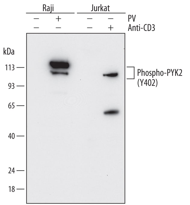
Western blot shows lysates of Raji human Burkitt's lymphoma cell line and Jurkat human acute T cell leukemia cell line untreated (-) or treated (+) with 1 mM Pervanadate (PV) for 30 minutes and 10 µg/mL Mouse Anti-Human CD3e Monoclonal Antibody (Catalog # MAB100) for 15 minutes. PVDF membrane was probed with 0.5 µg/mL of Mouse Anti-Human Phospho-PYK2/FAK2 (Y402) Monoclonal Antibody (Catalog # MAB6210) followed by HRP-conjugated Anti-Mouse IgG Secondary Antibody (Catalog # HAF018). Specific bands were detected for Phospho-PYK2/FAK2 (Y402) at approximately 105-115 kDa (as indicated). This experiment was conducted under reducing conditions and using Immunoblot Buffer Group 1.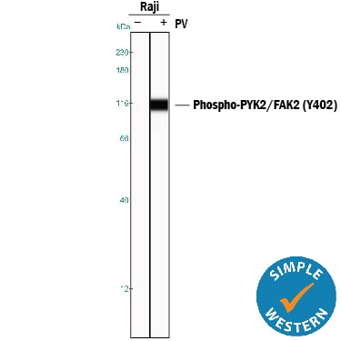
Detection of Human Phospho-PYK2/FAK2 (Y402) by Simple WesternTM.
Simple Western lane view shows lysates of Raji human Burkitt's lymphoma cell line untreated (-) or treated (+) with 0.2 mg/mL Pervanadate (PV) for 30 minutes, loaded at 0.2 mg/mL. A specific band was detected for Phospho-PYK2/FAK2 (Y402) at approximately 113 kDa (as indicated) using 5 µg/mL of Mouse Anti-Human Phospho-PYK2/FAK2 (Y402) Monoclonal Antibody (Catalog # MAB6210). This experiment was conducted under reducing conditions and using the 12-230 kDa separation system.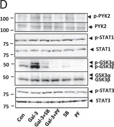
Detection of Phospho-PYK2/FAK2 (Y402) by Western Blot
Galectin-3 expression induces activation of PYK2, STAT1 and GSK3 alpha / beta signalling. Expression of 37 protein kinases in SW620 cells in response to 10 µg/ml galectin-3 or BSA for 0.5 h was assessed by Proteome Profiler Human Phospho-Kinase Array (A, Percentage changes of the kinases in cell response to galectin-3 in comparison to control are shown at the bottom panel). The presence of galectin-3 increases the phosphorylation of PYK2, GSK3 alpha / beta, and STAT1 and decreases phosphorylation of STAT3. SW620 cells treated with 10 µg/ml galectin-3 for different times were assessed by immunoblotting using antibodies against p-PYK2, p-STAT-1, p-GSK3 alpha / beta or p-STAT-3 (B). The blots were striped and reprobed with antibodies against PYK2, STAT-1, GSK3 alpha / beta or STAT-3. The band density was quantified and expressed as percentages of phospho-/non-phosphorylated proteins (C). In D and E, SW620 cells were treated with 10 µg/ml galectin-3 or BSA followed by introduction of GSK3 alpha / beta inhibitor SB 216763 (SB) or PKY2 inhibitor PF-431396 (PF) for 15 min and the levels of phosphorylated PYK2, STAT-1, GSK3 alpha / beta or STAT-3 were analysed by immunoblotting. The blots were striped and reprobed with antibodies against PYK2, STAT-1, GSK3 alpha / beta or STAT-3. The densities of the blots from three independent experiments were quantified and are expressed as the percentage of phosphorylated/non-phosphorylated levels of each protein. ***P < 0.001, **P < 0.01, *P < 0.05 (ANOVA). Image collected and cropped by CiteAb from the following open publication (https://pubmed.ncbi.nlm.nih.gov/37055381), licensed under a CC-BY license. Not internally tested by R&D Systems.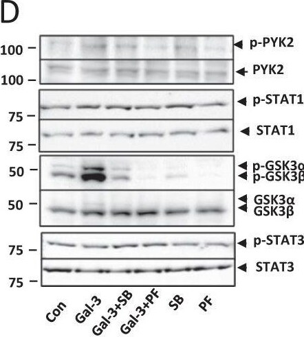
Detection of Phospho-PYK2/FAK2 (Y402) by Western Blot
Galectin-3 expression induces activation of PYK2, STAT1 and GSK3 alpha / beta signalling. Expression of 37 protein kinases in SW620 cells in response to 10 µg/ml galectin-3 or BSA for 0.5 h was assessed by Proteome Profiler Human Phospho-Kinase Array (A, Percentage changes of the kinases in cell response to galectin-3 in comparison to control are shown at the bottom panel). The presence of galectin-3 increases the phosphorylation of PYK2, GSK3 alpha / beta, and STAT1 and decreases phosphorylation of STAT3. SW620 cells treated with 10 µg/ml galectin-3 for different times were assessed by immunoblotting using antibodies against p-PYK2, p-STAT-1, p-GSK3 alpha / beta or p-STAT-3 (B). The blots were striped and reprobed with antibodies against PYK2, STAT-1, GSK3 alpha / beta or STAT-3. The band density was quantified and expressed as percentages of phospho-/non-phosphorylated proteins (C). In D and E, SW620 cells were treated with 10 µg/ml galectin-3 or BSA followed by introduction of GSK3 alpha / beta inhibitor SB 216763 (SB) or PKY2 inhibitor PF-431396 (PF) for 15 min and the levels of phosphorylated PYK2, STAT-1, GSK3 alpha / beta or STAT-3 were analysed by immunoblotting. The blots were striped and reprobed with antibodies against PYK2, STAT-1, GSK3 alpha / beta or STAT-3. The densities of the blots from three independent experiments were quantified and are expressed as the percentage of phosphorylated/non-phosphorylated levels of each protein. ***P < 0.001, **P < 0.01, *P < 0.05 (ANOVA). Image collected and cropped by CiteAb from the following open publication (https://pubmed.ncbi.nlm.nih.gov/37055381), licensed under a CC-BY license. Not internally tested by R&D Systems.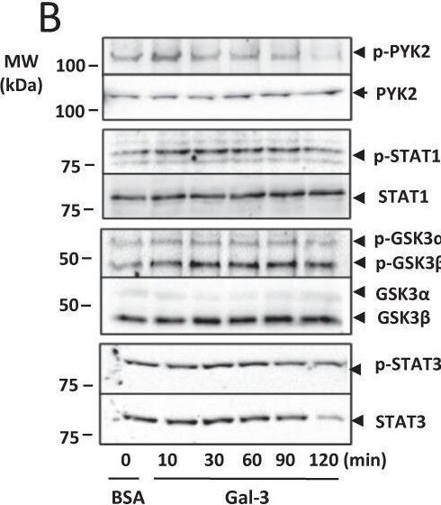
Detection of Phospho-PYK2/FAK2 (Y402) by Western Blot
Galectin-3 expression induces activation of PYK2, STAT1 and GSK3 alpha / beta signalling. Expression of 37 protein kinases in SW620 cells in response to 10 µg/ml galectin-3 or BSA for 0.5 h was assessed by Proteome Profiler Human Phospho-Kinase Array (A, Percentage changes of the kinases in cell response to galectin-3 in comparison to control are shown at the bottom panel). The presence of galectin-3 increases the phosphorylation of PYK2, GSK3 alpha / beta, and STAT1 and decreases phosphorylation of STAT3. SW620 cells treated with 10 µg/ml galectin-3 for different times were assessed by immunoblotting using antibodies against p-PYK2, p-STAT-1, p-GSK3 alpha / beta or p-STAT-3 (B). The blots were striped and reprobed with antibodies against PYK2, STAT-1, GSK3 alpha / beta or STAT-3. The band density was quantified and expressed as percentages of phospho-/non-phosphorylated proteins (C). In D and E, SW620 cells were treated with 10 µg/ml galectin-3 or BSA followed by introduction of GSK3 alpha / beta inhibitor SB 216763 (SB) or PKY2 inhibitor PF-431396 (PF) for 15 min and the levels of phosphorylated PYK2, STAT-1, GSK3 alpha / beta or STAT-3 were analysed by immunoblotting. The blots were striped and reprobed with antibodies against PYK2, STAT-1, GSK3 alpha / beta or STAT-3. The densities of the blots from three independent experiments were quantified and are expressed as the percentage of phosphorylated/non-phosphorylated levels of each protein. ***P < 0.001, **P < 0.01, *P < 0.05 (ANOVA). Image collected and cropped by CiteAb from the following open publication (https://pubmed.ncbi.nlm.nih.gov/37055381), licensed under a CC-BY license. Not internally tested by R&D Systems.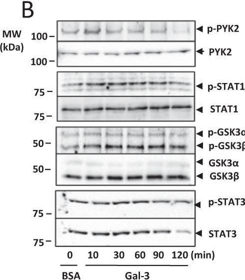
Detection of Phospho-PYK2/FAK2 (Y402) by Western Blot
Galectin-3 expression induces activation of PYK2, STAT1 and GSK3 alpha / beta signalling. Expression of 37 protein kinases in SW620 cells in response to 10 µg/ml galectin-3 or BSA for 0.5 h was assessed by Proteome Profiler Human Phospho-Kinase Array (A, Percentage changes of the kinases in cell response to galectin-3 in comparison to control are shown at the bottom panel). The presence of galectin-3 increases the phosphorylation of PYK2, GSK3 alpha / beta, and STAT1 and decreases phosphorylation of STAT3. SW620 cells treated with 10 µg/ml galectin-3 for different times were assessed by immunoblotting using antibodies against p-PYK2, p-STAT-1, p-GSK3 alpha / beta or p-STAT-3 (B). The blots were striped and reprobed with antibodies against PYK2, STAT-1, GSK3 alpha / beta or STAT-3. The band density was quantified and expressed as percentages of phospho-/non-phosphorylated proteins (C). In D and E, SW620 cells were treated with 10 µg/ml galectin-3 or BSA followed by introduction of GSK3 alpha / beta inhibitor SB 216763 (SB) or PKY2 inhibitor PF-431396 (PF) for 15 min and the levels of phosphorylated PYK2, STAT-1, GSK3 alpha / beta or STAT-3 were analysed by immunoblotting. The blots were striped and reprobed with antibodies against PYK2, STAT-1, GSK3 alpha / beta or STAT-3. The densities of the blots from three independent experiments were quantified and are expressed as the percentage of phosphorylated/non-phosphorylated levels of each protein. ***P < 0.001, **P < 0.01, *P < 0.05 (ANOVA). Image collected and cropped by CiteAb from the following open publication (https://pubmed.ncbi.nlm.nih.gov/37055381), licensed under a CC-BY license. Not internally tested by R&D Systems.Applications for Human phospho-PYK2/FAK2 (Y402) Antibody (592918)
Application
Recommended Usage
Simple Western
5 µg/mL
Sample: Raji human Burkitt's lymphoma cell line treated with Pervanadate (PV)
Sample: Raji human Burkitt's lymphoma cell line treated with Pervanadate (PV)
Western Blot
0.5 µg/mL
Sample: Raji human Burkitt's lymphoma cell line and Jurkat human acute T cell leukemia cell line treated with Pervanadate (PV) or Mouse Anti-Human CD3 epsilon Monoclonal Antibody (Catalog # MAB100)
Sample: Raji human Burkitt's lymphoma cell line and Jurkat human acute T cell leukemia cell line treated with Pervanadate (PV) or Mouse Anti-Human CD3 epsilon Monoclonal Antibody (Catalog # MAB100)
Reviewed Applications
Read 5 reviews rated 4.2 using MAB6210 in the following applications:
Formulation, Preparation, and Storage
Purification
Protein A or G purified from hybridoma culture supernatant
Reconstitution
Reconstitute at 0.5 mg/mL in sterile PBS. For liquid material, refer to CoA for concentration.
Loading...
Formulation
Lyophilized from a 0.2 μm filtered solution in PBS with Trehalose. *Small pack size (SP) is supplied either lyophilized or as a 0.2 µm filtered solution in PBS.
Shipping
Lyophilized product is shipped at ambient temperature. Liquid small pack size (-SP) is shipped with polar packs. Upon receipt, store immediately at the temperature recommended below.
Stability & Storage
Use a manual defrost freezer and avoid repeated freeze-thaw cycles.
- 12 months from date of receipt, -20 to -70 °C as supplied.
- 1 month, 2 to 8 °C under sterile conditions after reconstitution.
- 6 months, -20 to -70 °C under sterile conditions after reconstitution.
Calculators
Background: PYK2/FAK2
Long Name
Proline-rich Tyrosine Kinase 2
Alternate Names
CADTK, CAKB, FADK2, FAK2, FRNK, PTK2B, RAFTK
Gene Symbol
PTK2B
UniProt
Additional PYK2/FAK2 Products
Product Documents for Human phospho-PYK2/FAK2 (Y402) Antibody (592918)
Certificate of Analysis
To download a Certificate of Analysis, please enter a lot or batch number in the search box below.
Note: Certificate of Analysis not available for kit components.
Product Specific Notices for Human phospho-PYK2/FAK2 (Y402) Antibody (592918)
For research use only
Related Research Areas
Citations for Human phospho-PYK2/FAK2 (Y402) Antibody (592918)
 Powered by Bioz
Customer Reviews for Human phospho-PYK2/FAK2 (Y402) Antibody (592918) (5)
4.2 out of 5
5 Customer Ratings
Have you used Human phospho-PYK2/FAK2 (Y402) Antibody (592918)?
Submit a review and receive an Amazon gift card!
$25/€18/£15/$25CAN/¥2500 for a review with an image
$10/€7/£6/$10CAN/¥1110 for a review without an image
Submit a review
Customer Images
Showing
1
-
5 of
5 reviews
Showing All
Filter By:
-
Application: Western BlotSample Tested: MDA-MB-231 human breast cancer cell lineSpecies: HumanVerified Customer | Posted 04/18/2022It works well
-
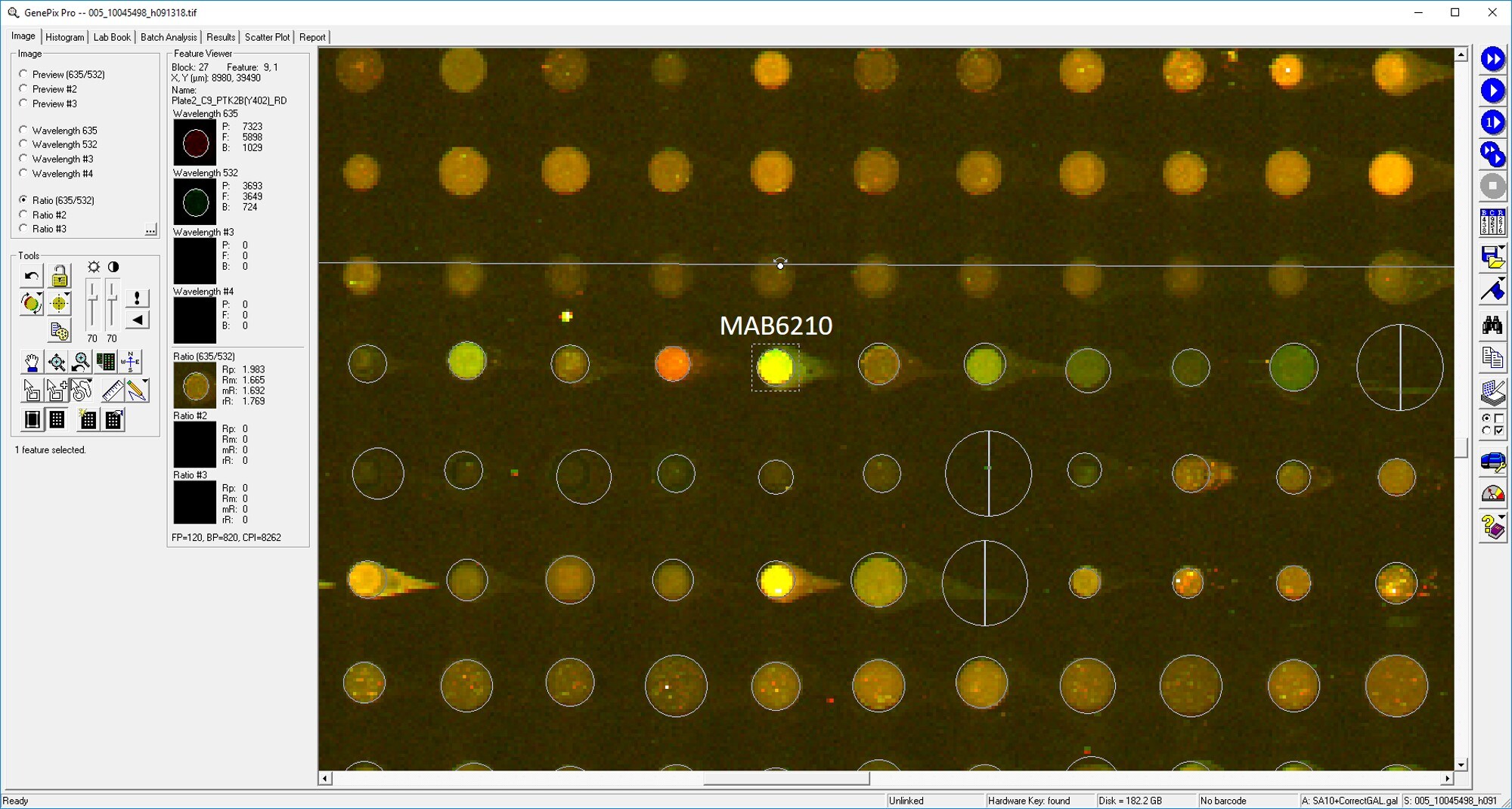
Application: MicroarraysSample Tested: EDTA PlasmaSpecies: HumanVerified Customer | Posted 01/03/2020

-
Application: ELISASample Tested: PlasmaSpecies: HumanVerified Customer | Posted 11/10/2018
-
Application: MicroarraySample Tested: EDTA PlasmaSpecies: HumanVerified Customer | Posted 11/09/2018
-
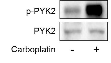
Application: Western BlotSample Tested: Breast cancer cellsSpecies: HumanVerified Customer | Posted 09/21/2018Human breast cancer cell line MDA-MB-231 was treated with carboplatin for 72 hours and the expression of phospho-PYK2 and total PYK2 were detected by western blot.
There are no reviews that match your criteria.
Protocols
Find general support by application which include: protocols, troubleshooting, illustrated assays, videos and webinars.
- Cellular Response to Hypoxia Protocols
- R&D Systems Quality Control Western Blot Protocol
- Troubleshooting Guide: Western Blot Figures
- Western Blot Conditions
- Western Blot Protocol
- Western Blot Protocol for Cell Lysates
- Western Blot Troubleshooting
- Western Blot Troubleshooting Guide
- View all Protocols, Troubleshooting, Illustrated assays and Webinars


