
PIKfyve (PhosphoInositide Kinase for five position containing a fyve finger (FAB1/YOTB/Vac1/EEA1) domain; also phosphatidylinositol 3-phosphate 5-kinase type III and Type III PIP kinase) is a 230-260 kDa cytoplasmic and endocytic member of an ancient family of phosphatidylinositol-3,5 biphosphate (PI[3,5]P2 synthesizing enzymes. It is associated with endosomes, particularly the microdomains of early endosomes that are EEA1-deficient. PIKfyve binds to membrane-embedded phosphatidylinositol and initially converts PI3P into PI(3,5)P2, which is then converted into PI5P. In conjunction with ArPIKfyve and Sac3, its actions are involved in the normal transport, fusion, and membrane export of endosomes. Notably, PIKfyve also appears to act as a protein kinase, apparently controlling its own activity through autophosphorylation. Human PIKfyve is 2098 amino acids (aa) in length. It contains one FYVE PI3P-binding zinc-finger domain (aa 154-219), a DEP domain (aa 365-440) and a C-terminal PIP kinase rehion (aa 1172-2085). There are no less than 29 utilized Ser/Thr phosphorylation sites, plus two utilized Tyr phosphorylation sites. Three potential isoform variants are reported, all which possess a three aa substitution for aa 546-2098. Two of these three also demonstrate a deletion of aa 108-204, and a nine aa substitution for aa 109-203, respectively. Over aa 215-361, human and mouse share 95% aa sequence identity.
Human PIKFyve Antibody
R&D Systems | Catalog # AF7885

Scientific images may be resized, cropped, or adjusted for brightness or contrast for presentation purposes. These updates are limited to presentation and do not affect the underlying scientific interpretation of the data. Where available, additional source data may be provided upon request.
Key Product Details
Species Reactivity
Validated:
Cited:
Applications
Validated:
Cited:
Label
Antibody Source
Product Specifications
Immunogen
Lys215-Lys361
Accession # Q9Y2I7
Specificity
Clonality
Host
Isotype
Scientific Data Images for Human PIKFyve Antibody
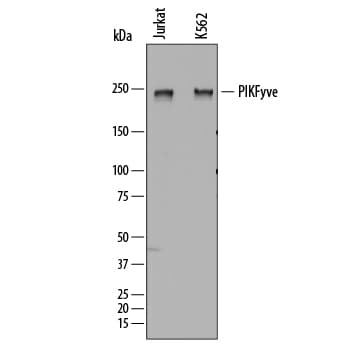
Detection of Human PIKFyve by Western Blot.
Western blot shows lysates of Jurkat human acute T cell leukemia cell line and K562 human chronic myelogenous leukemia cell line. PVDF membrane was probed with 0.25 µg/mL of Sheep Anti-Human PIKFyve Antigen Affinity-purified Polyclonal Antibody (Catalog # AF7885) followed by HRP-conjugated Anti-Sheep IgG Secondary Antibody (Catalog # HAF016). A specific band was detected for PIKFyve at approximately 240 kDa (as indicated). This experiment was conducted under reducing conditions and using Immunoblot Buffer Group 1.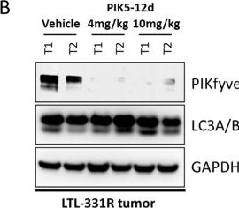
Detection of PIKFyve by Western Blot
PIK5-12d depleted PIKfyveprotein in vivo. (A) Studydesign of pharmacodynamic assessment on target engagement of PIKfyvePROTAC degrader in LTL-331R human prostate cancer patient-derivedxenograft model; (B) immunoblotting of PIKfyve, LC3A/B, and GAPDHin the whole cell lysate of human LTL-331R tumors treated with vehicleor various concentrations of PIK5-12d for 4 days; (C)representative images of TUNEL signal of LTL-331R tumors treated withvehicle or 10 mg/kg PIK5-12d for 4 days; and (D) tumorproliferation of LTL-331R tumors treated with vehicle or 15 mg/kg PIK5-12d at 5 days on and 2 days off regimen for 17 days,the p value was determined by two-tailed unpaired t test between vehicle and PIK5-12d groups. Image collected and cropped by CiteAb from the following open publication (https://pubmed.ncbi.nlm.nih.gov/37605297), licensed under a CC-BY license. Not internally tested by R&D Systems.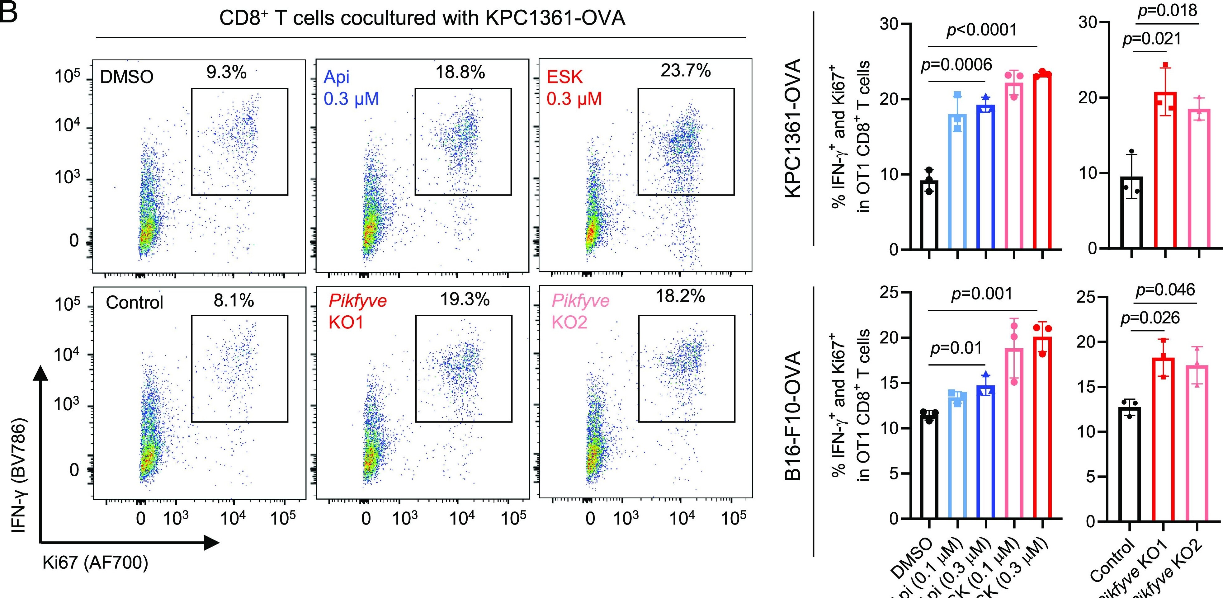
Detection of PIKFyve by Flow Cytometry
Genetic or pharmacologic inhibition of PIKfyve upregulates antigen presentation and enhances cancer cell killing by CD8+ T cells. (A) Left: Viability of cancer cells with or without OVA overexpression treated with the indicated agent for 24 h, followed by coculture with activated CD8+ T cells isolated from OT1 mice for 48 h. Right: Viability of control or Pikfyve-null cancer cells with or without OVA overexpression, cocultured with activated CD8+ T cells isolated from OT1 mice for 48 h. (B) Representative images (Left) and quantification (Right) of flow cytometry measuring the proportion of IFN-gamma + and Ki67+ cells in the activated CD8+ T cells cocultured with the indicated cancer cells in (A). (C) Representative images (Left) and quantification (Right) of flow cytometry measuring surface expression of an OVA-peptide (SIINFEKL) bound MHC-I (H-2Kb) in the control or Pikfyve-null cancer cells, treated with or without IFN-gamma at 10 ng/mL for 24 h. Data in A, B, and C were acquired with three independent biological replicates presented as mean ± SD. Statistics were acquired by two-tailed Student’s t test (A and B) or two-way ANOVA (C), with Bonferroni correction. MFI: mean fluorescence intensity. Control: non-targeting single-guide RNA. Pikfyve KO1 and Pikfyve KO2: independent single-guide RNAs depleting Pikfyve. Image collected and cropped by CiteAb from the following open publication (https://pubmed.ncbi.nlm.nih.gov/38011559), licensed under a CC-BY license. Not internally tested by R&D Systems.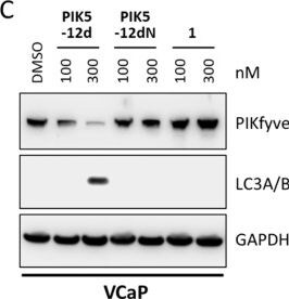
Detection of PIKFyve by Western Blot
PIK5-12d decreased prostatecancer cell proliferationand exerted prolonged suppression of PIKfyve downstream signaling.(A) Long-term cell viability is visualized by crystal violet stainingin VCaP either treated with PIK5-12d or 1 for 24 h and chased for 2 weeks, or continuous treatment for 2 weeks(top), IC50s were calculated for indicated conditions (bottom);(B) long-term cell viability is determined in VCaP cells with washoutin drug-free medium after 4 h treatment of PIK5-12d ornegative control PIK5-12dN; and (C) immunoblotting ofPIKfyve, LC3A/B, and GAPDH in whole cell lysate of VCaP cells treatedwith PIK5-12d, negative control PIK5-12dN, or inhibitor 1 for 4 h and chased in drug-free mediumfor 72 h. (D) Quantification of vacuole per cell in DU145-RFP cellsfor 4 h of treatment of PIK5-12d, negative control PIK5-12dN, or inhibitor 1. Image collected and cropped by CiteAb from the following open publication (https://pubmed.ncbi.nlm.nih.gov/37605297), licensed under a CC-BY license. Not internally tested by R&D Systems.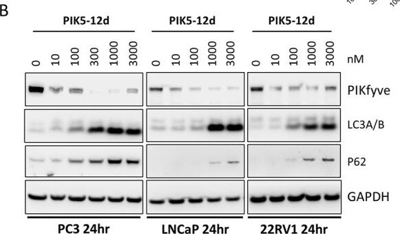
Detection of PIKFyve by Western Blot
PIK5-12d induced massive cytoplasmic vacuolizationand blocked autophagy. (A) Representative images of DU145-RFP cellswith treatments of DMSO, 300 nM PIK5-12d, or 1 for 24 h. The vacuole area per cell is quantified in indicated conditions;(B) immunoblotting of PIKfyve and autophagy markers in indicated humanprostate cancer cells with increasing concentrations of PIK5-12d for 24 h. Image collected and cropped by CiteAb from the following open publication (https://pubmed.ncbi.nlm.nih.gov/37605297), licensed under a CC-BY license. Not internally tested by R&D Systems.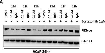
Detection of PIKFyve by Western Blot
PIK5-12d mediated-PIKfyve degradationwas VHL andproteasome-dependent. (A) Immunoblotting of PIKfyve and GAPDH in VCaPcells treated with increasing concentrations of 12d (PIK5-12d), 12f, or 12h in the conditionsof with or without 1 μM of proteasome inhibitor bortezomib for24 h; (B) immunoblotting of PIKfyve and GAPDH in VCaP cells treatedwith increasing concentration of PIK5-12d with or withoutwarhead 1 or VHL ligand VL285 for 24 h. Image collected and cropped by CiteAb from the following open publication (https://pubmed.ncbi.nlm.nih.gov/37605297), licensed under a CC-BY license. Not internally tested by R&D Systems.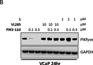
Detection of PIKFyve by Western Blot
PIK5-12d mediated-PIKfyve degradationwas VHL andproteasome-dependent. (A) Immunoblotting of PIKfyve and GAPDH in VCaPcells treated with increasing concentrations of 12d (PIK5-12d), 12f, or 12h in the conditionsof with or without 1 μM of proteasome inhibitor bortezomib for24 h; (B) immunoblotting of PIKfyve and GAPDH in VCaP cells treatedwith increasing concentration of PIK5-12d with or withoutwarhead 1 or VHL ligand VL285 for 24 h. Image collected and cropped by CiteAb from the following open publication (https://pubmed.ncbi.nlm.nih.gov/37605297), licensed under a CC-BY license. Not internally tested by R&D Systems.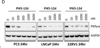
Detection of PIKFyve by Western Blot
PIK5-12d in a concentration-dependentfashion reducedPIKfyve protein in multiple human prostate cancer cells in vitro.(A) Immunoblotting of PIKfyve and GAPDH in VCaP cells treated withincreasing concentrations of PIK5-12d for 24 h (left),percent remaining PIKfyve protein was plotted for DC50 and Dmax determination (right); (B) immunoblottingof PIKfyve and GAPDH in VCaP with 100 nM PIK5-12d forvarious timepoints (left), percent remaining PIKfyve protein was quantified(right); (C) global proteomic analysis of PIK5-12d inVCaP cells after 4 h treatment of DMSO or 300 nM PIK5-12d (left), mass-spec quantification of PIKfyve protein (middle), lipidkinase changes (right); and (D) immunoblotting of PIKfyve and GAPDHin multiple human prostate cancer cell lines with increasing concentrationsof PIK5-12d for 24 h. Image collected and cropped by CiteAb from the following open publication (https://pubmed.ncbi.nlm.nih.gov/37605297), licensed under a CC-BY license. Not internally tested by R&D Systems.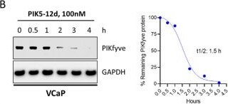
Detection of PIKFyve by Western Blot
PIK5-12d in a concentration-dependentfashion reducedPIKfyve protein in multiple human prostate cancer cells in vitro.(A) Immunoblotting of PIKfyve and GAPDH in VCaP cells treated withincreasing concentrations of PIK5-12d for 24 h (left),percent remaining PIKfyve protein was plotted for DC50 and Dmax determination (right); (B) immunoblottingof PIKfyve and GAPDH in VCaP with 100 nM PIK5-12d forvarious timepoints (left), percent remaining PIKfyve protein was quantified(right); (C) global proteomic analysis of PIK5-12d inVCaP cells after 4 h treatment of DMSO or 300 nM PIK5-12d (left), mass-spec quantification of PIKfyve protein (middle), lipidkinase changes (right); and (D) immunoblotting of PIKfyve and GAPDHin multiple human prostate cancer cell lines with increasing concentrationsof PIK5-12d for 24 h. Image collected and cropped by CiteAb from the following open publication (https://pubmed.ncbi.nlm.nih.gov/37605297), licensed under a CC-BY license. Not internally tested by R&D Systems.Applications for Human PIKFyve Antibody
Western Blot
Sample: Jurkat human acute T cell leukemia cell line and K562 human chronic myelogenous leukemia cell line
Formulation, Preparation, and Storage
Purification
Reconstitution
Sterile PBS to a final concentration of 0.2 mg/mL. For liquid material, refer to CoA for concentration.
Formulation
Shipping
Stability & Storage
- 12 months from date of receipt, -20 to -70 °C as supplied.
- 1 month, 2 to 8 °C under sterile conditions after reconstitution.
- 6 months, -20 to -70 °C under sterile conditions after reconstitution.
Calculators
Background: PIKFyve
Long Name
Alternate Names
Gene Symbol
UniProt
Additional PIKFyve Products
Product Documents for Human PIKFyve Antibody
Certificate of Analysis
To download a Certificate of Analysis, please enter a lot or batch number in the search box below.
Note: Certificate of Analysis not available for kit components.
Product Specific Notices for Human PIKFyve Antibody
For research use only
Citations for Human PIKFyve Antibody
 Powered by Bioz
Customer Reviews for Human PIKFyve Antibody
There are currently no reviews for this product. Be the first to review Human PIKFyve Antibody and earn rewards!
Have you used Human PIKFyve Antibody?
Submit a review and receive an Amazon gift card!
$25/€18/£15/$25CAN/¥2500 for a review with an image
$10/€7/£6/$10CAN/¥1110 for a review without an image
Submit a review
Protocols
Find general support by application which include: protocols, troubleshooting, illustrated assays, videos and webinars.
- Cellular Response to Hypoxia Protocols
- R&D Systems Quality Control Western Blot Protocol
- Troubleshooting Guide: Western Blot Figures
- Western Blot Conditions
- Western Blot Protocol
- Western Blot Protocol for Cell Lysates
- Western Blot Troubleshooting
- Western Blot Troubleshooting Guide
- View all Protocols, Troubleshooting, Illustrated assays and Webinars