
Photoreceptor-specific Nuclear Receptor (PNR, RNR; NR2E3) is a member of the orphan nuclear receptor superfamily. PNR is expressed in the retina and plays a role in retinal photoreceptor cell differentiation and degeneration.
Human PNR/NR2E3 Antibody (H7223)
R&D Systems | Catalog # PP-H7223-00

Scientific images may be resized, cropped, or adjusted for brightness or contrast for presentation purposes. These updates are limited to presentation and do not affect the underlying scientific interpretation of the data. Where available, additional source data may be provided upon request.
Loading...
Key Product Details
Species Reactivity
Validated:
Human
Cited:
Human, Mouse, Transgenic Mouse
Applications
Validated:
Immunohistochemistry, Western Blot, Direct ELISA
Cited:
Immunohistochemistry, Immunocytochemistry, Immunoprecipitation
Label
Unconjugated
Antibody Source
Monoclonal Mouse IgG2A Clone # H7223
Loading...
Product Specifications
Immunogen
Recombinant human PNR/NR2E3
Clonality
Monoclonal
Host
Mouse
Isotype
IgG2A
Scientific Data Images for Human PNR/NR2E3 Antibody (H7223)
Detection of Mouse PNR/NR2E3 by Immunocytochemistry/Immunofluorescence
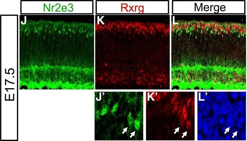
Coexpression of Rxrg and Nr2e3 in the embryonic mouse retina. (A-H) E14.5 mouse retinas electroporated with either the cNr2e3Enh2 or cNr2e3Enh3 plasmid, cultured ex vivo for 2 days, and processed for immunofluorescence confocal imaging of EGFP (green, chicken antibody), Nr2e3 (red, mouse antibody), Rxrg (white, rabbit antibody), and DAPI. Panels in A-H represent maximum projections of z-stacks with the depicted channel shown at the top of the column. The merge column has EGFP, Nr2e3, and Rxrg signals. (A’-H’) Magnified single z-plane images with the signals for (A’,E’) EGFP, (B’,F’) Nr2e3, (C’,G’) Rxrg, and (D’,H’) DAPI. White arrows point to GFP+ cells that also express Nr2e3 and Rxrg. Yellow arrows point to GFP+ cells that express Rxrg, but not Nr2e3. (I) A graph of the average percentage of GFP+ cells when driven by the cNr2e3Enh2 or cNr2e3Enh3 elements that express Nr2e3, Rxrg, or both Nr2e3 and Rxrg. (J-L) Maximum projection of a z-stack image of a E17.5 mouse retina processed for immunofluorescent detection of Nr2e3 (J, green) and Rxrg (K, red) or both (L, Merge). (J’-L’) Magnified single z-plane images of the same area visualized for signals for Nr2e3 (J’), Rxrg (K’), or DAPI (L’). (M) A graph of the average percentage of Nr2e3, Rxrg double-positive (D.P.) cells out of the total Nr2e3+ population (left bar) or the total Rxrg+ population (right bar). In both graphs N ≥ 3 biological replicates. Error bars represent standard error of the mean. All images are oriented with the scleral side of the retina at the top of the image. Scale bar in A represents 20 μm and applies to A-L. Image collected and cropped by CiteAb from the following publication (https://pubmed.ncbi.nlm.nih.gov/30466480), licensed under a CC-BY license. Not internally tested by R&D Systems.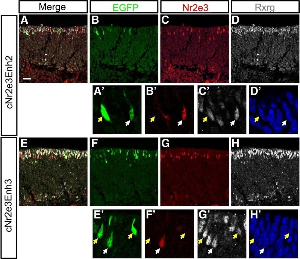
Detection of Mouse PNR/NR2E3 by Immunocytochemistry/Immunofluorescence
Coexpression of Rxrg and Nr2e3 in the embryonic mouse retina. (A-H) E14.5 mouse retinas electroporated with either the cNr2e3Enh2 or cNr2e3Enh3 plasmid, cultured ex vivo for 2 days, and processed for immunofluorescence confocal imaging of EGFP (green, chicken antibody), Nr2e3 (red, mouse antibody), Rxrg (white, rabbit antibody), and DAPI. Panels in A-H represent maximum projections of z-stacks with the depicted channel shown at the top of the column. The merge column has EGFP, Nr2e3, and Rxrg signals. (A’-H’) Magnified single z-plane images with the signals for (A’,E’) EGFP, (B’,F’) Nr2e3, (C’,G’) Rxrg, and (D’,H’) DAPI. White arrows point to GFP+ cells that also express Nr2e3 and Rxrg. Yellow arrows point to GFP+ cells that express Rxrg, but not Nr2e3. (I) A graph of the average percentage of GFP+ cells when driven by the cNr2e3Enh2 or cNr2e3Enh3 elements that express Nr2e3, Rxrg, or both Nr2e3 and Rxrg. (J-L) Maximum projection of a z-stack image of a E17.5 mouse retina processed for immunofluorescent detection of Nr2e3 (J, green) and Rxrg (K, red) or both (L, Merge). (J’-L’) Magnified single z-plane images of the same area visualized for signals for Nr2e3 (J’), Rxrg (K’), or DAPI (L’). (M) A graph of the average percentage of Nr2e3, Rxrg double-positive (D.P.) cells out of the total Nr2e3+ population (left bar) or the total Rxrg+ population (right bar). In both graphs N ≥ 3 biological replicates. Error bars represent standard error of the mean. All images are oriented with the scleral side of the retina at the top of the image. Scale bar in A represents 20 μm and applies to A-L. Image collected and cropped by CiteAb from the following publication (https://pubmed.ncbi.nlm.nih.gov/30466480), licensed under a CC-BY license. Not internally tested by R&D Systems.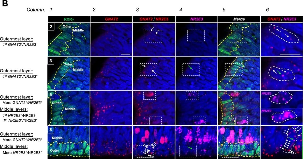
Detection of PNR/NR2E3 by Immunohistochemistry
NR2E3 and GNAT2 co-expression in photoreceptor precursors.(A) Composite images of combined RXR gamma and NR2E3 immunofluorescence staining and GNAT2 and NR2E3 RNA FISH in boxed regions 2–13 of Figure 7A. Scale bar = 50 µm. (B) Enlarged images of boxed regions in Figures 2, 3, 5 and 8 in panel A, as indicated at upper left of images in column 1. Dotted yellow lines demarcate the outermost and middle (sub-outermost) NBL cells analyzed separately in Figure 7C and D. White arrows = GNAT2 puncta in boxed regions of interest. Green arrows = NR2E3 puncta. Boxed regions in column 3 (GNAT2 RNA plus NR2E3 protein) and column 4 (NR2E3 RNA) are enlarged in column 6 (GNAT2 RNA plus NR2E3 RNA) and illustrate (region 2) initial expression of GNAT2 RNA without NR2E3 RNA in outermost NBL nascent cones; (region 3) initial co-expression of GNAT2 and NR2E3 RNA in early outermost NBL cones; (region 5) initial expression of NR2E3 RNA with or without co-expressed NR2E3 protein; and (region 8) co-expression of high GNAT2 and low NR2E3 RNA without NR2E3 protein in RXR gamma hi outermost NBL cones. Scale bars in columns 2 and 6 = 10 µm. Image collected and cropped by CiteAb from the following open publication (https://pubmed.ncbi.nlm.nih.gov/40767624), licensed under a CC-BY license. Not internally tested by R&D Systems.Applications for Human PNR/NR2E3 Antibody (H7223)
Application
Recommended Usage
Direct ELISA
Optimal dilutions of this antibody should be experimentally determined.
Immunohistochemistry
5-15 µg/mL
Sample: Perfusion fixed paraffin-embedded sections of rat retina (outer nuclear layer)
Sample: Perfusion fixed paraffin-embedded sections of rat retina (outer nuclear layer)
Western Blot
Optimal dilutions of this antibody should be experimentally determined.
Formulation, Preparation, and Storage
Formulation
A liquid formulation in physiologic saline with 0.1% NaN<sub>3</sub>.
Shipping
The product is shipped with dry ice or equivalent. Upon receipt, store it immediately at the temperature recommended below.
Stability & Storage
Store the unopened product at -70 °C. Use a manual defrost freezer and avoid repeated freeze-thaw cycles. Do not use past expiration date.
Background: PNR/NR2E3
Long Name
Photoreceptor-specific Nuclear Receptor
Alternate Names
NR2E3, RNR
Entrez Gene IDs
10002 (Human)
Gene Symbol
NR2E3
Additional PNR/NR2E3 Products
Product Documents for Human PNR/NR2E3 Antibody (H7223)
Certificate of Analysis
To download a Certificate of Analysis, please enter a lot or batch number in the search box below.
Note: Certificate of Analysis not available for kit components.
Product Specific Notices for Human PNR/NR2E3 Antibody (H7223)
For research use only
Related Research Areas
Citations for Human PNR/NR2E3 Antibody (H7223)
 Powered by Bioz
Customer Reviews for Human PNR/NR2E3 Antibody (H7223)
There are currently no reviews for this product. Be the first to review Human PNR/NR2E3 Antibody (H7223) and earn rewards!
Have you used Human PNR/NR2E3 Antibody (H7223)?
Submit a review and receive an Amazon gift card!
$25/€18/£15/$25CAN/¥2500 for a review with an image
$10/€7/£6/$10CAN/¥1110 for a review without an image
Submit a review
Protocols
Find general support by application which include: protocols, troubleshooting, illustrated assays, videos and webinars.
- Antigen Retrieval Protocol (PIER)
- Antigen Retrieval for Frozen Sections Protocol
- Appropriate Fixation of IHC/ICC Samples
- Cellular Response to Hypoxia Protocols
- Chromogenic IHC Staining of Formalin-Fixed Paraffin-Embedded (FFPE) Tissue Protocol
- Chromogenic Immunohistochemistry Staining of Frozen Tissue
- ClariTSA™ Fluorophore Kits
- Detection & Visualization of Antibody Binding
- ELISA Sample Preparation & Collection Guide
- ELISA Troubleshooting Guide
- Fluorescent IHC Staining of Frozen Tissue Protocol
- Graphic Protocol for Heat-induced Epitope Retrieval
- Graphic Protocol for the Preparation and Fluorescent IHC Staining of Frozen Tissue Sections
- Graphic Protocol for the Preparation and Fluorescent IHC Staining of Paraffin-embedded Tissue Sections
- Graphic Protocol for the Preparation of Gelatin-coated Slides for Histological Tissue Sections
- How to Run an R&D Systems DuoSet ELISA
- How to Run an R&D Systems Quantikine ELISA
- How to Run an R&D Systems Quantikine™ QuicKit™ ELISA
- IHC Sample Preparation (Frozen sections vs Paraffin)
- Immunofluorescent IHC Staining of Formalin-Fixed Paraffin-Embedded (FFPE) Tissue Protocol
- Immunohistochemistry (IHC) and Immunocytochemistry (ICC) Protocols
- Immunohistochemistry Frozen Troubleshooting
- Immunohistochemistry Paraffin Troubleshooting
- Preparing Samples for IHC/ICC Experiments
- Preventing Non-Specific Staining (Non-Specific Binding)
- Primary Antibody Selection & Optimization
- Protocol for Heat-Induced Epitope Retrieval (HIER)
- Protocol for Making a 4% Formaldehyde Solution in PBS
- Protocol for VisUCyte™ HRP Polymer Detection Reagent
- Protocol for the Preparation & Fixation of Cells on Coverslips
- Protocol for the Preparation and Chromogenic IHC Staining of Frozen Tissue Sections
- Protocol for the Preparation and Chromogenic IHC Staining of Frozen Tissue Sections - Graphic
- Protocol for the Preparation and Chromogenic IHC Staining of Paraffin-embedded Tissue Sections
- Protocol for the Preparation and Chromogenic IHC Staining of Paraffin-embedded Tissue Sections - Graphic
- Protocol for the Preparation and Fluorescent IHC Staining of Frozen Tissue Sections
- Protocol for the Preparation and Fluorescent IHC Staining of Paraffin-embedded Tissue Sections
- Protocol for the Preparation of Gelatin-coated Slides for Histological Tissue Sections
- Quantikine HS ELISA Kit Assay Principle, Alkaline Phosphatase
- Quantikine HS ELISA Kit Principle, Streptavidin-HRP Polymer
- R&D Systems Quality Control Western Blot Protocol
- Sandwich ELISA (Colorimetric) – Biotin/Streptavidin Detection Protocol
- Sandwich ELISA (Colorimetric) – Direct Detection Protocol
- TUNEL and Active Caspase-3 Detection by IHC/ICC Protocol
- The Importance of IHC/ICC Controls
- Troubleshooting Guide: ELISA
- Troubleshooting Guide: Immunohistochemistry
- Troubleshooting Guide: Western Blot Figures
- Western Blot Conditions
- Western Blot Protocol
- Western Blot Protocol for Cell Lysates
- Western Blot Troubleshooting
- Western Blot Troubleshooting Guide
- View all Protocols, Troubleshooting, Illustrated assays and Webinars
Loading...