
Protein Tyrosine Phosphatase, Non-receptor Type 13, also called PTPN13, PTPL1, FAP-1, hPTP1E and PTP-BAS, is a 260 kDa intracellular protein that removes phosphates from tyrosine residues. Genetic aberrations in PTPN13 have been reported in a wide variety of cancers, including, bone, colon, hepatocellular, and germ cell line. Elevated levels of PTPN13 in cancer cell lines have also been correlated with resistance to Fas-induced apoptosis, possibly due to prevention of CD95 reaching the cell surface.
Human PTPN13/PTPL1 Antibody
R&D Systems | Catalog # AF3577


Scientific images may be resized, cropped, or adjusted for brightness or contrast for presentation purposes. These updates are limited to presentation and do not affect the underlying scientific interpretation of the data. Where available, additional source data may be provided upon request.
Key Product Details
Species Reactivity
Human
Applications
Western Blot
Label
Unconjugated
Antibody Source
Polyclonal Goat IgG
Loading...
Product Specifications
Immunogen
E. coli-derived recombinant human PTPN13/PTPL1
Met1-Arg500
Accession # Q12923
Met1-Arg500
Accession # Q12923
Specificity
Detects endogenous human PTPN13/PTPL1 in Western blots.
Clonality
Polyclonal
Host
Goat
Isotype
IgG
Scientific Data Images for Human PTPN13/PTPL1 Antibody
Detection of Human PTPN13/PTPL1 by Western Blot.
Western blot shows lysates of HeLa human cervical epithelial carcinoma cell line. PVDF membrane was probed with 1 µg/mL of Human PTPN13/PTPL1 Antigen Affinity-purified Polyclonal Antibody (Catalog # AF3577) followed by HRP-conjugated Anti-Goat IgG Secondary Antibody (Catalog # HAF109). A specific band was detected for PTPN13/PTPL1 at approximately 260 kDa (as indicated). This experiment was conducted under reducing conditions and using Immunoblot Buffer Group 1.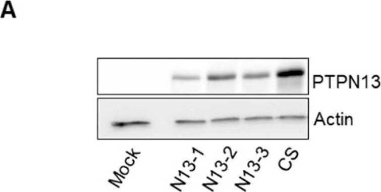
Detection of Human PTPN13/PTPL1 by Western Blot
PTPN13 regulates MDA-MB-231 cell motility and invasiveness. A: Expression of wt (N13-1, N13-2, N13-3) or catalytically inactive (CS) PTPN13 in the indicated cell clones was monitored by western blotting using anti-PTPN13 antibodies. Mock: control cells (vector alone); equal loading was verified by re-probing with an anti-actin antibody. B: Cell growth measured using the MTS assay. Results, expressed as % of Mock cells, are the mean ± s.d. of three (N13-1) four (N13-2, N13-3) or five (Mock, CS) independent experiments. C: Directional migration was assessed with the wound healing assay. C. Upper panel: Phase-contrast optical photomicrographs of the wounded area at 0 and 9h. C. Lower panel: Quantification of cell migration, expressed as % of Mock cells; mean ± s.d. of 3 (N13-1) or ≥5 (Mock, N13-2, N13-3, CS) independent experiments. **P<0.01, ****P<0.0001 versus Mock. D: Individual migration of the indicated cell clones was monitored by video microscopy and cell tracking on duplicate wells in three independent experiments. Graph represents the speed of about 1500 cells for each clone, Box-plot whiskers represent the 5 and 95 percentile values; ****P<0.0001 versus Mock and CS. E: Classification of cells (percentage) according to their migration speed (as in panel E): slow (<20µm/h), medium (20 to 40µm/h) and fast (>40µm/h). F: Cell invasiveness was evaluated with the Boyden chamber test. The percentage of cells that migrated through Matrigel-coated filters was quantified relative to the total number of seeded cells. Results, expressed as % of Mock cells, are the mean ± s.d. of five (N13-1), three (N13-2, N13-3) or two (CS) independent experiments *P<0.05 versus Mock. (C, D and F) two-tailed Student's t-test. Image collected and cropped by CiteAb from the following publication (https://pubmed.ncbi.nlm.nih.gov/31938048), licensed under a CC-BY license. Not internally tested by R&D Systems.Applications for Human PTPN13/PTPL1 Antibody
Application
Recommended Usage
Western Blot
1 µg/mL
Sample: HeLa human cervical epithelial carcinoma cell line
Sample: HeLa human cervical epithelial carcinoma cell line
Formulation, Preparation, and Storage
Purification
Antigen Affinity-purified
Reconstitution
Reconstitute at 0.2 mg/mL in sterile PBS. For liquid material, refer to CoA for concentration.
Loading...
Formulation
Lyophilized from a 0.2 μm filtered solution in PBS with Trehalose. *Small pack size (SP) is supplied either lyophilized or as a 0.2 µm filtered solution in PBS.
Shipping
Lyophilized product is shipped at ambient temperature. Liquid small pack size (-SP) is shipped with polar packs. Upon receipt, store immediately at the temperature recommended below.
Stability & Storage
Use a manual defrost freezer and avoid repeated freeze-thaw cycles.
- 12 months from date of receipt, -20 to -70 °C as supplied.
- 1 month, 2 to 8 °C under sterile conditions after reconstitution.
- 6 months, -20 to -70 °C under sterile conditions after reconstitution.
Calculators
Background: PTPN13/PTPL1
Long Name
Protein Tyrosine Phosphatase, Non-receptor Type 13
Alternate Names
EAP-1, FAP-1, PNP1, PTP-BAS, PTP-BL, PTP1E, PTPL1, PTPLE
Gene Symbol
PTPN13
UniProt
Additional PTPN13/PTPL1 Products
Product Documents for Human PTPN13/PTPL1 Antibody
Certificate of Analysis
To download a Certificate of Analysis, please enter a lot or batch number in the search box below.
Note: Certificate of Analysis not available for kit components.
Product Specific Notices for Human PTPN13/PTPL1 Antibody
For research use only
Related Research Areas
Citations for Human PTPN13/PTPL1 Antibody
 Powered by Bioz
Customer Reviews for Human PTPN13/PTPL1 Antibody
There are currently no reviews for this product. Be the first to review Human PTPN13/PTPL1 Antibody and earn rewards!
Have you used Human PTPN13/PTPL1 Antibody?
Submit a review and receive an Amazon gift card!
$25/€18/£15/$25CAN/¥2500 for a review with an image
$10/€7/£6/$10CAN/¥1110 for a review without an image
Submit a review
Protocols
Find general support by application which include: protocols, troubleshooting, illustrated assays, videos and webinars.
- Cellular Response to Hypoxia Protocols
- R&D Systems Quality Control Western Blot Protocol
- Troubleshooting Guide: Western Blot Figures
- Western Blot Conditions
- Western Blot Protocol
- Western Blot Protocol for Cell Lysates
- Western Blot Troubleshooting
- Western Blot Troubleshooting Guide
- View all Protocols, Troubleshooting, Illustrated assays and Webinars
Loading...