
The Ras proteins (H-Ras, N-Ras, and two splice variants of K-Ras) define a closely related family of 21 kDa GTPases that cycle between GTP-bound active and GDP-bound inactive states at the cytoplasmic face of the plasma membrane. Receptor tyrosine kinases and G-protein coupled receptors activate Ras, and effectors include Raf and PI 3-kinase. Activating mutations of Ras, which impair GTPase activity and stabilize the active state, are found in nearly one-third of all human cancers.
Human Ras Antibody (342404)
R&D Systems | Catalog # MAB3429


Scientific images may be resized, cropped, or adjusted for brightness or contrast for presentation purposes. These updates are limited to presentation and do not affect the underlying scientific interpretation of the data. Where available, additional source data may be provided upon request.
Key Product Details
Species Reactivity
Validated:
Human
Cited:
Human
Applications
Validated:
Western Blot
Cited:
Western Blot
Label
Unconjugated
Antibody Source
Monoclonal Rat IgG2B Clone # 342404
Loading...
Product Specifications
Immunogen
E. coli-derived recombinant human K-Ras2B
Met1-Met188 (Gln61His)
Accession # P01116-2
Met1-Met188 (Gln61His)
Accession # P01116-2
Specificity
Detects human Ras (H-Ras, N-Ras, and K-Ras) in Western blots. Does not detect more distantly related Ras superfamily members, including R-Ras, Ral, Rap, and Rheb.
Clonality
Monoclonal
Host
Rat
Isotype
IgG2B
Scientific Data Images for Human Ras Antibody (342404)
Detection of Human Ras by Western Blot.
Western blot shows lysates of K562 human chronic myelo-genous leukemia cell line and Ramos human Burkitt's lymphoma cell line. PVDF membrane was probed with 5 µg/mL Rat Anti-Human Ras Monoclonal Antibody (Catalog # MAB3429) followed by HRP-conjugated Anti-Rat IgG Secondary Antibody (Catalog # HAF005). For additional refer-ence, recombinant human H-Ras, N Ras, K-Ras2A and 2B, R-Ras2 and 3, Rap2A and 2B, RalA and B, and Rheb (5 ng/lane) were included. A specific band for Ras was detected at approximately 21 kDa (as indicated). This experiment was conducted under reducing conditions and using Immunoblot Buffer Group 3.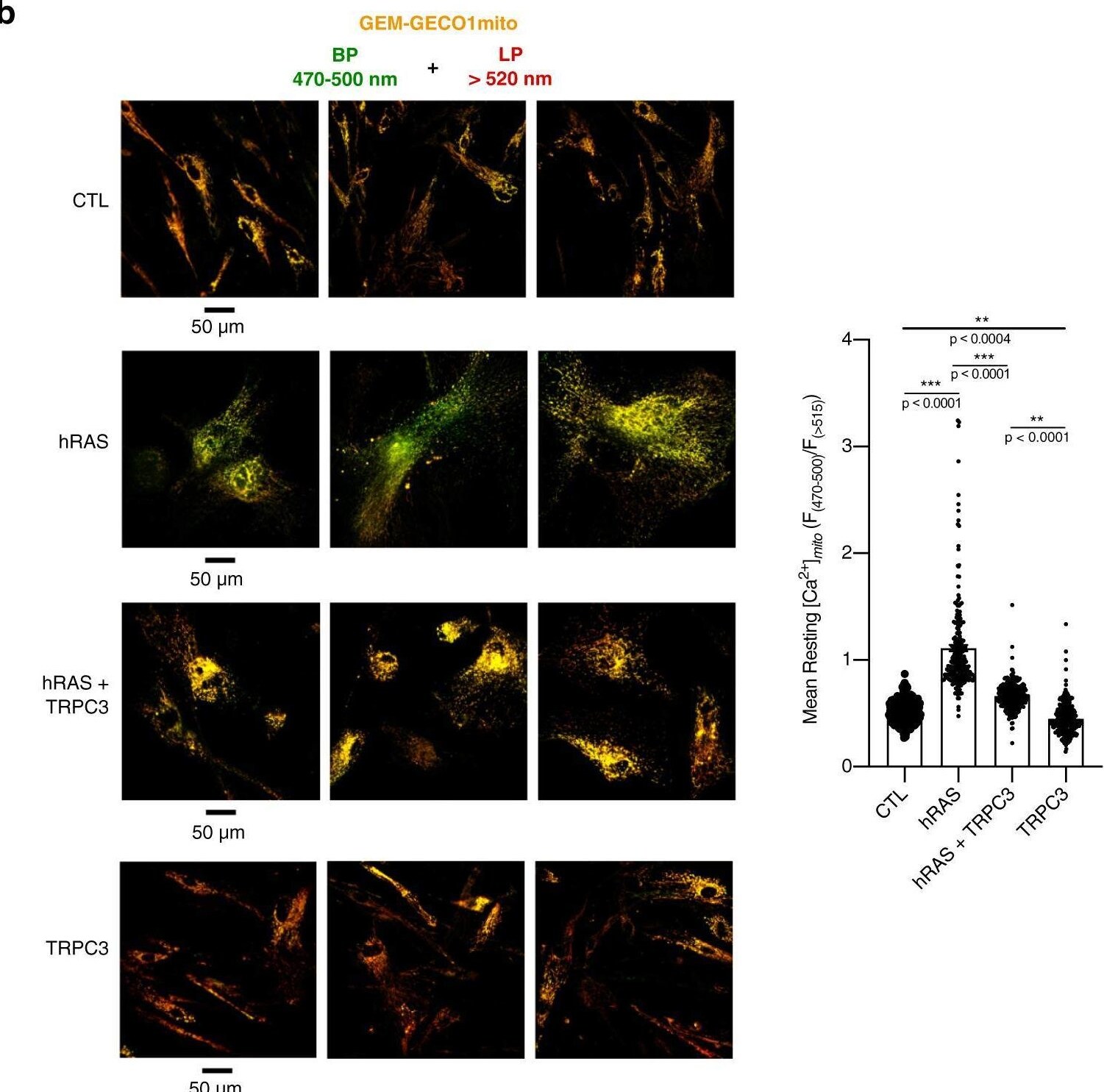
Detection of Ras by Immunocytochemistry/ Immunofluorescence
The level of TRPC3 in fibroblasts affects basal mitochondrial Ca2+ load.a The gallery (left) shows (from top to bottom) the transmitted light image of a single fibroblast, confocal image of GEM-GECO1mito fluorescence captured at wavelengths 470–500 nm, at wavelengths >520 nm and their overlay. The gallery (middle) compares confocal images (overlay of 2 channels: BP 470–500 nm plus LP > 520 nm) of GEM-GECO1mito fluorescence from the fibroblasts transduced with shCTL and shTRPC3, as indicated. The bar diagram plot (right) compares mean resting [Ca2+]mito (estimated as F470–500/F>520) in the fibroblasts transduced with shCTL (n = 96) and shTRPC3 (n = 96). ***P < 0.001 relative to shCTL (Student’s t test, two-sided). b Elevation of mitochondrial Ca2+ load upon hRAS overexpression is markedly prevented by simultaneous overexpression of TRPC3. The gallery (left) compares confocal images (overlay of 2 channels: BP 470–500 nm plus LP > 520 nm) of GEM-GECO1mito fluorescence from control MRC5, MRC5 with hRAS-induced senescence (hRAS) and MRC5 overexpressing hRAS and TRPC3 simultaneously (hRAS + TRPC3) or TRPC3 only (TRPC3). Images of three representative fields of view per condition are shown. The bar diagram plot (right) compares mean resting [Ca2+]mito (estimated as F470–500/F>520) in control MRC5 (Control: n = 390) with that in MRC5 with hRAS-induced senescence (+hRAS: n = 232) and MRC5 overexpressing hRAS and TRPC3 simultaneously (+hRAS + TRPC3: n = 266) or TRPC3 only (+TRPC3: n = 251). ***P < 0.001 relative to control, unless stated otherwise. One-way ANOVA, Tukey’s multiple comparisons test. Image collected and cropped by CiteAb from the following open publication (https://www.nature.com/articles/s41467-022-28597-x), licensed under a CC-BY license. Not internally tested by R&D Systems.Applications for Human Ras Antibody (342404)
Application
Recommended Usage
Western Blot
5 µg/mL
Sample: K562 human chronic myelogenous leukemia cell line and Ramos human Burkitt's lymphoma cell line
Sample: K562 human chronic myelogenous leukemia cell line and Ramos human Burkitt's lymphoma cell line
Formulation, Preparation, and Storage
Purification
Protein A or G purified from hybridoma culture supernatant
Reconstitution
Reconstitute at 0.5 mg/mL in sterile PBS. For liquid material, refer to CoA for concentration.
Loading...
Formulation
Lyophilized from a 0.2 μm filtered solution in PBS with Trehalose. *Small pack size (SP) is supplied either lyophilized or as a 0.2 µm filtered solution in PBS.
Shipping
Lyophilized product is shipped at ambient temperature. Liquid small pack size (-SP) is shipped with polar packs. Upon receipt, store immediately at the temperature recommended below.
Stability & Storage
Use a manual defrost freezer and avoid repeated freeze-thaw cycles.
- 12 months from date of receipt, -20 to -70 °C as supplied.
- 1 month, 2 to 8 °C under sterile conditions after reconstitution.
- 6 months, -20 to -70 °C under sterile conditions after reconstitution.
Calculators
Background: Ras
Long Name
v-Ki-ras2 Kirsten Rat Sarcoma Viral Oncogene Jomolog
Alternate Names
c-K-ras, K-RAS2A, K-RAS2B, K-RAS4A, K-RAS4B, Ki-Ras, KRAS, KRAS1, KRAS2, Oncogene Ras, RASK2
Gene Symbol
KRAS
UniProt
Additional Ras Products
Product Documents for Human Ras Antibody (342404)
Certificate of Analysis
To download a Certificate of Analysis, please enter a lot or batch number in the search box below.
Note: Certificate of Analysis not available for kit components.
Product Specific Notices for Human Ras Antibody (342404)
For research use only
Related Research Areas
Citations for Human Ras Antibody (342404)
 Powered by Bioz
Customer Reviews for Human Ras Antibody (342404)
There are currently no reviews for this product. Be the first to review Human Ras Antibody (342404) and earn rewards!
Have you used Human Ras Antibody (342404)?
Submit a review and receive an Amazon gift card!
$25/€18/£15/$25CAN/¥2500 for a review with an image
$10/€7/£6/$10CAN/¥1110 for a review without an image
Submit a review
Protocols
Find general support by application which include: protocols, troubleshooting, illustrated assays, videos and webinars.
- Cellular Response to Hypoxia Protocols
- R&D Systems Quality Control Western Blot Protocol
- Troubleshooting Guide: Western Blot Figures
- Western Blot Conditions
- Western Blot Protocol
- Western Blot Protocol for Cell Lysates
- Western Blot Troubleshooting
- Western Blot Troubleshooting Guide
- View all Protocols, Troubleshooting, Illustrated assays and Webinars
Loading...
Associated Pathways


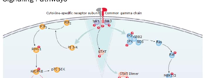
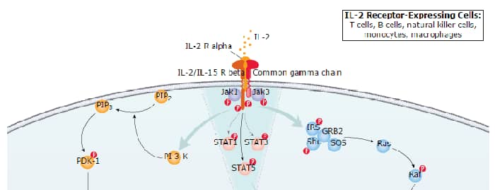
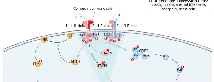
IL-7 Signaling Pathways
 IL-9 Signaling Pathways
 IL-15 Signaling Pathways
 IL-21 Signaling Pathways
 MAPK Signaling: Oxidative Stress Pathway
 MAPK Signaling Pathway: Mitogen Stimulation Pathway
 mTOR Signaling Pathway
 Pathogen or Damage-activated C-Type Lectin Receptor Signaling Pathways
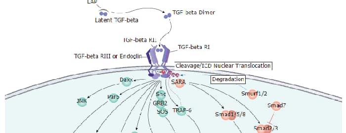
 TGF-beta Signaling Pathways
 VEGF - VEGF R2 Signaling Pathways
