
Human SCP3/SYCP3 Antibody
R&D Systems | Catalog # AF3750

Scientific images may be resized, cropped, or adjusted for brightness or contrast for presentation purposes. These updates are limited to presentation and do not affect the underlying scientific interpretation of the data. Where available, additional source data may be provided upon request.
Key Product Details
Species Reactivity
Validated:
Cited:
Applications
Validated:
Cited:
Label
Antibody Source
Product Specifications
Immunogen
Met1-Phe236
Accession # Q8IZU3
Specificity
Clonality
Host
Isotype
Scientific Data Images for Human SCP3/SYCP3 Antibody
Detection of Mouse SCP3/SYCP3 by Immunocytochemistry/ Immunofluorescence
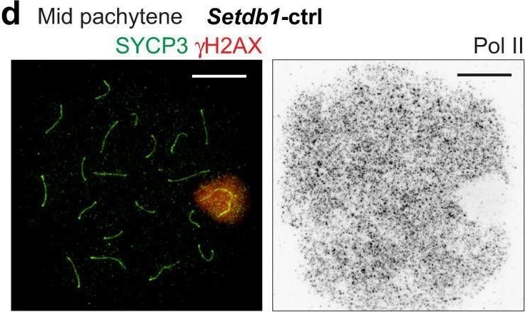
SETDB1-dependent H3K9me3 is dispensable for the initiation of MSCI.a Schematic of the strategy for generating Setdb1-cKO mice. b Evaluation of the efficiency of SETDB1 deletion shown as the mean and individual values for 3 independent samples. c Chromosome spreads of early and mid pachytene spermatocytes, distinguished by the absence (early) or the presence (mid) of H1T, respectively (not shown), immunostained with antibodies raised against SYCP3 and H3K9me3. XY chromosomes are indicated with dashed squares and are magnified in the panels to the right. Putative regions of XY chromatin are indicated by dashed circles. d Chromosome spreads of mid pachytene spermatocytes immunostained with antibodies raised against SYCP3, gamma H2AX, and Pol II. e A population of pachytene spermatocytes with a normal gamma H2AX domain and normal MSCI, as defined by gamma H2AX and Pol II, respectively. The mean and individual values for 3 independent samples are shown. EP early pachytene, MP mid pachytene, X-PCH X-pericentromeric heterochromatin. Scale bars: 10 μm. Source data are provided as a Source Data file. Image collected and cropped by CiteAb from the following open publication (https://pubmed.ncbi.nlm.nih.gov/36443288), licensed under a CC-BY license. Not internally tested by R&D Systems.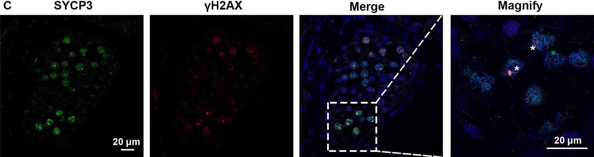
Detection of SCP3/SYCP3 by Immunohistochemistry
Expression of SYCP3 and gamma H2AX in the testis of the patient with SYCE1 CNVs and OA-affected patient as a positive control. A–C Immunofluorescence staining showed the expression of SYCP3 (Green) and gamma H2AX (Red) in the testis of the patient P6326 (A), P10377 (B), and the patient with OA as a positive control (C) Scale bars = 20 μm in A–D. And the arrowhead indicated the XY body in the spermatocytes in the testis of the patient with OA Image collected and cropped by CiteAb from the following open publication (https://pubmed.ncbi.nlm.nih.gov/35718780), licensed under a CC-BY license. Not internally tested by R&D Systems.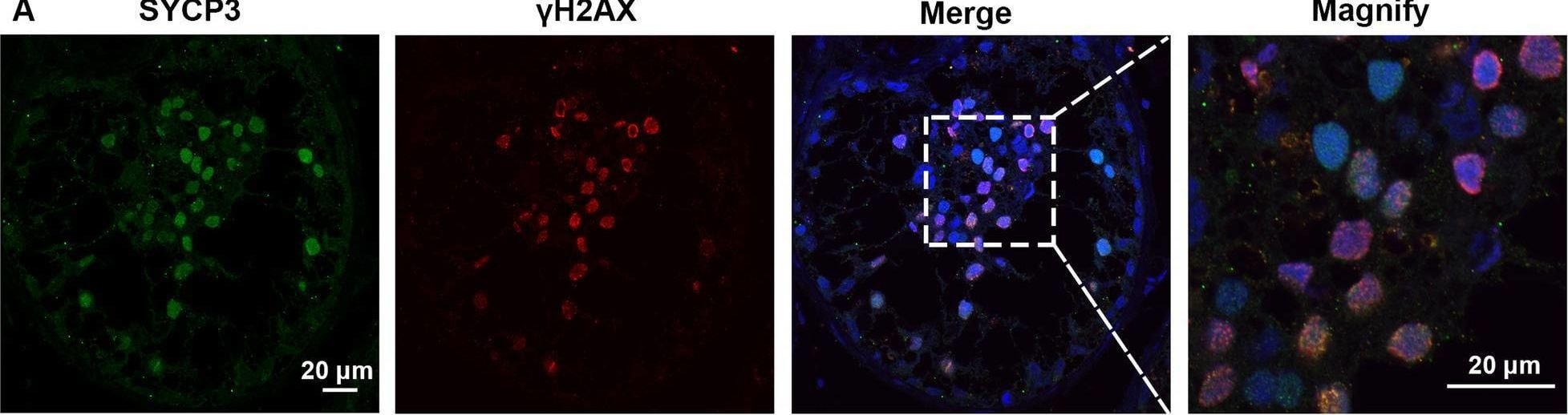
Detection of SCP3/SYCP3 by Immunohistochemistry
Expression of SYCP3 and gamma H2AX in the testis of the patient with SYCE1 CNVs and OA-affected patient as a positive control. A–C Immunofluorescence staining showed the expression of SYCP3 (Green) and gamma H2AX (Red) in the testis of the patient P6326 (A), P10377 (B), and the patient with OA as a positive control (C) Scale bars = 20 μm in A–D. And the arrowhead indicated the XY body in the spermatocytes in the testis of the patient with OA Image collected and cropped by CiteAb from the following open publication (https://pubmed.ncbi.nlm.nih.gov/35718780), licensed under a CC-BY license. Not internally tested by R&D Systems.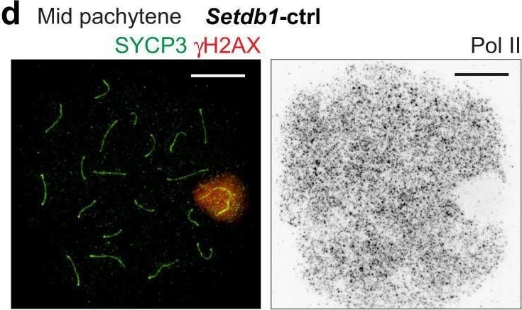
Detection of Mouse SCP3/SYCP3 by Immunocytochemistry/ Immunofluorescence
SETDB1-dependent H3K9me3 is dispensable for the initiation of MSCI.a Schematic of the strategy for generating Setdb1-cKO mice. b Evaluation of the efficiency of SETDB1 deletion shown as the mean and individual values for 3 independent samples. c Chromosome spreads of early and mid pachytene spermatocytes, distinguished by the absence (early) or the presence (mid) of H1T, respectively (not shown), immunostained with antibodies raised against SYCP3 and H3K9me3. XY chromosomes are indicated with dashed squares and are magnified in the panels to the right. Putative regions of XY chromatin are indicated by dashed circles. d Chromosome spreads of mid pachytene spermatocytes immunostained with antibodies raised against SYCP3, gamma H2AX, and Pol II. e A population of pachytene spermatocytes with a normal gamma H2AX domain and normal MSCI, as defined by gamma H2AX and Pol II, respectively. The mean and individual values for 3 independent samples are shown. EP early pachytene, MP mid pachytene, X-PCH X-pericentromeric heterochromatin. Scale bars: 10 μm. Source data are provided as a Source Data file. Image collected and cropped by CiteAb from the following open publication (https://pubmed.ncbi.nlm.nih.gov/36443288), licensed under a CC-BY license. Not internally tested by R&D Systems.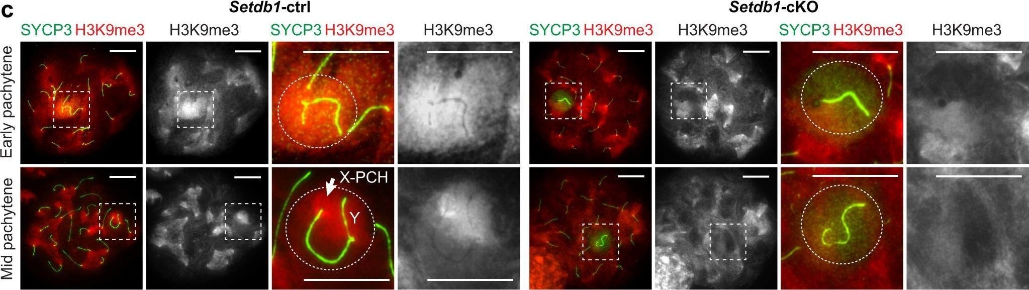
Detection of Mouse SCP3/SYCP3 by Immunocytochemistry/ Immunofluorescence
SETDB1-dependent H3K9me3 is dispensable for the initiation of MSCI.a Schematic of the strategy for generating Setdb1-cKO mice. b Evaluation of the efficiency of SETDB1 deletion shown as the mean and individual values for 3 independent samples. c Chromosome spreads of early and mid pachytene spermatocytes, distinguished by the absence (early) or the presence (mid) of H1T, respectively (not shown), immunostained with antibodies raised against SYCP3 and H3K9me3. XY chromosomes are indicated with dashed squares and are magnified in the panels to the right. Putative regions of XY chromatin are indicated by dashed circles. d Chromosome spreads of mid pachytene spermatocytes immunostained with antibodies raised against SYCP3, gamma H2AX, and Pol II. e A population of pachytene spermatocytes with a normal gamma H2AX domain and normal MSCI, as defined by gamma H2AX and Pol II, respectively. The mean and individual values for 3 independent samples are shown. EP early pachytene, MP mid pachytene, X-PCH X-pericentromeric heterochromatin. Scale bars: 10 μm. Source data are provided as a Source Data file. Image collected and cropped by CiteAb from the following open publication (https://pubmed.ncbi.nlm.nih.gov/36443288), licensed under a CC-BY license. Not internally tested by R&D Systems.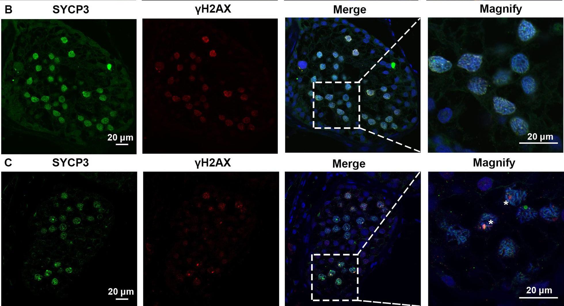
Detection of SCP3/SYCP3 by Immunocytochemistry/ Immunofluorescence
Expression of SYCP3 and gamma H2AX in the testis of the patient with SYCE1 CNVs and OA-affected patient as a positive control. A–C Immunofluorescence staining showed the expression of SYCP3 (Green) and gamma H2AX (Red) in the testis of the patient P6326 (A), P10377 (B), and the patient with OA as a positive control (C) Scale bars = 20 μm in A–D. And the arrowhead indicated the XY body in the spermatocytes in the testis of the patient with OA Image collected and cropped by CiteAb from the following open publication (https://pubmed.ncbi.nlm.nih.gov/35718780), licensed under a CC-BY license. Not internally tested by R&D Systems.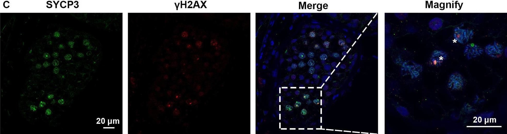
Detection of SCP3/SYCP3 by Immunohistochemistry
Expression of SYCP3 and gamma H2AX in the testis of the patient with SYCE1 CNVs and OA-affected patient as a positive control. A–C Immunofluorescence staining showed the expression of SYCP3 (Green) and gamma H2AX (Red) in the testis of the patient P6326 (A), P10377 (B), and the patient with OA as a positive control (C) Scale bars = 20 μm in A–D. And the arrowhead indicated the XY body in the spermatocytes in the testis of the patient with OA Image collected and cropped by CiteAb from the following open publication (https://pubmed.ncbi.nlm.nih.gov/35718780), licensed under a CC-BY license. Not internally tested by R&D Systems.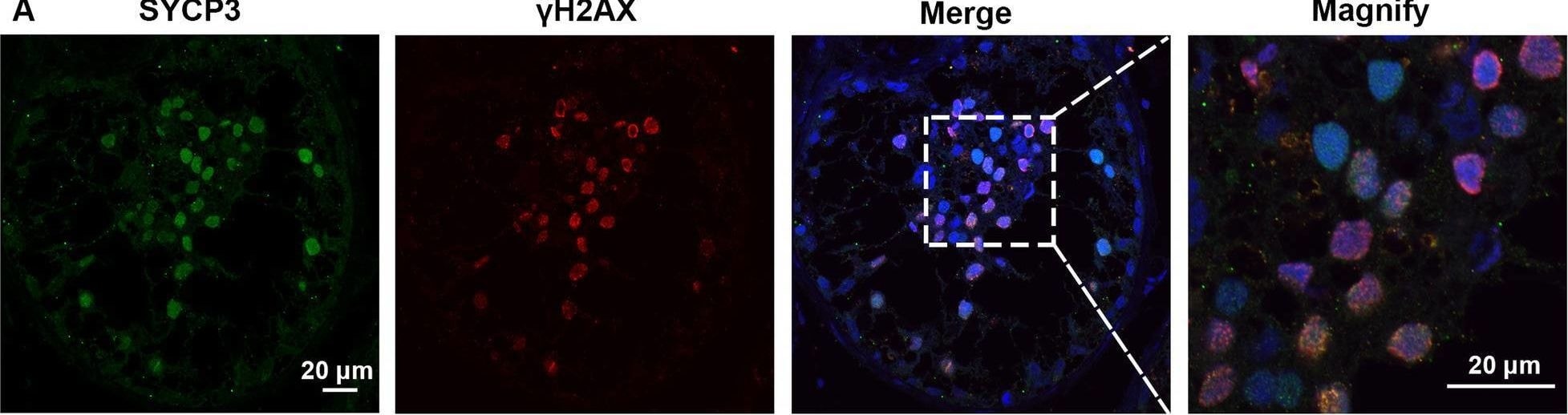
Detection of SCP3/SYCP3 by Immunohistochemistry
Expression of SYCP3 and gamma H2AX in the testis of the patient with SYCE1 CNVs and OA-affected patient as a positive control. A–C Immunofluorescence staining showed the expression of SYCP3 (Green) and gamma H2AX (Red) in the testis of the patient P6326 (A), P10377 (B), and the patient with OA as a positive control (C) Scale bars = 20 μm in A–D. And the arrowhead indicated the XY body in the spermatocytes in the testis of the patient with OA Image collected and cropped by CiteAb from the following open publication (https://pubmed.ncbi.nlm.nih.gov/35718780), licensed under a CC-BY license. Not internally tested by R&D Systems.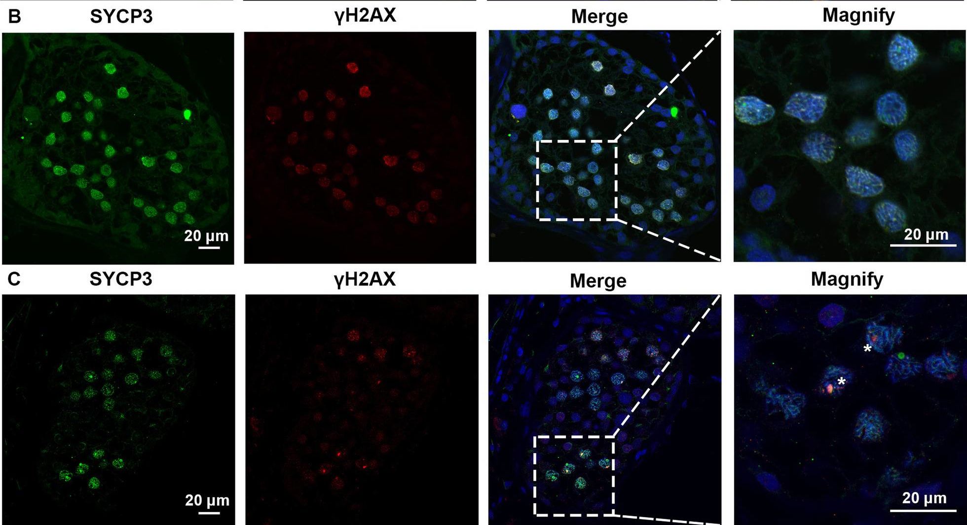
Detection of SCP3/SYCP3 by Immunocytochemistry/ Immunofluorescence
Expression of SYCP3 and gamma H2AX in the testis of the patient with SYCE1 CNVs and OA-affected patient as a positive control. A–C Immunofluorescence staining showed the expression of SYCP3 (Green) and gamma H2AX (Red) in the testis of the patient P6326 (A), P10377 (B), and the patient with OA as a positive control (C) Scale bars = 20 μm in A–D. And the arrowhead indicated the XY body in the spermatocytes in the testis of the patient with OA Image collected and cropped by CiteAb from the following open publication (https://pubmed.ncbi.nlm.nih.gov/35718780), licensed under a CC-BY license. Not internally tested by R&D Systems.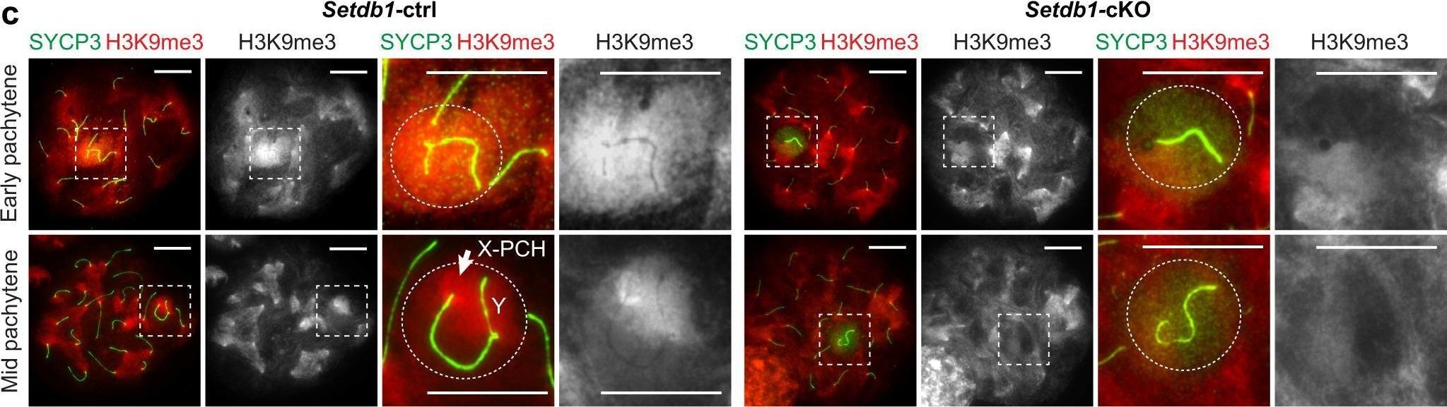
Detection of Mouse SCP3/SYCP3 by Immunocytochemistry/ Immunofluorescence
SETDB1-dependent H3K9me3 is dispensable for the initiation of MSCI.a Schematic of the strategy for generating Setdb1-cKO mice. b Evaluation of the efficiency of SETDB1 deletion shown as the mean and individual values for 3 independent samples. c Chromosome spreads of early and mid pachytene spermatocytes, distinguished by the absence (early) or the presence (mid) of H1T, respectively (not shown), immunostained with antibodies raised against SYCP3 and H3K9me3. XY chromosomes are indicated with dashed squares and are magnified in the panels to the right. Putative regions of XY chromatin are indicated by dashed circles. d Chromosome spreads of mid pachytene spermatocytes immunostained with antibodies raised against SYCP3, gamma H2AX, and Pol II. e A population of pachytene spermatocytes with a normal gamma H2AX domain and normal MSCI, as defined by gamma H2AX and Pol II, respectively. The mean and individual values for 3 independent samples are shown. EP early pachytene, MP mid pachytene, X-PCH X-pericentromeric heterochromatin. Scale bars: 10 μm. Source data are provided as a Source Data file. Image collected and cropped by CiteAb from the following open publication (https://pubmed.ncbi.nlm.nih.gov/36443288), licensed under a CC-BY license. Not internally tested by R&D Systems.Applications for Human SCP3/SYCP3 Antibody
Western Blot
Sample: Recombinant Human SCP3/SYCP3
Formulation, Preparation, and Storage
Purification
Reconstitution
Reconstitute at 0.2 mg/mL in sterile PBS. For liquid material, refer to CoA for concentration.
Formulation
Shipping
Stability & Storage
- 12 months from date of receipt, -20 to -70 °C as supplied.
- 1 month, 2 to 8 °C under sterile conditions after reconstitution.
- 6 months, -20 to -70 °C under sterile conditions after reconstitution.
Calculators
Background: SCP3/SYCP3
Long Name
Alternate Names
Gene Symbol
UniProt
Additional SCP3/SYCP3 Products
Product Documents for Human SCP3/SYCP3 Antibody
Certificate of Analysis
To download a Certificate of Analysis, please enter a lot or batch number in the search box below.
Note: Certificate of Analysis not available for kit components.
Product Specific Notices for Human SCP3/SYCP3 Antibody
For research use only
Citations for Human SCP3/SYCP3 Antibody
 Powered by Bioz
Customer Reviews for Human SCP3/SYCP3 Antibody
There are currently no reviews for this product. Be the first to review Human SCP3/SYCP3 Antibody and earn rewards!
Have you used Human SCP3/SYCP3 Antibody?
Submit a review and receive an Amazon gift card!
$25/€18/£15/$25CAN/¥2500 for a review with an image
$10/€7/£6/$10CAN/¥1110 for a review without an image
Submit a review
Protocols
Find general support by application which include: protocols, troubleshooting, illustrated assays, videos and webinars.
- Cellular Response to Hypoxia Protocols
- R&D Systems Quality Control Western Blot Protocol
- Troubleshooting Guide: Western Blot Figures
- Western Blot Conditions
- Western Blot Protocol
- Western Blot Protocol for Cell Lysates
- Western Blot Troubleshooting
- Western Blot Troubleshooting Guide
- View all Protocols, Troubleshooting, Illustrated assays and Webinars