
ID4 Antibody - Azide and BSA Free
Novus Biologicals | Catalog # H00003400-B01P

![Western Blot: ID4 Antibody [H00003400-B01P]](https://resources.rndsystems.com/images/products/ID4-Antibody-Western-Blot-H00003400-B01P-img0002.jpg "Western Blot: ID4 Antibody [H00003400-B01P]")
Scientific images may be resized, cropped, or adjusted for brightness or contrast for presentation purposes. These updates are limited to presentation and do not affect the underlying scientific interpretation of the data. Where available, additional source data may be provided upon request.
Key Product Details
Species Reactivity
Validated:
Cited:
Applications
Validated:
Cited:
Label
Antibody Source
Format
Product Specifications
Immunogen
Specificity
Clonality
Host
Isotype
Description
Scientific Data Images for ID4 Antibody - Azide and BSA Free
Western Blot: ID4 Antibody [H00003400-B01P]
Western Blot: ID4 Antibody [H00003400-B01P] - Analysis of ID4 expression in transfected 293T cell line by ID4 polyclonal antibody. Lane 1: ID4 transfected lysate(17.71 KDa). Lane 2: Non-transfected lysate.Applications for ID4 Antibody - Azide and BSA Free
Immunocytochemistry/ Immunofluorescence
Western Blot
Reviewed Applications
Read 1 review rated 1 using H00003400-B01P in the following applications:
Formulation, Preparation, and Storage
Purification
Formulation
Format
Preservative
Concentration
Shipping
Stability & Storage
Background: ID4
Long Name
Alternate Names
Entrez Gene IDs
Gene Symbol
UniProt
Additional ID4 Products
Product Documents for ID4 Antibody - Azide and BSA Free
Certificate of Analysis
To download a Certificate of Analysis, please enter a lot or batch number in the search box below.
Product Specific Notices for ID4 Antibody - Azide and BSA Free
This product is produced by and distributed for Abnova, a company based in Taiwan.
This product is for research use only and is not approved for use in humans or in clinical diagnosis. Primary Antibodies are guaranteed for 1 year from date of receipt.
Citations for ID4 Antibody - Azide and BSA Free
 Powered by Bioz
Customer Reviews for ID4 Antibody - Azide and BSA Free (1)
Have you used ID4 Antibody - Azide and BSA Free?
Submit a review and receive an Amazon gift card!
$25/€18/£15/$25CAN/¥2500 for a review with an image
$10/€7/£6/$10CAN/¥1110 for a review without an image
Submit a review
Customer Images
-
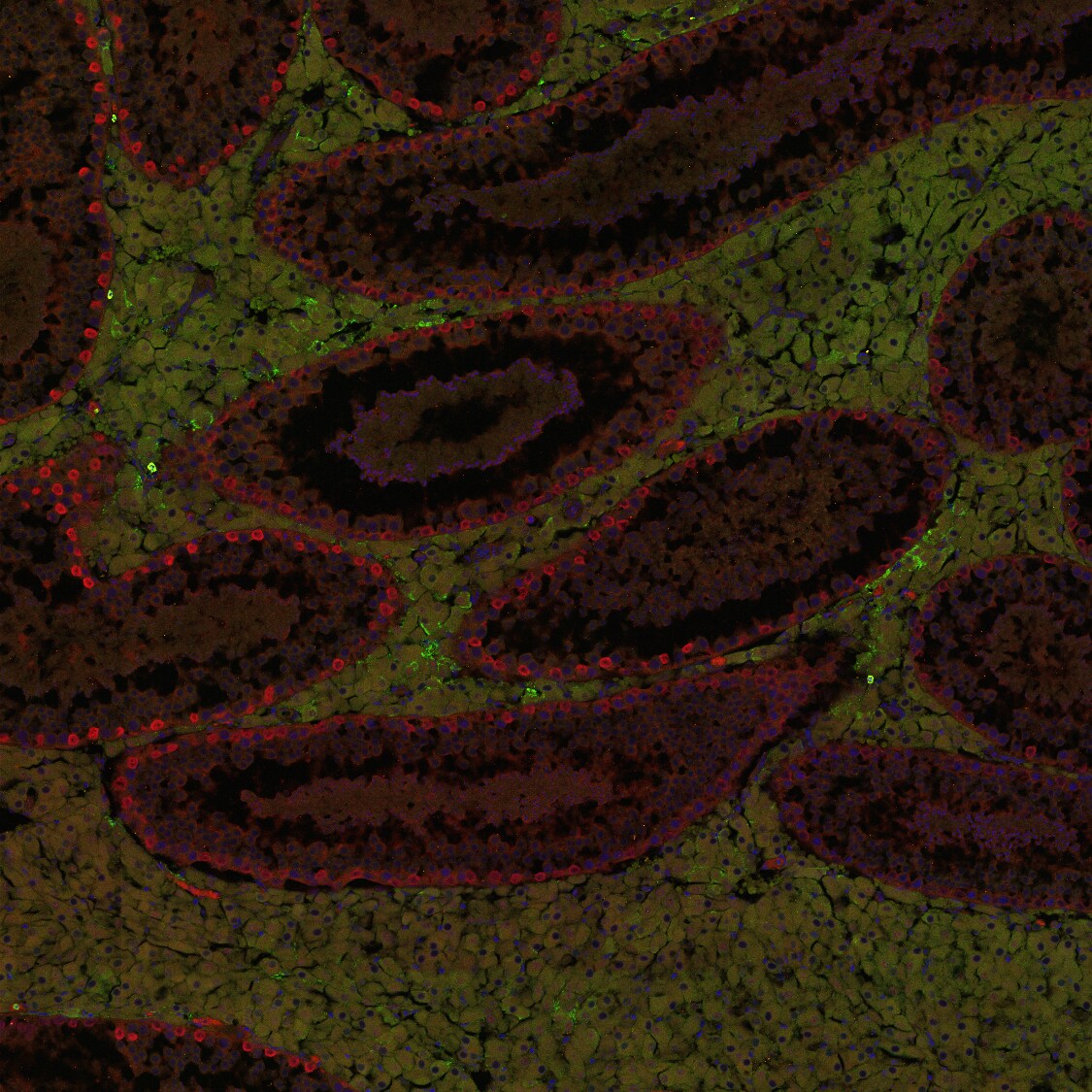
Application: Immunohistochemistry-FrozenSample Tested: TestisSpecies: OpossumVerified Customer | Posted 01/05/2017Tested in gray short-tailed opossum (Monodelphis domestica) in testis from 8-month animal. Tissue was fixed in 4% Paraformaldehyde overnight, incubated in 30% sucrose/PBS overnight and embedded in OCT. Sections were cut on a cryostat at 10um. For immunofluorescence staining: Slides were dried 15 minutes, washed 3x3 min TBST (0.1% Triton X-100), permeabilized for 10 minutes in 2% Triton X-100 in TBS, rinsed 3x3 min TBST, blocked in 10% heat inactivated goat serum in TBST for 30 min, and primary added at 1:100 in 10% blocking solution. Slides were coverslipped with parafilm overnight at 4C. The next day slides were washed with TBST 4x5 min, secondaries (AlexaFluor 568 goat anti rabbit and AlexaFluor 488 goat anti mouse 1:500) for 1 hour, 3x3 washes TBST, and mounted with VectaShield HardSet (+ DAPI). Intense staining at periphery of seminiferous tubules of Uchl-1 (red in image) with these conditions. 1:200 also worked though staining was less intense (not shown). The same conditions with 1% Triton X-100 for permeabiliziation resulted in weak staining at 1:100 and 1:200. However, ID-4 did not work well under any conditions. Shown in green is some faint staining in interstitial cells but this was not consistent between samples and did not appear to be specific. ID-4 was tested in 4 and 8 month opossum testes, and at dilutions ranging from 1:25 to 1:500. Single-antibody staining was also performed for each antibody and successful in Uchl-1, not ID-4.

There are no reviews that match your criteria.
Protocols
Find general support by application which include: protocols, troubleshooting, illustrated assays, videos and webinars.
- Appropriate Fixation of IHC/ICC Samples
- Cellular Response to Hypoxia Protocols
- ClariTSA™ Fluorophore Kits
- Detection & Visualization of Antibody Binding
- ICC Cell Smear Protocol for Suspension Cells
- ICC Immunocytochemistry Protocol Videos
- ICC for Adherent Cells
- Immunocytochemistry (ICC) Protocol
- Immunocytochemistry Troubleshooting
- Immunofluorescence of Organoids Embedded in Cultrex Basement Membrane Extract
- Immunohistochemistry (IHC) and Immunocytochemistry (ICC) Protocols
- Preparing Samples for IHC/ICC Experiments
- Preventing Non-Specific Staining (Non-Specific Binding)
- Primary Antibody Selection & Optimization
- Protocol for VisUCyte™ HRP Polymer Detection Reagent
- Protocol for the Fluorescent ICC Staining of Cell Smears - Graphic
- Protocol for the Fluorescent ICC Staining of Cultured Cells on Coverslips - Graphic
- Protocol for the Preparation and Fluorescent ICC Staining of Cells on Coverslips
- Protocol for the Preparation and Fluorescent ICC Staining of Non-adherent Cells
- Protocol for the Preparation and Fluorescent ICC Staining of Stem Cells on Coverslips
- Protocol for the Preparation of a Cell Smear for Non-adherent Cell ICC - Graphic
- R&D Systems Quality Control Western Blot Protocol
- TUNEL and Active Caspase-3 Detection by IHC/ICC Protocol
- The Importance of IHC/ICC Controls
- Troubleshooting Guide: Western Blot Figures
- Western Blot Conditions
- Western Blot Protocol
- Western Blot Protocol for Cell Lysates
- Western Blot Troubleshooting
- Western Blot Troubleshooting Guide
- View all Protocols, Troubleshooting, Illustrated assays and Webinars