
INSTA-Blot Colon Tissue OncoPair
Novus Biologicals | Catalog # NBP2-30129

![SDS-PAGE: INSTA-Blot Colon Tissue OncoPair [NBP2-30129]](https://resources.rndsystems.com/images/products/INSTA-Blot-Colon-Tissue-OncoPair-SDS-Page-NBP2-30129-img0001.jpg "SDS-PAGE: INSTA-Blot Colon Tissue OncoPair [NBP2-30129]")
Loading...
Key Product Details
Applications
Western Blot
Product Summary for INSTA-Blot Colon Tissue OncoPair
Tissue
| Lane | Tissue Type | Cat. No. | Grade | Stage | Sex | Age | Diagnosis |
| MWM | |||||||
| 2 | Colon | NBP2-28142 | 1 | I | M | 75 | Adenocarcinoma |
| 3 | Colon | NBP2-28140 | Normal adjacent | ||||
| 4 | Colon | NBP2-28146 | 1 | I | M | 25 | Adenocarcinoma |
| 5 | Colon | NBP2-28144 | Normal adjacent | ||||
| 6 | Colon | NBP2-28184 | 1 | II | M | 77 | Adenocarcinoma |
| 7 | Colon | NBP2-28182 | Normal adjacent | ||||
| 8 | Colon | NBP2-28263 | 1 | II | F | 38 | Adenocarcinoma |
| 9 | Colon | NBP2-28261 | Normal adjacent | ||||
| 10 | Colon | NBP2-28273 | 1 | II | F | 46 | Adenocarcinoma |
| 11 | Colon | NBP2-28271 | Normal adjacent | ||||
| 12 | Colon | NBP2-28306. | 1 | IIIA | F | 66 | Adenocarcinoma |
| 13 | Colon | NBP2-28304 | Normal adjacent | ||||
| 14 | Colon | NBP2-28311 | 1 | IIIA | F | 43 | Adenocarcinoma |
| 15 | Colon | NBP2-28309 | Normal adjacent |
The development of biomarkers has enormous potential for revolutionizing the diagnosis and treatment of disease. Western blot analysis is an integral technique for biomarker profiling. While key for biomarker discovery, the existing body of western blot data has primarily been defined from studies of tumor, immortal, and primary cells growing in vitro. Collectively, results obtained over decades have been integral to the dogma that up- and down-regulation of proteins can be leveraged as biomarkers of normal development, homeostasis, and disease. However, data from human tissue-derived products is key for generating biomarker validation data and progressing the most promising biomarker targets towards the clinic. The OncoPair INSTA-Blot™ product line addresses the increasing demand for the inclusion of human tissue-derived products in biomarker profiling.
The OncoPair INSTA-Blot™ is a ready-to-use PVDF western blot membrane containing denatured protein lysates manufactured from Novus's extensive Human Clinical Tissue Lysate collection. Each blot contains 14 lanes of alternating tumor (T) and matched normal adjacent (NA) tissue lysates from 7 patient donors. Incorporating both T and NA on the blot enables protein expression analysis of the tumor and microenvironment. The tumor microenvironment is increasingly being recognized as a major factor in influencing malignant progression and metastatic potential. Cancer is a heterogeneous disease and the presence of multiple donors on a single blot enables rapid screening of protein expression variability between T/NA from different individuals and at different disease stages. Clinical diagnosis and histopathology reports from board-certified pathologists are available for every patient sample.
OncoPair INSTA-Blots™ are available for a wide range of tumor types and stages. The product line is manufactured from donor patient tissue obtained from established hospital-based collection sites around the world. Tissues are collected under strict bioethical standards using IRB and HIPAA approved protocols. These protocols ensure patient confidentiality, safety, and informed consent. Tissues are flash frozen at collection sites within minutes of removal, maintained in liquid nitrogen and processed into lysates using protocols optimized for extracting proteins from tissues. Lysates are denatured for OncoPair INSTA-Blot™ production; native lysates are available for follow up proteomic studies. OncoPair INSTA-Blots™ and lysates are also useful in conjunction with the Cell Line and Normal Tissue INSTA-Blots™ and lysates for comprehensive antibody validation and to profile antibody reactivity and protein expression patterns across species and between tissues and cells lines.
Loading...
Scientific Data Images for INSTA-Blot Colon Tissue OncoPair
SDS-PAGE: INSTA-Blot Colon Tissue OncoPair [NBP2-30129]
SDS-Page: INSTA-Blot Colon Tissue OncoPair [NBP2-30129] - Samples on the blot are layed out in pairs of tumor and normal tissue lysate from the same patient. There are seven pairs of samples, representing seven different patients, on each OncoPair blot.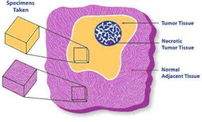
INSTA-Blot Colon Tissue OncoPair [NBP2-30129] - Tumor tissue is taken from healthy tumor tissue while normal tissue is taken from an area adjacent to the tumor tissue.
Formulation, Preparation, and Storage
Preparation Method
Tissue specimens were homogenized in modified RIPA buffer to obtain the soluble proteins and centrifuged to clarify. Sample buffer was added to the soluble fraction and approximately 14 ug/lane of protein was run and then transfered to PVDF membrane.
-Soluble fraction extraction buffer (extract 1): PBS at pH 7.4, 1 ug/ml Aprotinin, 1 mM NaF -Modified RIPA buffer: 1 mM EDTA, 1 ug/ml Pepstatin-A, 0.1% SDS, 0.25% Na deoxycholate, 1 ug/ml Leupeptin, 1 mM PMSF, 1 mM Na3VO4 -5X Sample buffer: 50 ml glycerol, 15 g SDS, 3.819 g Tris, pH 6.8, 25 ml 2-ME, 100 mg bromophenol blue, final volume of 100 ml with DI H2O
Concentration
Concentration is not relevant for this product. Please see the protocols for proper use of this product.
Shipping
The product is shipped at ambient temperature. Upon receipt, store it immediately at the temperature recommended below.
Storage
Store at room temperature.
Background: INSTA-Blot Colon Tissue OncoPair
Alternate Names
INSTA Blot Colon Tissue OncoPair, INSTA-Blot Colon Tissue, INSTABlot Colon Tissue OncoPair
Additional INSTA-Blot Colon Tissue OncoPair Products
Product Documents for INSTA-Blot Colon Tissue OncoPair
Certificate of Analysis
To download a Certificate of Analysis, please enter a lot or batch number in the search box below.
Product Specific Notices for INSTA-Blot Colon Tissue OncoPair
This product is for research use only and is not approved for use in humans or in clinical diagnosis. Western Blot Membranes are guaranteed for 1 year from date of receipt.
Citations for INSTA-Blot Colon Tissue OncoPair
 Powered by Bioz
Customer Reviews for INSTA-Blot Colon Tissue OncoPair
There are currently no reviews for this product. Be the first to review INSTA-Blot Colon Tissue OncoPair and earn rewards!
Have you used INSTA-Blot Colon Tissue OncoPair?
Submit a review and receive an Amazon gift card!
$25/€18/£15/$25CAN/¥2500 for a review with an image
$10/€7/£6/$10CAN/¥1110 for a review without an image
Submit a review
Protocols
View specific protocols for INSTA-Blot Colon Tissue OncoPair (NBP2-30129):
INSTA-Blot Colon Tissue OncoPair:
Material Safety Data Sheet for Non Hazardous Products
Hazard Information
Chemical Name: Non hazardous products.
Chemical Formula: N/A
CAS Number: N/A
EEC-No: N/A
Hazard Identification
None
First Aid Measures
Eye Contact: None
Skin Contact: None
Inhalation: None
Ingestion: None
Accidental Release Measures
This product either does not contain hazardous constituents or the concentration of all chemical constituents are below the regulatory threshold limits described by Occupational Safety Health Administration Hazard Communication Standard 29 CFR 1910.1200 and the European Directive 91/155/EEC. 88/379/EEC, and 67/546/EEC.
Handling and Storage
Exposure Controls / Personal Protection
Other Precautions: None
Physical and Chemical Properties
Form: N/A
Color: N/A
Odor: N/A
Melting Point: N/A
Boiling Temperature: N/A
Density: N/A
Vapor Pressure: N/A
Solubility in Water: N/A
Flash Point: N/A
Explosion limits: N/A
Ignition Temperature: N/A
Material Safety Data Sheet for Non Hazardous Products
Hazard Information
Chemical Name: Non hazardous products.
Chemical Formula: N/A
CAS Number: N/A
EEC-No: N/A
Hazard Identification
None
First Aid Measures
Eye Contact: None
Skin Contact: None
Inhalation: None
Ingestion: None
Accidental Release Measures
This product either does not contain hazardous constituents or the concentration of all chemical constituents are below the regulatory threshold limits described by Occupational Safety Health Administration Hazard Communication Standard 29 CFR 1910.1200 and the European Directive 91/155/EEC. 88/379/EEC, and 67/546/EEC.
Handling and Storage
Exposure Controls / Personal Protection
Other Precautions: None
Physical and Chemical Properties
Form: N/A
Color: N/A
Odor: N/A
Melting Point: N/A
Boiling Temperature: N/A
Density: N/A
Vapor Pressure: N/A
Solubility in Water: N/A
Flash Point: N/A
Explosion limits: N/A
Ignition Temperature: N/A
INSTA-Blot Colon Tissue OncoPair:
Note: The INSTA-Blot(TM) PVDF membrane has been dried and must be rehydrated (Step one) prior to use.
Before wetting, it is suggested that the top of the lanes be marked with an ink pen that will not wash off in methanol.
1. Wet the blots with 100% methanol then thoroughly wash with TBST (25 mM Tris-Cl, pH 8.0; 125 mM NaCl; 0.1% Tween 20) twice to remove residual methanol.
2. Incubate the blot for 1 hr with 5% Carnation nonfat dry milk in TBST to block non-specific antibody binding.
3. Incubate the blots with primary antibody in 1% milk/TBST for 1-2 h at RT or overnight at 4 degrees C.
4. After incubation with the primary antibody, wash the blots five times in TBST then incubate with a secondary antibody conjugated to horseradish peroxidase (HRP-conjugated secondary antibodies) for 1-2 h at RT.
5. After five washes with TBST, develop the blots for 5 min using the PicoTect(TM) Western Blot Chemiluminescent Substrate.
6. Expose the blots to photographic film for an appropriate time period. We normally use Hyper-film(TM)-ECL films (Amersham Life Science Inc.) and expose for various periods ranging from 10 s to 20 min to visualize the chemiluminescence signal corresponding to the specific antibody-antigen reaction.
Note: The INSTA-Blot(TM) PVDF membrane has been dried and must be rehydrated (Step one) prior to use.
Before wetting, it is suggested that the top of the lanes be marked with an ink pen that will not wash off in methanol.
1. Wet the blots with 100% methanol then thoroughly wash with TBST (25 mM Tris-Cl, pH 8.0; 125 mM NaCl; 0.1% Tween 20) twice to remove residual methanol.
2. Incubate the blot for 1 hr with 5% Carnation nonfat dry milk in TBST to block non-specific antibody binding.
3. Incubate the blots with primary antibody in 1% milk/TBST for 1-2 h at RT or overnight at 4 degrees C.
4. After incubation with the primary antibody, wash the blots five times in TBST then incubate with a secondary antibody conjugated to horseradish peroxidase (HRP-conjugated secondary antibodies) for 1-2 h at RT.
5. After five washes with TBST, develop the blots for 5 min using the PicoTect(TM) Western Blot Chemiluminescent Substrate.
6. Expose the blots to photographic film for an appropriate time period. We normally use Hyper-film(TM)-ECL films (Amersham Life Science Inc.) and expose for various periods ranging from 10 s to 20 min to visualize the chemiluminescence signal corresponding to the specific antibody-antigen reaction.
Find general support by application which include: protocols, troubleshooting, illustrated assays, videos and webinars.
- Cellular Response to Hypoxia Protocols
- R&D Systems Quality Control Western Blot Protocol
- Troubleshooting Guide: Western Blot Figures
- Western Blot Conditions
- Western Blot Protocol
- Western Blot Protocol for Cell Lysates
- Western Blot Troubleshooting
- Western Blot Troubleshooting Guide
- View all Protocols, Troubleshooting, Illustrated assays and Webinars
FAQs
No product specific FAQs exist for this product.
View all FAQs for Supplemental Kits and ReagentsLoading...