
Ki67/MKI67 Antibody (6P3K5)
Novus Biologicals | Catalog # NBP3-57376
Recombinant Monoclonal Antibody

Scientific images may be resized, cropped, or adjusted for brightness or contrast for presentation purposes. These updates are limited to presentation and do not affect the underlying scientific interpretation of the data. Where available, additional source data may be provided upon request.
Loading...
Key Product Details
Species Reactivity
Human, Mouse, Rat
Applications
Knockout Validated, Immunohistochemistry-Paraffin, Immunohistochemistry-Frozen, Western Blot, ELISA, Immunocytochemistry/ Immunofluorescence, Multiplex Immunoassay
Label
Unconjugated
Antibody Source
Recombinant Monoclonal Rabbit IgG Clone # 6P3K5
Loading...
Product Specifications
Immunogen
Synthetic peptide. This information is considered to be commercially sensitive. (Swissprot# P46013)
Clonality
Monoclonal
Host
Rabbit
Isotype
IgG
Description
This antibody is highly specific and optimized for use in Immunohistochemistry.
Normal Tissues Tested
Pancreas, Lung, Tonsil, Cervix, Placenta, Colon, Kidney, Spleen, Liver, esophagus
Cancer Tissues Tested
Breast cancer, Colon cancer, Thyroid cancer, Hepatocellular Carcinoma, Hepatocholangio carcinoma, Lung adenocarcinoma, Lung squamous carcinoma
Positive Controls
Preferentially during late G1, S, G2 and M phases of the cell cycle tonsil (A moderate to strong, distinct nuclear staining reaction in 80-90% of the germinal centre B-cells in both the light and the dark zone and in the vast majority of the suprabasal squamous epithelial cells), esophagus (squamous epithelia)
Negative Controls
Liver (Hepatocytes, 1%)
Scientific Data Images for Ki67/MKI67 Antibody (6P3K5)
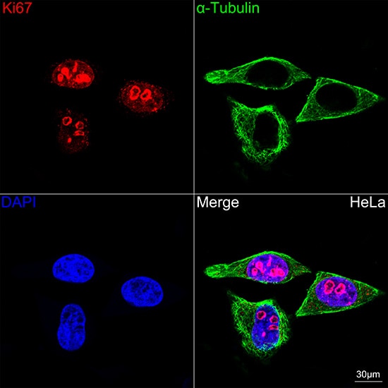
Immunocytochemistry/ Immunofluorescence: Ki67/MKI67 Antibody (6P3K5) [NBP3-57376]
Confocal imaging of HeLa cells using NBP3-57376 (dilution 1:100)(Red) followed by a further incubation with Cy3-conjugated Goat Anti-Rabbit IgG (H+L) (dilution 1:500) (Red). The cells were counterstained with alpha-Tubulin Mouse mAb (dilution 1:400) followed by incubation with conjugated Goat Anti-Mouse IgG (H+L) (dilution 1:500) (Green). DAPI was used for nuclear staining (Blue). Objective: 100x.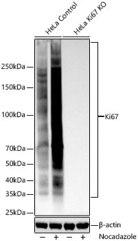
Western Blot: Ki67/MKI67 Antibody (6P3K5) [NBP3-57376]
Western blot analysis of lysates from wild type (WT) and Ki67 knockout (KO) HeLa cells using NBP3-57376 at 1:5000 dilution incubated overnight at 4C. HeLa cells were treated with Nocadazole (1 ug/mL) at 37C for 16 hours Secondary antibody: HRP-conjugated Goat anti-Rabbit IgG (H+L) at 1:10000 dilution. Lysates/proteins: 25 ug per lane. Blocking buffer: 3% nonfat dry milk in TBST. Exposure time: 45 s.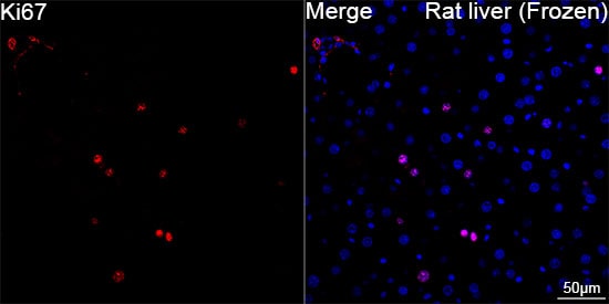
Immunohistochemistry-Frozen: Ki67/MKI67 Antibody (6P3K5) [NBP3-57376]
Confocal imaging of frozen sections of Rat liver tissue using NBP3-57376 (dilution 1:200) followed by a further incubation with Cy3-conjugated Goat anti-Rabbit IgG (H+L) (dilution 1:500) (Red). DAPI was used for nuclear staining (Blue). Microwave antigen retrieval performed with 0.01M Citrate Buffer (pH 6.0) prior to IF staining. Objective: 40x.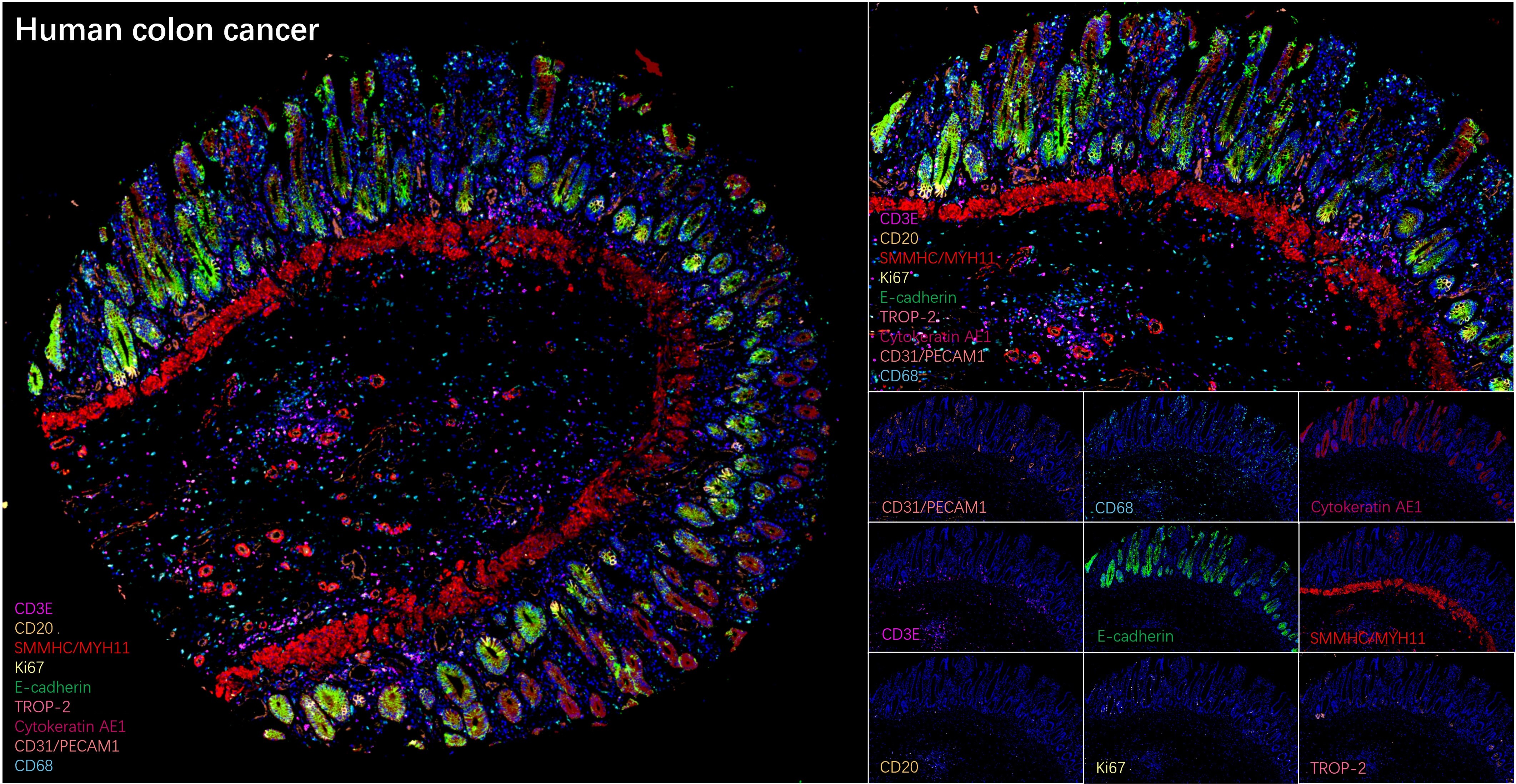
Multiplex Immunofluorescence: Ki67/MKI67 Antibody (6P3K5) [NBP3-57376]
The multiplex IHC analysis on paraffin-embedded Human colon cancer tissue using tyramide signal amplification (TSA) reagents with SMMHC/MYH11 Rabbit mAb (1:4000) (Red), and CD3 epsilon mAb (NBP3-57370, 1:40000) (Magenta), and CD20 Rabbit mAb (1:2000) (Pale Yellow), andE-Cadherin mAb (NBP3-57377, 1:1000) (Green), and CD31/PECAM-1 mAb (NBP3-57369, 1:200) (Light Coral), and CD68 Rabbit mAb (1:20000) (Cyan), and Ki67/MKI67 mAb (NBP3-57376, 1:500) (Light Yellow), and Cytokeratin, Acidic (Type I or LMW) mAb (NBP3-57402, 1:2000) (Magenta), and TROP-2 Rabbit mAb (1:10000) (Magenta). DAPI (Blue) was used for nuclear staining. Prior to multiplex IHC staining, high-pressure antigen retrieval was performed using 0.01M citrate buffer at pH 6.0. The analysis was completed using a 40x objective lens.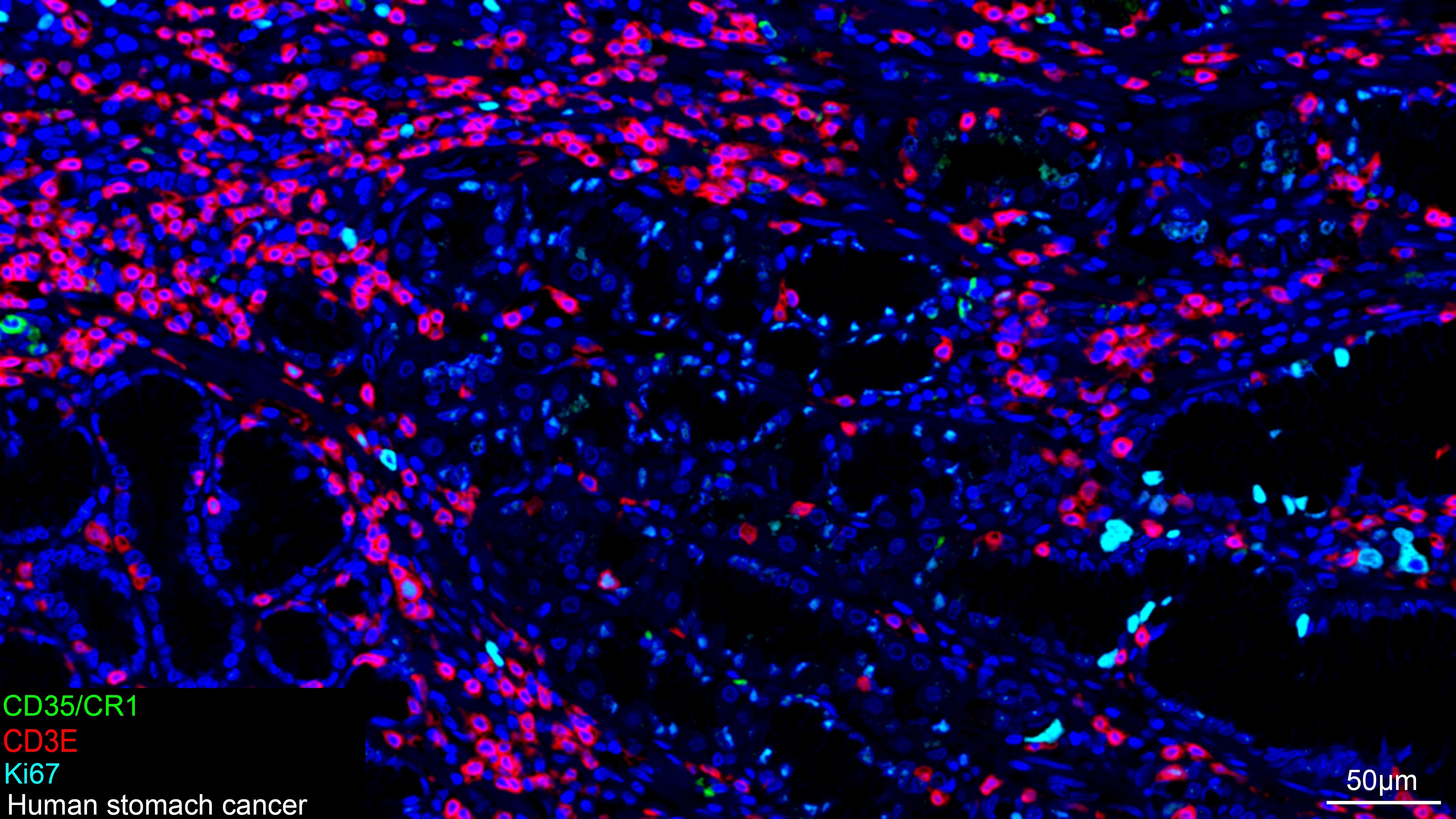
Multiplex Immunofluorescence: Ki67/MKI67 Antibody (6P3K5) [NBP3-57376]
The multiplex IHC analysis on paraffin-embedded Human stomach cancer tissue using tyramide signal amplification (TSA) reagents with CD35/CR1 Rabbit mAb (1:100) (Green), CD3 epsilon mAb (NBP3-57370, 1:2000) (Red), and Ki67/MKI67 mAb (NBP3-57376, 1:500) (cyan). DAPI (Blue) was used for nuclear staining. Prior to multiplex IHC staining, high-pressure antigen retrieval was performed using 0.01M citrate buffer at pH 6.0. The analysis was completed using a 20x objective lens.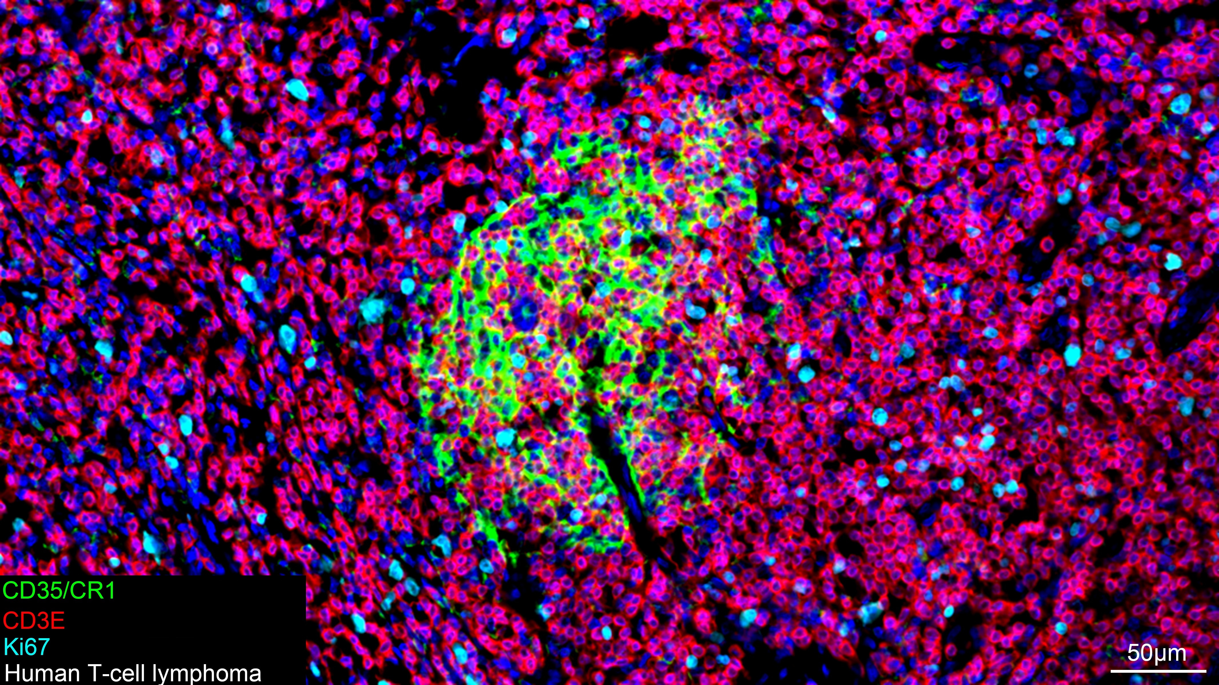
Multiplex Immunofluorescence: Ki67/MKI67 Antibody (6P3K5) [NBP3-57376]
The multiplex IHC analysis on paraffin-embedded Human T-cell lymphoma tissue using tyramide signal amplification (TSA) reagents with CD35/CR1 Rabbit mAb (1:100) (Green), CD3 epsilon mAb (NBP3-57370, 1:2000) (Red), and Ki67/MKI67 mAb (NBP3-57376, 1:500) (cyan). DAPI (Blue) was used for nuclear staining. Prior to multiplex IHC staining, high-pressure antigen retrieval was performed using 0.01M citrate buffer at pH 6.0. The analysis was completed using a 20x objective lens.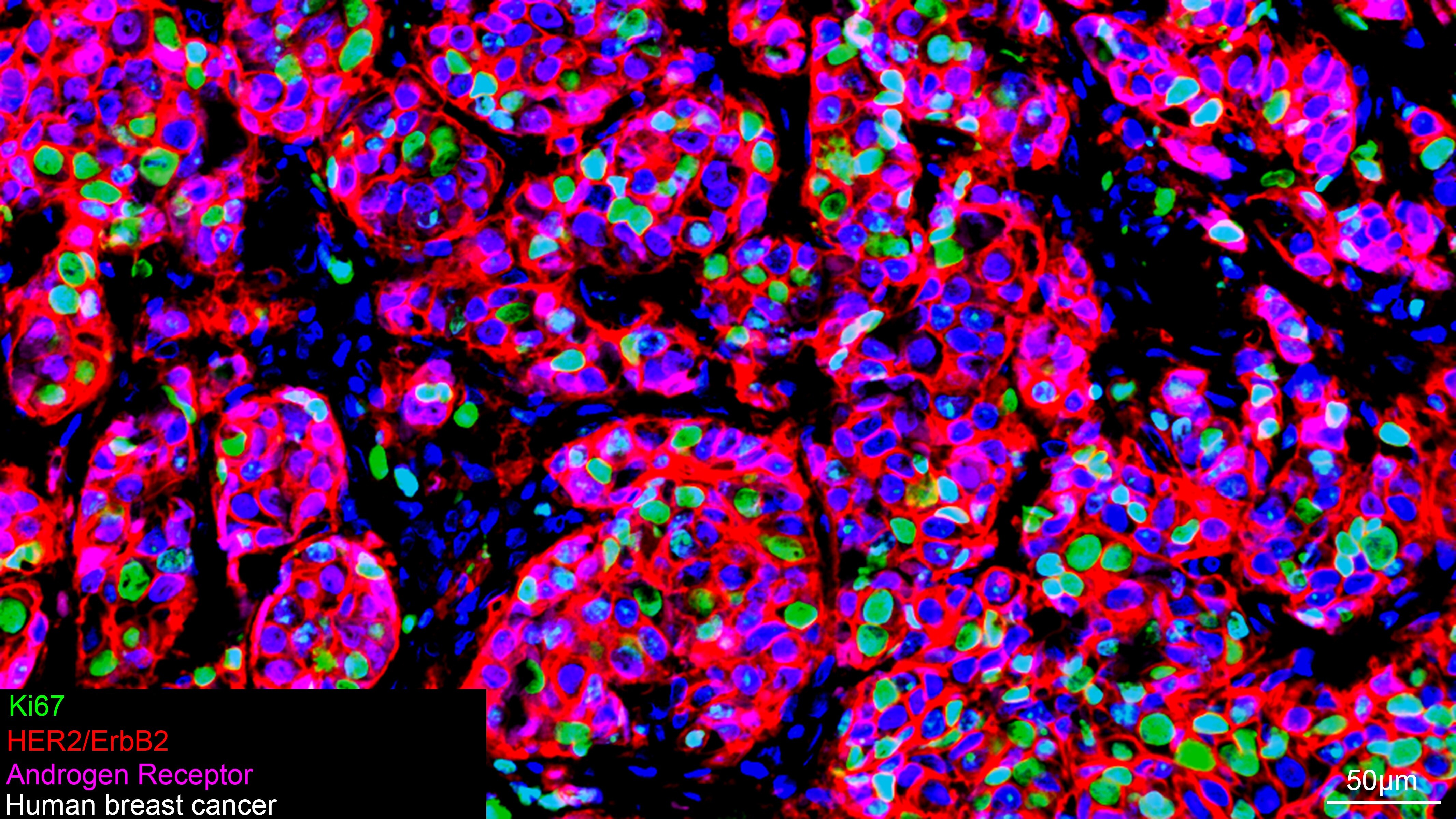
Multiplex Immunofluorescence: Ki67/MKI67 Antibody (6P3K5) [NBP3-57376]
The multiplex IHC analysis on paraffin-embedded Human breast cancer tissue using tyramide signal amplification (TSA) reagents with Ki67/MKI67 mAb (NBP3-57376, 1:500) (Green), HER2/ErbB2 Rabbit mAb (1:200) (Red), and Androgen R/NR3C4 mAb (NBP3-57373, 1:400) (Magenta). DAPI (Blue) was used for nuclear staining. Prior to multiplex IHC staining, high-pressure antigen retrieval was performed using 0.01M citrate buffer at pH 6.0. The analysis was completed using a 20x objective lens.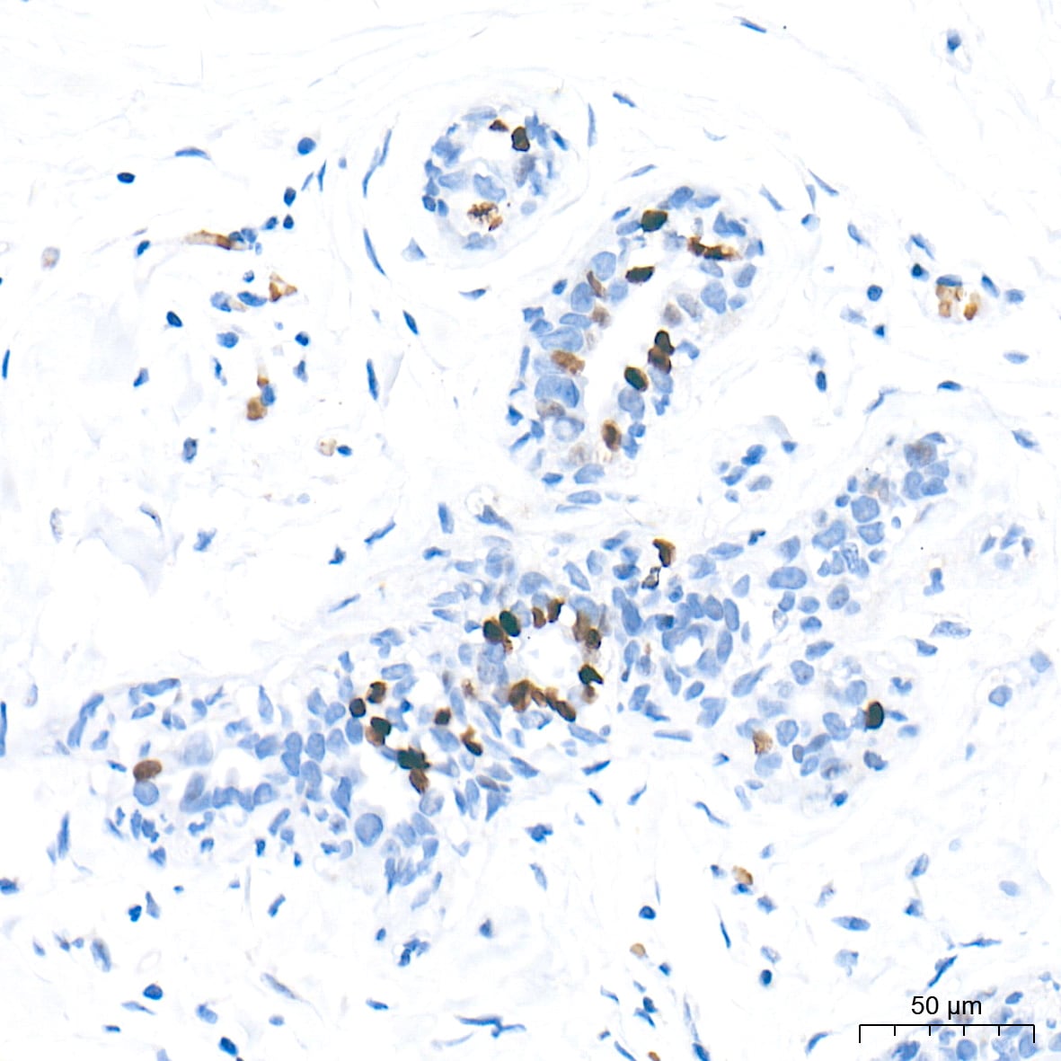
Immunohistochemistry-Paraffin: Ki67/MKI67 Antibody (6P3K5) [NBP3-57376]
Immunohistochemistry analysis of paraffin-embedded Human breast cancer tissue using NBP3-57376 at a dilution of 1:1000 (40x lens). High pressure antigen retrieval performed with 0.01M Tris-EDTA Buffer (pH 9.0) prior to IHC staining.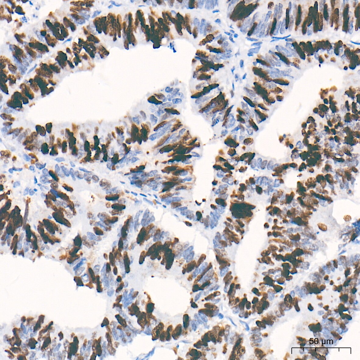
Immunohistochemistry-Paraffin: Ki67/MKI67 Antibody (6P3K5) [NBP3-57376]
Immunohistochemistry analysis of paraffin-embedded Human colon carcinoma tissue using NBP3-57376 at a dilution of 1:1000 (40x lens). High pressure antigen retrieval performed with 0.01M Tris-EDTA Buffer (pH 9.0) prior to IHC staining.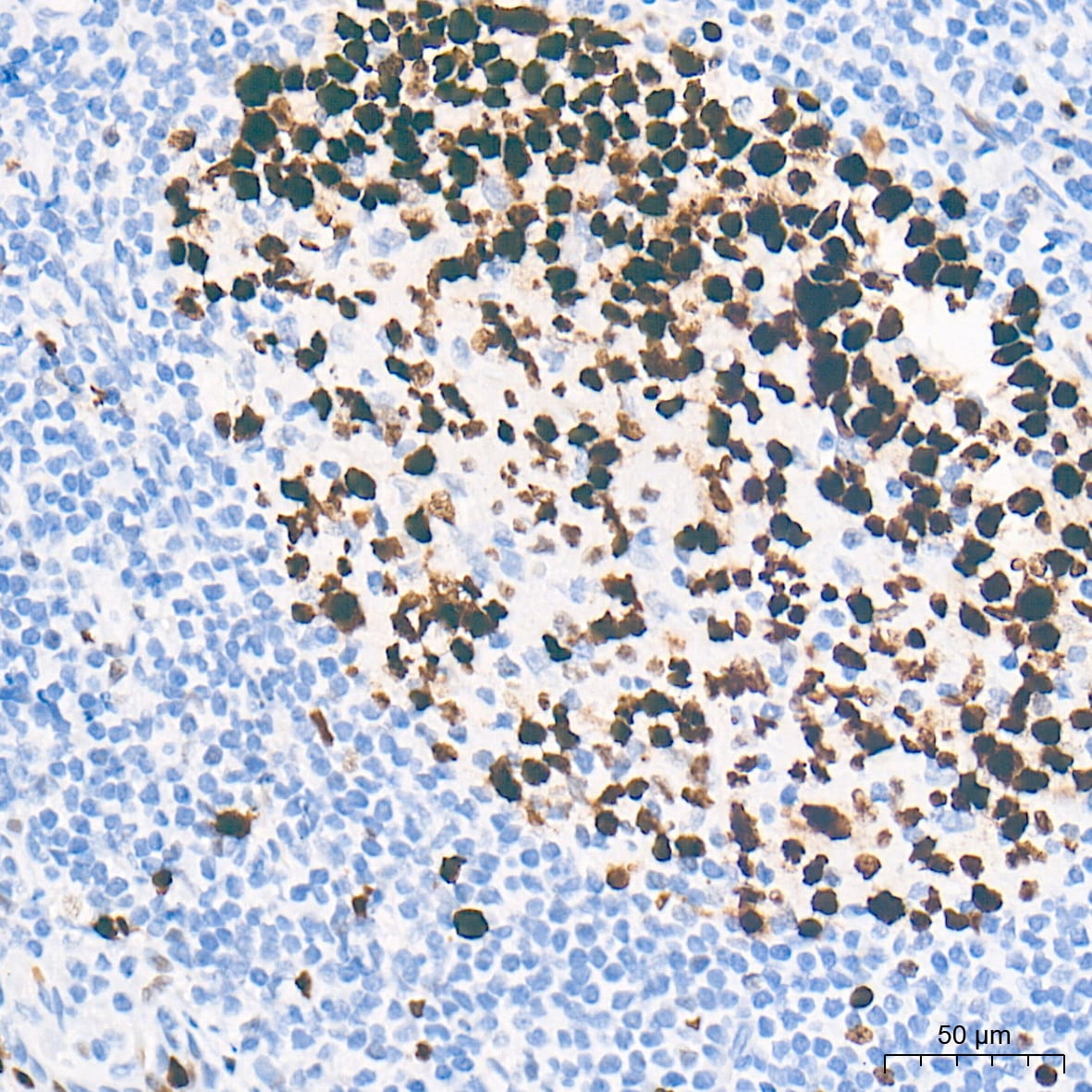
Immunohistochemistry-Paraffin: Ki67/MKI67 Antibody (6P3K5) [NBP3-57376]
Immunohistochemistry analysis of paraffin-embedded Human tonsil tissue using NBP3-57376 at a dilution of 1:1000 (40x lens). High pressure antigen retrieval performed with 0.01M Tris-EDTA Buffer (pH 9.0) prior to IHC staining.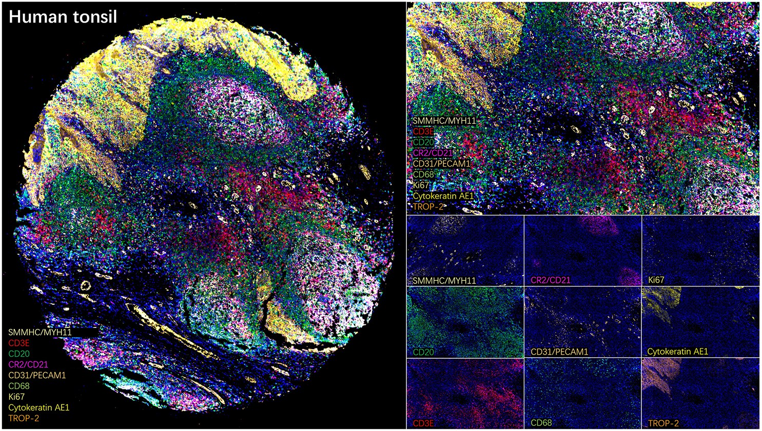
Multiplex Immunofluorescence: Ki67/MKI67 Antibody (6P3K5) [NBP3-57376]
The multiplex IHC analysis on paraffin-embedded Human tonsil tissue using tyramide signal amplification (TSA) reagents with Myosin heavy chain 11 mAb (NBP3-16323, 1:4000) (Pale Yellow), and CD3 epsilon mAb (NBP3-57370, 1:4000) (Red), and CD20 Rabbit mAb (1:2000) (Green), and CR2/CD21 Rabbit mAb (1:2000) (Magenta), and CD31/PECAM-1 mAb (NBP3-57369, 1:200) (Beige), and CD68 Rabbit mAb (1:20000) (Cyan), and Ki67/MKI67 mAb (NBP3-57376, 1:500) (Light Yellow), and Cytokeratin, Acidic (Type I or LMW) mAb (NBP3-57402, 1:2000) (Yellow), and TROP-2 Rabbit mAb (1:10000) (Orange). DAPI (Blue) was used for nuclear staining. Prior to multiplex IHC staining, high-pressure antigen retrieval was performed using 0.01M citrate buffer at pH 6.0. The analysis was completed using a 40x objective lens.Applications for Ki67/MKI67 Antibody (6P3K5)
Application
Recommended Usage
ELISA
1 ug/mL
Immunocytochemistry/ Immunofluorescence
1:50 - 1:200
Immunohistochemistry-Frozen
1:200 - 1:800
Immunohistochemistry-Paraffin
1:200 - 1:800
Multiplex Immunoassay
1:200 - 1:800
Western Blot
1:2000 - 1:10000
Application Notes
This product can be used in Multiplex Immunohistochemistry at a recommended concentration of 1:200 - 1:800.
Formulation, Preparation, and Storage
Purification
Affinity purified
Formulation
PBS, 50% glycerol, 0.05% BSA, (pH 7.3)
Preservative
0.09% Sodium Azide
Concentration
Please see the vial label for concentration. If unlisted please contact technical services.
Shipping
The product is shipped with polar packs. Upon receipt, store it immediately at the temperature recommended below.
Stability & Storage
Store at -20C. Avoid freeze-thaw cycles.
Background: Ki67/MKI67
Detection of Ki67 by immunostaining is commonly used as a proliferation marker in solid tumors, as well as certain hematological malignancies (3-5). The Ki67 index, which reports on positive Ki67 stained tumor cell nuclei, has been extensively studied as a prognostic biomarker in cancers such as breast cancer and cervical cancer.
References
1. Gerdes J, Schwab U, Lemke H, Stein H. (1983) Production of a mouse monoclonal antibody reactive with a human nuclear antigen associated with cell proliferation. Int J Cancer. 31:13-20. PMID: 6339421
2. Starborg M, Gell K, Brundell E and Hoog C. (1996) The murine Ki-67 cell proliferation antigen accumulates in the nucleolar and heterochromatic regions of interphase cells and at the periphery of the mitotic chromosomes in a process essential for cell cycle progression. J Cell Sci. 109:143-153. 1996
3. Karamitopoulou E, Perentes E, Tolnay M, Probst A. (1998) Prognostic significance of MIB-1, p53, and bcl-2 immunoreactivity in meningiomas. Hum Pathol. 29(2):140-5. PMID: 9490273
4. Geyer FC, Rodrigues DN, Weigelt B and Reis-Filho JS. (2012) Molecular classification of estrogen receptor-positive/luminal breast cancers. Adv Anat Pathol. 19(1):39-53. PMID: 22156833
5. Ikenberg H, Bergeron C, Schmidt D, Griesser H, Alameda F, Angeloni C, Bogers J, Dachez R, Denton K, Hariri J, Keller T, von Knebel Doeberitz M, Neumann HH, Puig-Tintore LM, Sideri M, Rehm S, Ridder R; PALMS Study Group. (2013) Screening for cervical cancer precursors with p16/Ki-67 dual-stained cytology: results of the PALMS study. J Natl Cancer Inst. 105(20):1550-7. PMID: 24096620
Long Name
Antigen Identified by Monoclonal Antibody Ki67
Alternate Names
Ki-67, KIA, MIB-1, MKI67, PPP1R105, TSG126
Gene Symbol
MKI67
Additional Ki67/MKI67 Products
Product Documents for Ki67/MKI67 Antibody (6P3K5)
Certificate of Analysis
To download a Certificate of Analysis, please enter a lot or batch number in the search box below.
Product Specific Notices for Ki67/MKI67 Antibody (6P3K5)
This product is for research use only and is not approved for use in humans or in clinical diagnosis. Primary Antibodies are guaranteed for 1 year from date of receipt.
Related Research Areas
Customer Reviews for Ki67/MKI67 Antibody (6P3K5)
There are currently no reviews for this product. Be the first to review Ki67/MKI67 Antibody (6P3K5) and earn rewards!
Have you used Ki67/MKI67 Antibody (6P3K5)?
Submit a review and receive an Amazon gift card!
$25/€18/£15/$25CAN/¥2500 for a review with an image
$10/€7/£6/$10CAN/¥1110 for a review without an image
Submit a review
Protocols
Find general support by application which include: protocols, troubleshooting, illustrated assays, videos and webinars.
- Antigen Retrieval Protocol (PIER)
- Antigen Retrieval for Frozen Sections Protocol
- Appropriate Fixation of IHC/ICC Samples
- Cellular Response to Hypoxia Protocols
- Chromogenic IHC Staining of Formalin-Fixed Paraffin-Embedded (FFPE) Tissue Protocol
- Chromogenic Immunohistochemistry Staining of Frozen Tissue
- ClariTSA™ Fluorophore Kits
- Detection & Visualization of Antibody Binding
- ELISA Sample Preparation & Collection Guide
- ELISA Troubleshooting Guide
- Fluorescent IHC Staining of Frozen Tissue Protocol
- Graphic Protocol for Heat-induced Epitope Retrieval
- Graphic Protocol for the Preparation and Fluorescent IHC Staining of Frozen Tissue Sections
- Graphic Protocol for the Preparation and Fluorescent IHC Staining of Paraffin-embedded Tissue Sections
- Graphic Protocol for the Preparation of Gelatin-coated Slides for Histological Tissue Sections
- How to Run an R&D Systems DuoSet ELISA
- How to Run an R&D Systems Quantikine ELISA
- How to Run an R&D Systems Quantikine™ QuicKit™ ELISA
- ICC Cell Smear Protocol for Suspension Cells
- ICC Immunocytochemistry Protocol Videos
- ICC for Adherent Cells
- IHC Sample Preparation (Frozen sections vs Paraffin)
- Immunocytochemistry (ICC) Protocol
- Immunocytochemistry Troubleshooting
- Immunofluorescence of Organoids Embedded in Cultrex Basement Membrane Extract
- Immunofluorescent IHC Staining of Formalin-Fixed Paraffin-Embedded (FFPE) Tissue Protocol
- Immunohistochemistry (IHC) and Immunocytochemistry (ICC) Protocols
- Immunohistochemistry Frozen Troubleshooting
- Immunohistochemistry Paraffin Troubleshooting
- Preparing Samples for IHC/ICC Experiments
- Preventing Non-Specific Staining (Non-Specific Binding)
- Primary Antibody Selection & Optimization
- Protocol for Heat-Induced Epitope Retrieval (HIER)
- Protocol for Making a 4% Formaldehyde Solution in PBS
- Protocol for VisUCyte™ HRP Polymer Detection Reagent
- Protocol for the Fluorescent ICC Staining of Cell Smears - Graphic
- Protocol for the Fluorescent ICC Staining of Cultured Cells on Coverslips - Graphic
- Protocol for the Preparation & Fixation of Cells on Coverslips
- Protocol for the Preparation and Chromogenic IHC Staining of Frozen Tissue Sections
- Protocol for the Preparation and Chromogenic IHC Staining of Frozen Tissue Sections - Graphic
- Protocol for the Preparation and Chromogenic IHC Staining of Paraffin-embedded Tissue Sections
- Protocol for the Preparation and Chromogenic IHC Staining of Paraffin-embedded Tissue Sections - Graphic
- Protocol for the Preparation and Fluorescent ICC Staining of Cells on Coverslips
- Protocol for the Preparation and Fluorescent ICC Staining of Non-adherent Cells
- Protocol for the Preparation and Fluorescent ICC Staining of Stem Cells on Coverslips
- Protocol for the Preparation and Fluorescent IHC Staining of Frozen Tissue Sections
- Protocol for the Preparation and Fluorescent IHC Staining of Paraffin-embedded Tissue Sections
- Protocol for the Preparation of Gelatin-coated Slides for Histological Tissue Sections
- Protocol for the Preparation of a Cell Smear for Non-adherent Cell ICC - Graphic
- Quantikine HS ELISA Kit Assay Principle, Alkaline Phosphatase
- Quantikine HS ELISA Kit Principle, Streptavidin-HRP Polymer
- R&D Systems Quality Control Western Blot Protocol
- Sandwich ELISA (Colorimetric) – Biotin/Streptavidin Detection Protocol
- Sandwich ELISA (Colorimetric) – Direct Detection Protocol
- TUNEL and Active Caspase-3 Detection by IHC/ICC Protocol
- The Importance of IHC/ICC Controls
- Troubleshooting Guide: ELISA
- Troubleshooting Guide: Immunohistochemistry
- Troubleshooting Guide: Western Blot Figures
- Western Blot Conditions
- Western Blot Protocol
- Western Blot Protocol for Cell Lysates
- Western Blot Troubleshooting
- Western Blot Troubleshooting Guide
- View all Protocols, Troubleshooting, Illustrated assays and Webinars
FAQs for Ki67/MKI67 Antibody (6P3K5)
Showing
1
-
2 of
2 FAQs
Showing All
-
Q: Whats the concentration of this Ki67/MKI67 Antibody?
A: The concentration is lot specific available upon request.
-
Q: Whats the concentration of this Ki67/MKI67 Antibody?
A: Optimal concentrations and conditions for each application should be determined by the user.
-
Q: Whats the concentration of this Ki67/MKI67 Antibody?
A: The concentration is lot specific available upon request.
-
Q: Whats the concentration of this Ki67/MKI67 Antibody?
A: Optimal concentrations and conditions for each application should be determined by the user.
Loading...