
Mitofilin Antibody - BSA Free
Novus Biologicals | Catalog # NB100-1919

![Western Blot: Mitofilin Antibody [NB100-1919]](https://resources.rndsystems.com/images/products/Mitofilin-Antibody-Western-Blot-NB100-1919-img0006.jpg "Western Blot: Mitofilin Antibody [NB100-1919]")
Scientific images may be resized, cropped, or adjusted for brightness or contrast for presentation purposes. These updates are limited to presentation and do not affect the underlying scientific interpretation of the data. Where available, additional source data may be provided upon request.
Key Product Details
Species Reactivity
Validated:
Cited:
Applications
Validated:
Cited:
Label
Antibody Source
Format
Product Specifications
Immunogen
Reactivity Notes
Localization
Marker
Clonality
Host
Isotype
Scientific Data Images for Mitofilin Antibody - BSA Free
Western Blot: Mitofilin Antibody [NB100-1919]
Western Blot: Mitofilin Antibody [NB100-1919] - Western blot of dog testis. Total protein extract 1:2000 dilution of antibody.![Immunocytochemistry/ Immunofluorescence: Mitofilin Antibody [NB100-1919]](https://resources.rndsystems.com/images/products/Mitofilin-Antibody-Immunocytochemistry-Immunofluorescence-NB100-1919-img0004.jpg "Immunocytochemistry/ Immunofluorescence: Mitofilin Antibody [NB100-1919]")
Immunocytochemistry/ Immunofluorescence: Mitofilin Antibody [NB100-1919]
Immunocytochemistry/Immunofluorescence: Mitofilin Antibody [NB100-1919] - HeLa cells with antibody 1:500 dilution labeling mitochondria.![Immunohistochemistry-Paraffin: Mitofilin Antibody [NB100-1919]](https://resources.rndsystems.com/images/products/Mitofilin-Antibody-Immunohistochemistry-Paraffin-NB100-1919-img0009.jpg "Immunohistochemistry-Paraffin: Mitofilin Antibody [NB100-1919]")
Immunohistochemistry-Paraffin: Mitofilin Antibody [NB100-1919]
Immunohistochemistry-Paraffin: Mitofilin Antibody [NB100-1919] - IHC-P detection of Mitofilin/Mitochondrial inner membrane protein in a section of normal human skin using Mitofilin antibody at 1:200 dilution. The representative image shows a specific and expected granular staining of Mitofilin in the keratinocytes as well as the other cells of epidermal layer with no staining in basal cells. [60X Magnification]![Western Blot: Mitofilin Antibody [NB100-1919]](https://resources.rndsystems.com/images/products/Mitofilin-Antibody-Western-Blot-NB100-1919-img0012.jpg "Western Blot: Mitofilin Antibody [NB100-1919]")
Western Blot: Mitofilin Antibody [NB100-1919]
Mitofilin-Antibody-Western-Blot-NB100-1919-img0012.jpg![Immunohistochemistry-Paraffin: Mitofilin Antibody [NB100-1919]](https://resources.rndsystems.com/images/products/Mitofilin-Antibody-Immunohistochemistry-Paraffin-NB100-1919-img0011.jpg "Immunohistochemistry-Paraffin: Mitofilin Antibody [NB100-1919]")
Immunohistochemistry-Paraffin: Mitofilin Antibody [NB100-1919]
Immunohistochemistry-Paraffin: Mitofilin Antibody [NB100-1919] - IHC-P analysis of Mitofilin/Mitochondrial inner membrane protein in a transverse section of normal human skeletal muscle using Mitofilin antibody at 1:200 dilution. The myocytes showed an expected and very specific granular staining in the cytoplasm. [10X Magnification]![Immunohistochemistry-Paraffin: Mitofilin Antibody [NB100-1919]](https://resources.rndsystems.com/images/products/Mitofilin-Antibody-Immunohistochemistry-Paraffin-NB100-1919-img0010.jpg "Immunohistochemistry-Paraffin: Mitofilin Antibody [NB100-1919]")
Immunohistochemistry-Paraffin: Mitofilin Antibody [NB100-1919]
Immunohistochemistry-Paraffin: Mitofilin Antibody [NB100-1919] - IHC-P detection of Mitofilin/Mitochondrial inner membrane protein in a section of normal human skin using Mitofilin antibody at 1:200 dilution. Specific cytoplasmic positivity of Mitofilin was observed in the keratinocytes as well as other cells of the epidermal layer except for basal cells and dermal connective tissue. [10X Magnification]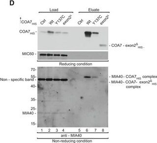
Western Blot: Mitofilin Antibody [NB100-1919] -
Western Blot: Mitofilin Antibody [NB100-1919] - COA7 pathological variants are import‐defectiveASchematic representation of wild‐type & mutant COA7.BCellular protein extracts were isolated from HEK293 cells that were transfected with a plasmid that encoded wild‐type or mutant COA7. The samples were analyzed by reducing SDS–PAGE & Western blot.CCellular fractions were prepared from HEK293 cells that were transfected with a plasmid that encoded wild‐type or mutant COA7. The fractions were analyzed by reducing SDS–PAGE & Western blot. T, total; C, cytosol; M, mitochondria.DHEK293 cells that transiently expressed wild‐type or mutant COA7HIS were solubilized, & the affinity purification of COA7HIS was performed. The samples were analyzed by reducing & non‐reducing SDS–PAGE & Western blot. Load: 3%. Eluate: 100%.EEqual amounts of wild‐type & mutant radiolabeled [35S]COA7 precursors were imported into mitochondria isolated from HEK293 cells. The samples were analyzed by reducing SDS–PAGE & autoradiography. The results of three biological replicates were analyzed, quantified, & normalized to wild‐type COA7 at 45 min. The data are expressed as a mean ± SEM (n = 3). IAA, iodoacetamide.Source data are available online for this figure. Image collected & cropped by CiteAb from the following publication (https://pubmed.ncbi.nlm.nih.gov/30885959), licensed under a CC-BY license. Not internally tested by Novus Biologicals.Applications for Mitofilin Antibody - BSA Free
Immunocytochemistry/ Immunofluorescence
Immunohistochemistry
Immunohistochemistry-Paraffin
Western Blot
Reviewed Applications
Read 3 reviews rated 4.3 using NB100-1919 in the following applications:
Formulation, Preparation, and Storage
Purification
Formulation
Format
Preservative
Concentration
Shipping
Stability & Storage
Background: Mitofilin
Alternate Names
Gene Symbol
UniProt
Additional Mitofilin Products
Product Documents for Mitofilin Antibody - BSA Free
Certificate of Analysis
To download a Certificate of Analysis, please enter a lot or batch number in the search box below.
Product Specific Notices for Mitofilin Antibody - BSA Free
This product is for research use only and is not approved for use in humans or in clinical diagnosis. Primary Antibodies are guaranteed for 1 year from date of receipt.
Citations for Mitofilin Antibody - BSA Free
 Powered by Bioz
Customer Reviews for Mitofilin Antibody - BSA Free (3)
Have you used Mitofilin Antibody - BSA Free?
Submit a review and receive an Amazon gift card!
$25/€18/£15/$25CAN/¥2500 for a review with an image
$10/€7/£6/$10CAN/¥1110 for a review without an image
Submit a review
Customer Images
-
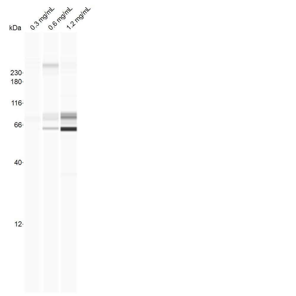
Application: Simple WesternSample Tested: cell line, whole cell lysateSpecies: RatVerified Customer | Posted 08/18/2017B103 whole-cell lysates (concentrations indicated), probed with 1:25 dilution of antibody on the standard separation matrix (12-230 kDa).

-
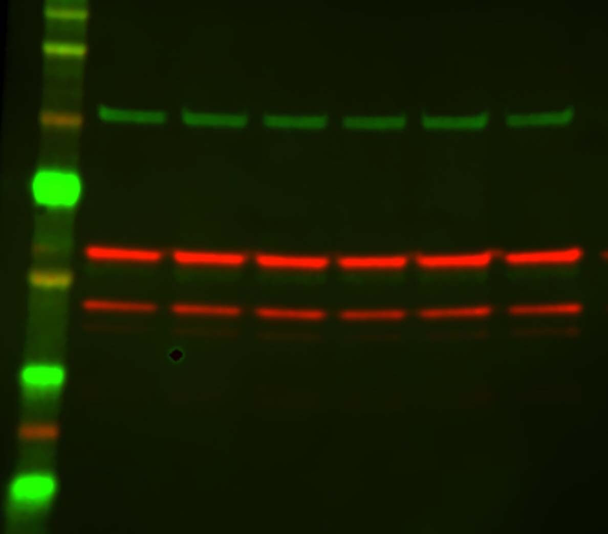
Application: Western BlotSample Tested: Protein lysate from mitochondria isolated from SH-SY5Y cellsSpecies: HumanVerified Customer | Posted 06/14/2016Neuroblastoma SH-SY5Y mitochondrial protein lysate probed for mitofillin: Each lane run at 12ug of protein per lane
-
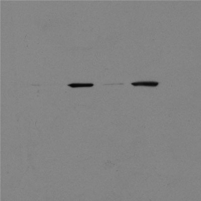
Application: ImmunoprecipitationSample Tested: Mouse heartSpecies: MouseVerified Customer | Posted 08/27/2013
There are no reviews that match your criteria.
Protocols
Find general support by application which include: protocols, troubleshooting, illustrated assays, videos and webinars.
- Antigen Retrieval Protocol (PIER)
- Antigen Retrieval for Frozen Sections Protocol
- Appropriate Fixation of IHC/ICC Samples
- Cellular Response to Hypoxia Protocols
- Chromogenic IHC Staining of Formalin-Fixed Paraffin-Embedded (FFPE) Tissue Protocol
- Chromogenic Immunohistochemistry Staining of Frozen Tissue
- ClariTSA™ Fluorophore Kits
- Detection & Visualization of Antibody Binding
- Fluorescent IHC Staining of Frozen Tissue Protocol
- Graphic Protocol for Heat-induced Epitope Retrieval
- Graphic Protocol for the Preparation and Fluorescent IHC Staining of Frozen Tissue Sections
- Graphic Protocol for the Preparation and Fluorescent IHC Staining of Paraffin-embedded Tissue Sections
- Graphic Protocol for the Preparation of Gelatin-coated Slides for Histological Tissue Sections
- ICC Cell Smear Protocol for Suspension Cells
- ICC Immunocytochemistry Protocol Videos
- ICC for Adherent Cells
- IHC Sample Preparation (Frozen sections vs Paraffin)
- Immunocytochemistry (ICC) Protocol
- Immunocytochemistry Troubleshooting
- Immunofluorescence of Organoids Embedded in Cultrex Basement Membrane Extract
- Immunofluorescent IHC Staining of Formalin-Fixed Paraffin-Embedded (FFPE) Tissue Protocol
- Immunohistochemistry (IHC) and Immunocytochemistry (ICC) Protocols
- Immunohistochemistry Frozen Troubleshooting
- Immunohistochemistry Paraffin Troubleshooting
- Immunoprecipitation Protocol
- Preparing Samples for IHC/ICC Experiments
- Preventing Non-Specific Staining (Non-Specific Binding)
- Primary Antibody Selection & Optimization
- Protocol for Heat-Induced Epitope Retrieval (HIER)
- Protocol for Making a 4% Formaldehyde Solution in PBS
- Protocol for VisUCyte™ HRP Polymer Detection Reagent
- Protocol for the Fluorescent ICC Staining of Cell Smears - Graphic
- Protocol for the Fluorescent ICC Staining of Cultured Cells on Coverslips - Graphic
- Protocol for the Preparation & Fixation of Cells on Coverslips
- Protocol for the Preparation and Chromogenic IHC Staining of Frozen Tissue Sections
- Protocol for the Preparation and Chromogenic IHC Staining of Frozen Tissue Sections - Graphic
- Protocol for the Preparation and Chromogenic IHC Staining of Paraffin-embedded Tissue Sections
- Protocol for the Preparation and Chromogenic IHC Staining of Paraffin-embedded Tissue Sections - Graphic
- Protocol for the Preparation and Fluorescent ICC Staining of Cells on Coverslips
- Protocol for the Preparation and Fluorescent ICC Staining of Non-adherent Cells
- Protocol for the Preparation and Fluorescent ICC Staining of Stem Cells on Coverslips
- Protocol for the Preparation and Fluorescent IHC Staining of Frozen Tissue Sections
- Protocol for the Preparation and Fluorescent IHC Staining of Paraffin-embedded Tissue Sections
- Protocol for the Preparation of Gelatin-coated Slides for Histological Tissue Sections
- Protocol for the Preparation of a Cell Smear for Non-adherent Cell ICC - Graphic
- R&D Systems Quality Control Western Blot Protocol
- TUNEL and Active Caspase-3 Detection by IHC/ICC Protocol
- The Importance of IHC/ICC Controls
- Troubleshooting Guide: Immunohistochemistry
- Troubleshooting Guide: Western Blot Figures
- Western Blot Conditions
- Western Blot Protocol
- Western Blot Protocol for Cell Lysates
- Western Blot Troubleshooting
- Western Blot Troubleshooting Guide
- View all Protocols, Troubleshooting, Illustrated assays and Webinars
FAQs for Mitofilin Antibody - BSA Free
-
Q: One of our customers is interested in testing your Anti-Mitofilin antibody (NB100-1919) with IHC. Do you know how likely this Ab will be in succeeding to this attempt?
A: This Ab has been validated for use in IHC-P in Canine, Feline, Mouse, Rat and Human.