
Axl (Ufo, Ark), Dtk (Sky, Tyro3, Rse, Brt), and Mer (human and mouse homologues of chicken c-Eyk) constitute a subfamily of the receptor tyrosine kinases (1, 2). The extracellular domains of these proteins contain two Ig-like motifs and two fibronectin type III motifs. This characteristic topology is also found in neural cell adhesion molecules and in receptor tyrosine phosphatases. The mouse Axl cDNA encodes an 888 amino acid (aa) precursor that includes an 18 aa signal sequence, a 427 aa extracellular domain, a 21 aa transmembrane segment, and a 422 aa cytoplasmic domain. The extracellular domains of mouse and human Axl share 81% aa sequence identity. These receptors bind the vitamin K-dependent protein growth arrest specific gene 6 (Gas6) which is structurally related to the anticoagulation factor protein S. Binding of Gas6 induces receptor autophosphorylation and downstream signaling pathways that can lead to cell proliferation, migration, or the prevention of apoptosis (3). This family of tyrosine kinase receptors is involved in hematopoiesis, embryonic development, tumorigenesis, and regulation of testicular functions.

Scientific images may be resized, cropped, or adjusted for brightness or contrast for presentation purposes. These updates are limited to presentation and do not affect the underlying scientific interpretation of the data. Where available, additional source data may be provided upon request.
Key Product Details
Species Reactivity
Validated:
Cited:
Applications
Validated:
Cited:
Label
Antibody Source
Product Specifications
Immunogen
His20-Pro443
Accession # Q00993
Specificity
Clonality
Host
Isotype
Scientific Data Images for Mouse Axl Antibody (107332)
Detection of Mouse Axl by Western Blot
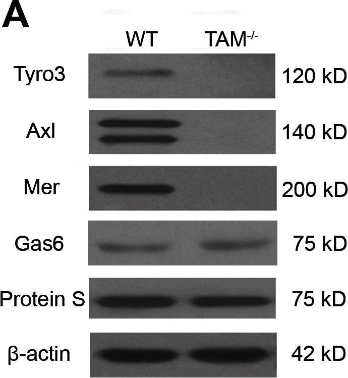
Expression of TAM RTKs, Gas6 and Protein S.(A) Western blot analyses of the liver lysates for the examination of TAM RTKs, Gas6 and Protein S. (B) Expression of TAM RTKs, Gas6 and Protein S in isolated liver cells: parenchymal cells (PCs), Kupffer cells(KCs) and sinusoidal endothelial cells (SECs). The primary cells were subjected to Western blotting. (C) Immunohistochemistry for the detection of TAM RTKs, Gas6 and Protein S. Arrowheads indicate PCs, and arrows indicate spindle-shaped sinusoidal cells corresponding to KCs and SECs. In negative controls (islets), sections were incubated with primary antibodies pre-incubated with an excess of blocking peptide. The livers of 15-week-old WT mice were used for the protein analyses. The images are representatives of at least three experiments. Scale bar = 20 µm. Image collected and cropped by CiteAb from the following open publication (https://pubmed.ncbi.nlm.nih.gov/23799121), licensed under a CC-BY license. Not internally tested by R&D Systems.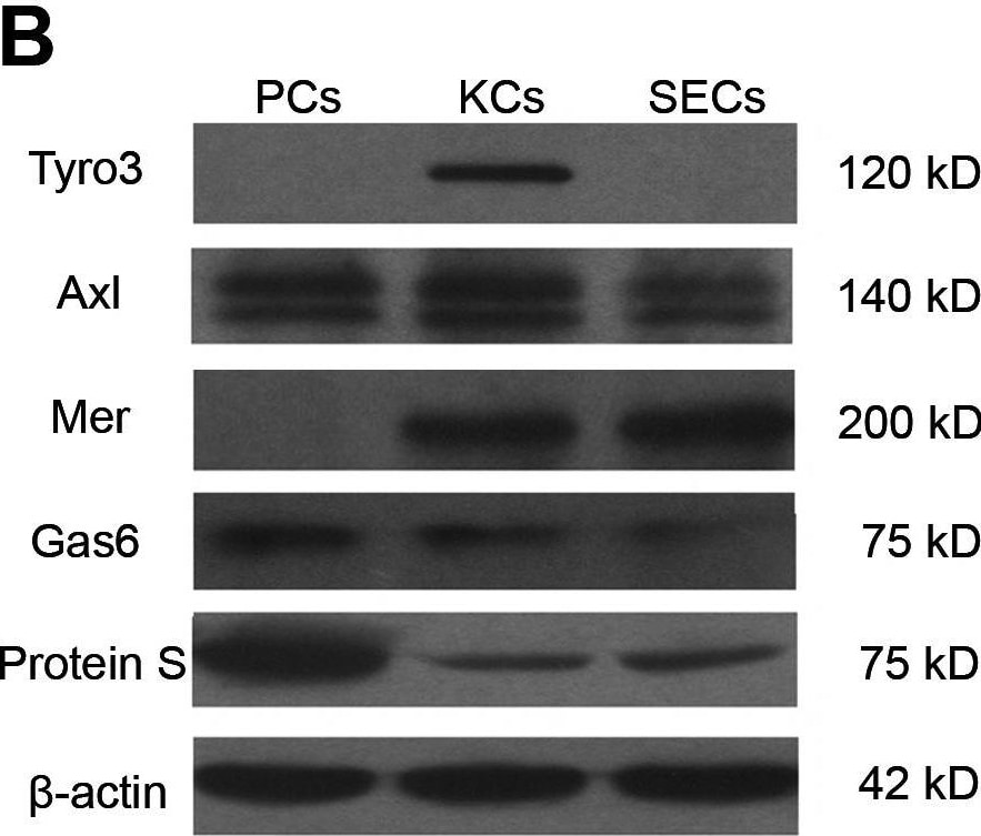
Detection of Mouse Axl by Western Blot
Expression of TAM RTKs, Gas6 and Protein S.(A) Western blot analyses of the liver lysates for the examination of TAM RTKs, Gas6 and Protein S. (B) Expression of TAM RTKs, Gas6 and Protein S in isolated liver cells: parenchymal cells (PCs), Kupffer cells(KCs) and sinusoidal endothelial cells (SECs). The primary cells were subjected to Western blotting. (C) Immunohistochemistry for the detection of TAM RTKs, Gas6 and Protein S. Arrowheads indicate PCs, and arrows indicate spindle-shaped sinusoidal cells corresponding to KCs and SECs. In negative controls (islets), sections were incubated with primary antibodies pre-incubated with an excess of blocking peptide. The livers of 15-week-old WT mice were used for the protein analyses. The images are representatives of at least three experiments. Scale bar = 20 µm. Image collected and cropped by CiteAb from the following open publication (https://pubmed.ncbi.nlm.nih.gov/23799121), licensed under a CC-BY license. Not internally tested by R&D Systems.
Mouse Axl ELISA Standard Curve
Recombinant Mouse Axl Fc Chimera (Catalog # 854-AX) was serially diluted and captured by Rat Anti-Mouse Axl Monoclonal Antibody (Catalog # MAB854) coated on a Clear Polystyrene Microplate (Catalog # DY990). Goat Anti-Mouse Axl Antigen Affinity-purified Polyclonal Antibody (Catalog # AF854) was biotinylated and incubated with the protein captured on the plate. Detection of the standard curve was achieved by incubating Streptavidin-HRP (Catalog # DY998)Applications for Mouse Axl Antibody (107332)
Western Blot
Sample: Recombinant Mouse Axl Fc Chimera (Catalog # 854-AX)
Mouse Axl Sandwich Immunoassay
Reviewed Applications
Read 7 reviews rated 3.9 using MAB854 in the following applications:
Formulation, Preparation, and Storage
Purification
Reconstitution
Reconstitute at 0.5 mg/mL in sterile PBS. For liquid material, refer to CoA for concentration.
Formulation
Shipping
Stability & Storage
- 12 months from date of receipt, -20 to -70 °C as supplied.
- 1 month, 2 to 8 °C under sterile conditions after reconstitution.
- 6 months, -20 to -70 °C under sterile conditions after reconstitution.
Calculators
Background: Axl
References
- Yanagita, M. (2004) Curr. Opin. Nephrol. Hypertens. 13:465.
- Nagata, K. et al. (1996) J. Biol. Chem. 22:30022.
- Holland, S. et al. (2005) Canc. Res. 65:9294.
Long Name
Alternate Names
Gene Symbol
UniProt
Additional Axl Products
Product Documents for Mouse Axl Antibody (107332)
Certificate of Analysis
To download a Certificate of Analysis, please enter a lot or batch number in the search box below.
Note: Certificate of Analysis not available for kit components.
Product Specific Notices for Mouse Axl Antibody (107332)
For research use only
Related Research Areas
Citations for Mouse Axl Antibody (107332)
 Powered by Bioz
Customer Reviews for Mouse Axl Antibody (107332) (7)
Have you used Mouse Axl Antibody (107332)?
Submit a review and receive an Amazon gift card!
$25/€18/£15/$25CAN/¥2500 for a review with an image
$10/€7/£6/$10CAN/¥1110 for a review without an image
Submit a review
Customer Images
_0_0_0.png)
-
Application: Western BlotSample Tested: bone marrowSpecies: MouseVerified Customer | Posted 09/29/2021Mouse Axl Antibody. Bone marrow.

-
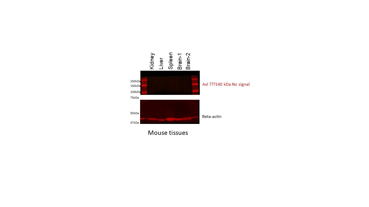
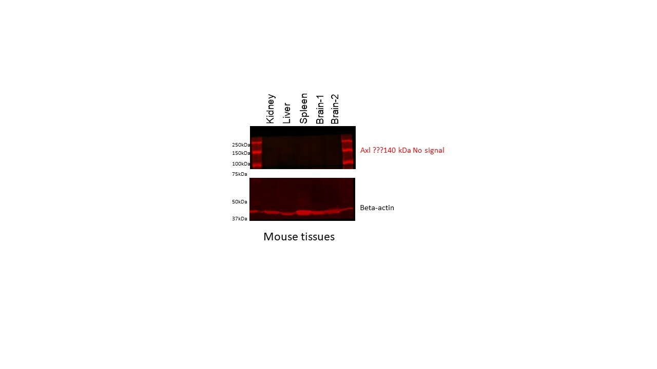
Application: Western BlotSample Tested: Adult kidney, Liver, Mouse Spleen and BrainSpecies: MouseVerified Customer | Posted 02/24/2020Mouse tissue lysate were separated on 12% SDS-PAGE. Western blotting was performed on PVDF membrane and b-actin was used as normalizer.No signal at all in any of he tissues.
-
Application: Western BlotSample Tested: Adult kidney, Adult brain, Liver tissue and Spleen tissueSpecies: MouseVerified Customer | Posted 02/24/2020
-
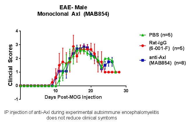
Application: Functional AssaySample Tested: Mouse brainSpecies: MouseVerified Customer | Posted 09/25/2018Mice were injected with MAB854 during EAE to activate or inhibit Axl receptor. No changes were observed.
-
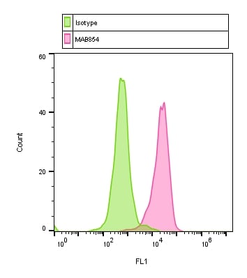
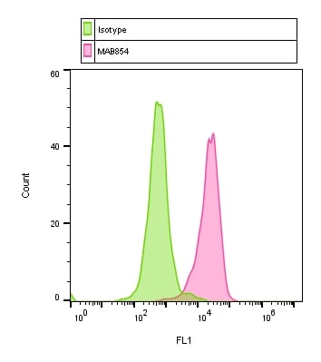
Application: Flow CytometrySample Tested: 4T1 mouse breast cancer cell lineSpecies: MouseVerified Customer | Posted 04/12/2018Staining on live cells with MAB854 and a secondary antibody.
-
Application: Flow CytometrySample Tested: Mouse cell lineSpecies: MouseVerified Customer | Posted 03/15/2018
-
Application: Flow CytometrySample Tested: Mouse CellsSpecies: MouseVerified Customer | Posted 03/13/2018
There are no reviews that match your criteria.
Protocols
Find general support by application which include: protocols, troubleshooting, illustrated assays, videos and webinars.
- Cellular Response to Hypoxia Protocols
- R&D Systems Quality Control Western Blot Protocol
- Troubleshooting Guide: Western Blot Figures
- Western Blot Conditions
- Western Blot Protocol
- Western Blot Protocol for Cell Lysates
- Western Blot Troubleshooting
- Western Blot Troubleshooting Guide
- View all Protocols, Troubleshooting, Illustrated assays and Webinars