
Cyr61, also known as IGFBP-10 and CCN1, is a 50 kDa secreted matrix- and cell-associated protein that regulates the growth and adhesion of vascular endothelial cells, fibroblasts, and monocytes. Cyr61 interacts with cells that express integrins alpha V beta 3, alpha V beta 5, alpha M beta 2, and alpha 6 beta 1. Cyr61 is cleaved by plasmin within its VWF domain which generates an N-terminal fragment that is not associated with the matrix but retains the ability to induce endothelial cell migration. Cyr61 induces VEGF upregulation, angiogenesis, and tumorigenesis. Between amino acids 176‑281, mouse Cyr61 shares 87% and 97% amino acid sequence identity with human and rat Cyr61, respectively.


Scientific images may be resized, cropped, or adjusted for brightness or contrast for presentation purposes. These updates are limited to presentation and do not affect the underlying scientific interpretation of the data. Where available, additional source data may be provided upon request.
Key Product Details
Species Reactivity
Validated:
Mouse
Cited:
Mouse, Transgenic Mouse
Applications
Validated:
Immunohistochemistry, Western Blot, Simple Western
Cited:
Immunohistochemistry, Immunohistochemistry-Frozen, Western Blot, Direct ELISA, Neutralization, Immunoprecipitation
Label
Unconjugated
Antibody Source
Polyclonal Sheep IgG
Loading...
Product Specifications
Immunogen
E. coli-derived recombinant mouse Cyr61/CCN1
Asp176-Gly281
Accession # NP_034646
Asp176-Gly281
Accession # NP_034646
Specificity
Detects mouse Cyr61/CCN1 in direct ELISAs and Western blots. In direct ELISAs, this antibody shows approximately 10% cross-reactivity with recombinant human Cyr61/CCN1.
Clonality
Polyclonal
Host
Sheep
Isotype
IgG
Scientific Data Images for Mouse Cyr61/CCN1 Antibody
Detection of Mouse Cyr61/CCN1 by Western Blot.
Western blot shows lysates of mouse spleenocyte cell, RAW 264.7 mouse monocyte/macrophage cell line, NIH-3T3 mouse embryonic fibroblast cell line, and A20 mouse B cell lymphoma cell line. PVDF membrane was probed with 1 µg/mL of Mouse Cyr61/CCN1 Antigen Affinity-purified Polyclonal Antibody (Catalog # AF4055) followed by HRP-conjugated Anti-Sheep IgG Secondary Antibody (Catalog # HAF016). A specific band was detected for Cyr61/CCN1 at approximately 50 kDa (as indicated). This experiment was conducted under reducing conditions and using Immunoblot Buffer Group 1.
Cyr61/CCN1 in Mouse Embryo.
Cyr61/CCN1 was detected in immersion fixed frozen sections of mouse embryo (13 d.p.c.) using 15 µg/mL Mouse Cyr61/CCN1 Antigen Affinity-purified Polyclonal Antibody (Catalog # AF4055) overnight at 4 °C. Tissue was stained with the Anti-Sheep HRP-DAB Cell & Tissue Staining Kit (brown; Catalog # CTS019) and counterstained with hematoxylin (blue). Specific labeling was localized to the cytoplasm of muscle cells. View our protocol for Chromogenic IHC Staining of Frozen Tissue Sections.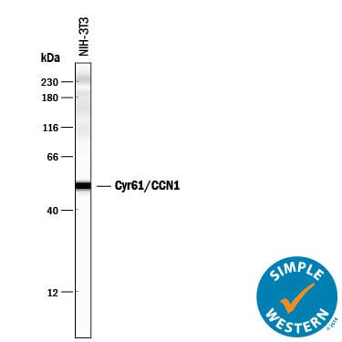
Detection of Mouse Cyr61/CCN1 by Simple WesternTM.
Simple Western lane view shows lysates of NIH-3T3 mouse embryonic fibroblast cell line, loaded at 0.2 mg/mL. A specific band was detected for Cyr61/CCN1 at approximately 51 kDa (as indicated) using 10 µg/mL of Sheep Anti-Mouse Cyr61/CCN1 Antigen Affinity-purified Polyclonal Antibody (Catalog # AF4055) followed by 1:50 dilution of HRP-conjugated Anti-Sheep IgG Secondary Antibody (Catalog # HAF016). This experiment was conducted under reducing conditions and using the 12-230 kDa separation system.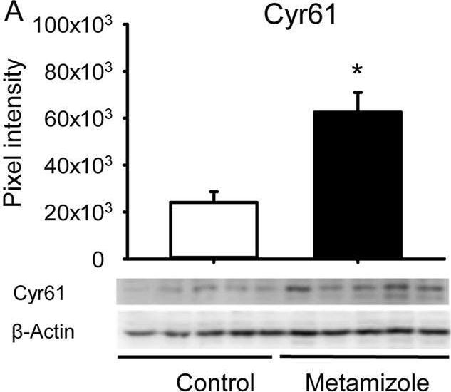
Detection of Cyr61/CCN1 by Western Blot
Western blot analysis of callus tissue. (A–F): Representative Western blots and expression of Cyr61 (A), CD31 (B), OPG (C), RANKL (D), RUNX2 (E), PCNA (F) and beta -actin (A–F) within the callus tissue of control (white; n = 5) and metamizole-treated (black; n = 5) femurs at 2 weeks after surgery. Mean ± SEM; *p < 0.05 vs. control. Image collected and cropped by CiteAb from the following open publication (https://www.frontiersin.org/articles/10.3389/fphys.2025.1630268/full), licensed under a CC-BY license. Not internally tested by R&D Systems.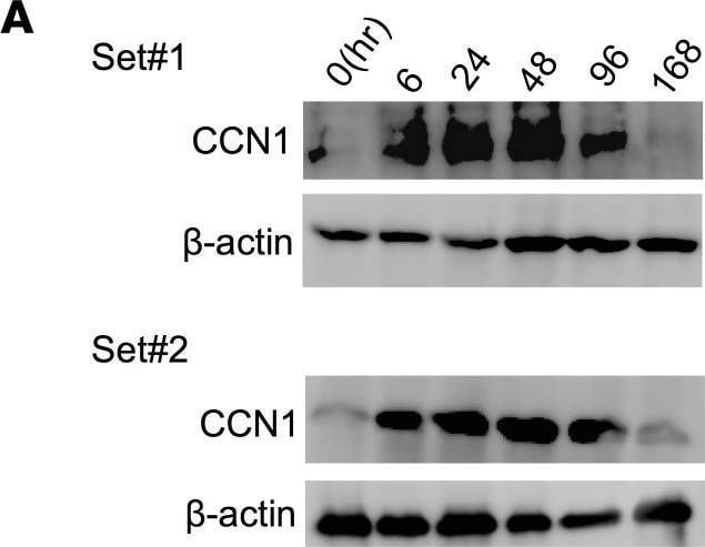
Detection of Cyr61/CCN1 by Western Blot
CCN1 induces senescence in HSCs and contributes to liver regeneration.(A) Liver protein extracts from WT mice at the indicated times (hours) after PHx were immunoblotted and probed with anti-CCN1 and anti– beta -actin antibodies. (B) The remnant liver recovery rates were assessed for WT and Ccn1DM/DM (DM) mice at indicated times after PHx (day 0, n = 3; day 2 and 7, n = 4; day 4, n = 8). (C) SA-beta -Gal staining of frozen liver sections from WT and DM mice 2 days after PHx. Liver sections were also double immunostained for p16 (red) and desmin (green) and counterstained with DAPI (blue). Yellow arrowheads are pointing to p16+ cells. The number of SA-beta -Gal+ cells (n = 6) per microscopic field and percentage of p16+ cells (n = 4) were quantified by cell counting. (D) Expression of genes related to cell cycle arrest and the SASP in WT and Ccn1DM/DM mouse livers 2 days after PHx was measured by qRT-PCR (n = 3). (E) Schematic diagram illustrating proposed SASP signaling pathways contributing to liver regeneration. The matricellular protein CCN1 is released upon injury and induces cellular senescence in HSCs through integrin alpha 6 beta 1 within 2 days after PHx. Senescent cells secrete proteins of the SASP critical to liver regeneration, including IL-6, which engages IL-6R and gp130 to induce hepatocyte proliferation through activation of STAT3 and SFK-mediated phosphorylation of YAP. IL-6 also synergizes with CXCR2 ligands secreted as part of the SASP, including CXCL1, CXCL2, and CXCL5, to induce a robust activation of ERK1/2 that promotes hepatocyte proliferation. Scale bars: 50 μm. Data expressed as mean ± SD. *P < 0.033, **P < 0.002, ***P < 0.001 assessed by Student’s t test (C and D) or 2-way ANOVA (B). Image collected and cropped by CiteAb from the following open publication (https://insight.jci.org/articles/view/158207), licensed under a CC-BY license. Not internally tested by R&D Systems.Applications for Mouse Cyr61/CCN1 Antibody
Application
Recommended Usage
Immunohistochemistry
5-15 µg/mL
Sample: Immersion fixed frozen sections of mouse embryo (13 d.p.c.)
Sample: Immersion fixed frozen sections of mouse embryo (13 d.p.c.)
Simple Western
10 µg/mL
Sample: NIH‑3T3 mouse embryonic fibroblast cell line
Sample: NIH‑3T3 mouse embryonic fibroblast cell line
Western Blot
1 µg/mL
Sample: Mouse spleenocyte cell, RAW 264.7 mouse monocyte/macrophage cell line, NIH‑3T3 mouse embryonic fibroblast cell line, and A20 mouse B cell lymphoma cell line
Sample: Mouse spleenocyte cell, RAW 264.7 mouse monocyte/macrophage cell line, NIH‑3T3 mouse embryonic fibroblast cell line, and A20 mouse B cell lymphoma cell line
Reviewed Applications
Read 2 reviews rated 5 using AF4055 in the following applications:
Formulation, Preparation, and Storage
Purification
Antigen Affinity-purified
Reconstitution
Reconstitute at 0.2 mg/mL in sterile PBS. For liquid material, refer to CoA for concentration.
Loading...
Formulation
Lyophilized from a 0.2 μm filtered solution in PBS with Trehalose. *Small pack size (SP) is supplied either lyophilized or as a 0.2 µm filtered solution in PBS.
Shipping
Lyophilized product is shipped at ambient temperature. Liquid small pack size (-SP) is shipped with polar packs. Upon receipt, store immediately at the temperature recommended below.
Stability & Storage
Use a manual defrost freezer and avoid repeated freeze-thaw cycles.
- 12 months from date of receipt, -20 to -70 °C as supplied.
- 1 month, 2 to 8 °C under sterile conditions after reconstitution.
- 6 months, -20 to -70 °C under sterile conditions after reconstitution.
Calculators
Background: Cyr61/CCN1
Long Name
Cysteine-Rich, Angiogenic Inducer 61
Alternate Names
CCN1, GIG1, IGFBP-10
Gene Symbol
CCN1
UniProt
Additional Cyr61/CCN1 Products
Product Documents for Mouse Cyr61/CCN1 Antibody
Certificate of Analysis
To download a Certificate of Analysis, please enter a lot or batch number in the search box below.
Note: Certificate of Analysis not available for kit components.
Product Specific Notices for Mouse Cyr61/CCN1 Antibody
For research use only
Related Research Areas
Citations for Mouse Cyr61/CCN1 Antibody
 Powered by Bioz
Customer Reviews for Mouse Cyr61/CCN1 Antibody (2)
5 out of 5
2 Customer Ratings
Have you used Mouse Cyr61/CCN1 Antibody?
Submit a review and receive an Amazon gift card!
$25/€18/£15/$25CAN/¥2500 for a review with an image
$10/€7/£6/$10CAN/¥1110 for a review without an image
Submit a review
Showing
1
-
2 of
2 reviews
Showing All
Filter By:
-
Application: Immunocytochemistry/ImmunofluorescenceSample Tested: A549 human lung carcinoma cell lineSpecies: MouseVerified Customer | Posted 05/20/2019
-
Application: Western BlotSample Tested: 3T3-L1 mouse embryonic fibroblast adipose-like cell lineSpecies: MouseVerified Customer | Posted 09/30/2018
There are no reviews that match your criteria.
Protocols
Find general support by application which include: protocols, troubleshooting, illustrated assays, videos and webinars.
- Antigen Retrieval Protocol (PIER)
- Antigen Retrieval for Frozen Sections Protocol
- Appropriate Fixation of IHC/ICC Samples
- Cellular Response to Hypoxia Protocols
- Chromogenic IHC Staining of Formalin-Fixed Paraffin-Embedded (FFPE) Tissue Protocol
- Chromogenic Immunohistochemistry Staining of Frozen Tissue
- ClariTSA™ Fluorophore Kits
- Detection & Visualization of Antibody Binding
- Fluorescent IHC Staining of Frozen Tissue Protocol
- Graphic Protocol for Heat-induced Epitope Retrieval
- Graphic Protocol for the Preparation and Fluorescent IHC Staining of Frozen Tissue Sections
- Graphic Protocol for the Preparation and Fluorescent IHC Staining of Paraffin-embedded Tissue Sections
- Graphic Protocol for the Preparation of Gelatin-coated Slides for Histological Tissue Sections
- IHC Sample Preparation (Frozen sections vs Paraffin)
- Immunofluorescent IHC Staining of Formalin-Fixed Paraffin-Embedded (FFPE) Tissue Protocol
- Immunohistochemistry (IHC) and Immunocytochemistry (ICC) Protocols
- Immunohistochemistry Frozen Troubleshooting
- Immunohistochemistry Paraffin Troubleshooting
- Preparing Samples for IHC/ICC Experiments
- Preventing Non-Specific Staining (Non-Specific Binding)
- Primary Antibody Selection & Optimization
- Protocol for Heat-Induced Epitope Retrieval (HIER)
- Protocol for Making a 4% Formaldehyde Solution in PBS
- Protocol for VisUCyte™ HRP Polymer Detection Reagent
- Protocol for the Preparation & Fixation of Cells on Coverslips
- Protocol for the Preparation and Chromogenic IHC Staining of Frozen Tissue Sections
- Protocol for the Preparation and Chromogenic IHC Staining of Frozen Tissue Sections - Graphic
- Protocol for the Preparation and Chromogenic IHC Staining of Paraffin-embedded Tissue Sections
- Protocol for the Preparation and Chromogenic IHC Staining of Paraffin-embedded Tissue Sections - Graphic
- Protocol for the Preparation and Fluorescent IHC Staining of Frozen Tissue Sections
- Protocol for the Preparation and Fluorescent IHC Staining of Paraffin-embedded Tissue Sections
- Protocol for the Preparation of Gelatin-coated Slides for Histological Tissue Sections
- R&D Systems Quality Control Western Blot Protocol
- TUNEL and Active Caspase-3 Detection by IHC/ICC Protocol
- The Importance of IHC/ICC Controls
- Troubleshooting Guide: Immunohistochemistry
- Troubleshooting Guide: Western Blot Figures
- Western Blot Conditions
- Western Blot Protocol
- Western Blot Protocol for Cell Lysates
- Western Blot Troubleshooting
- Western Blot Troubleshooting Guide
- View all Protocols, Troubleshooting, Illustrated assays and Webinars
Loading...