Glypican 6 (glypiated proteoglycan 6; also GPC6) is an 85-95 kDa heparan sulfate proteoglycan member of the GPI-linked glypican family of molecules. It is expressed by postnatal chrondrocytes and embryonic mesenchyme (connective tissue plus smooth muscle cells), and potentially modulates the availability, or signaling, of heparin-binding growth factors. Mouse Glypican 6 is synthesized as a 532 amino acid (aa) proprecursor that contains a 507 mature region (aa 4-530) and a 25 aa C-terminal prosegment. There is one potential splice variant that shows a 10 aa insertion after Leu384. Over aa 4-513, mouse Glypican 6 shares 99% aa identity with both human and rat Glypican 6.
Mouse Glypican 6 Antibody
R&D Systems | Catalog # AF1053

Scientific images may be resized, cropped, or adjusted for brightness or contrast for presentation purposes. These updates are limited to presentation and do not affect the underlying scientific interpretation of the data. Where available, additional source data may be provided upon request.
Key Product Details
Species Reactivity
Validated:
Mouse
Cited:
Human, Mouse, Rat
Applications
Validated:
Immunohistochemistry, Western Blot
Cited:
Immunohistochemistry, Western Blot
Label
Unconjugated
Antibody Source
Polyclonal Goat IgG
Loading...
Product Specifications
Immunogen
Mouse myeloma cell line NS0-derived recombinant mouse Glypican 6
Asp24-Val513
Accession # Q9R087
Asp24-Val513
Accession # Q9R087
Specificity
Detects mouse Glypican 6 in direct ELISAs and Western blots.
Clonality
Polyclonal
Host
Goat
Isotype
IgG
Scientific Data Images for Mouse Glypican 6 Antibody
Detection of Mouse Glypican 6 by Western Blot
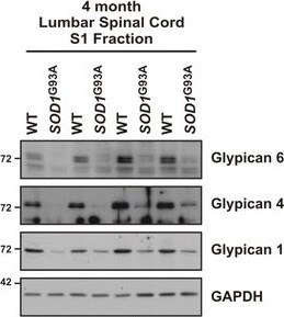
GDE2 substrates show impaired release in SOD1G93A mice. a Schematic of the sequential solubilization protocol used to separate hydrophilic non-membrane proteins (S1) from hydrophobic membrane proteins (S2). b Western blot demonstrating the successful enrichment of non-membrane protein (GAPDH) in the S1 fraction and membrane protein (Na/K ATPase) in the S2 fraction for WT and SOD1G93A transgenic mice. c Western blots visualizing the impaired production of non-membrane bound (S1) Glypican 6, Glypican 4, and Glypican 1 in the lumbar spinal cord of 4 month SOD1G93A mice. d Quantification of the reduced membrane release of Glypican 6, *p = 0.038; Glypican 4, *p = 0.016; and Glypican 1, *p = 0.030 relative to GAPDH. Graph represents mean ± SEM. Student’s t test, n = 4 WT, 4 SOD1G93A. Image collected and cropped by CiteAb from the following open publication (https://molecularneurodegeneration.biomedcentral.com/articles/10.1186/s…), licensed under a CC-BY license. Not internally tested by R&D Systems.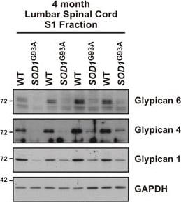
Detection of Mouse Glypican 6 by Western Blot
GDE2 substrates show impaired release in SOD1G93A mice. a Schematic of the sequential solubilization protocol used to separate hydrophilic non-membrane proteins (S1) from hydrophobic membrane proteins (S2). b Western blot demonstrating the successful enrichment of non-membrane protein (GAPDH) in the S1 fraction and membrane protein (Na/K ATPase) in the S2 fraction for WT and SOD1G93A transgenic mice. c Western blots visualizing the impaired production of non-membrane bound (S1) Glypican 6, Glypican 4, and Glypican 1 in the lumbar spinal cord of 4 month SOD1G93A mice. d Quantification of the reduced membrane release of Glypican 6, *p = 0.038; Glypican 4, *p = 0.016; and Glypican 1, *p = 0.030 relative to GAPDH. Graph represents mean ± SEM. Student’s t test, n = 4 WT, 4 SOD1G93A Image collected and cropped by CiteAb from the following open publication (https://molecularneurodegeneration.biomedcentral.com/articles/10.1186/s…), licensed under a CC-BY license. Not internally tested by R&D Systems.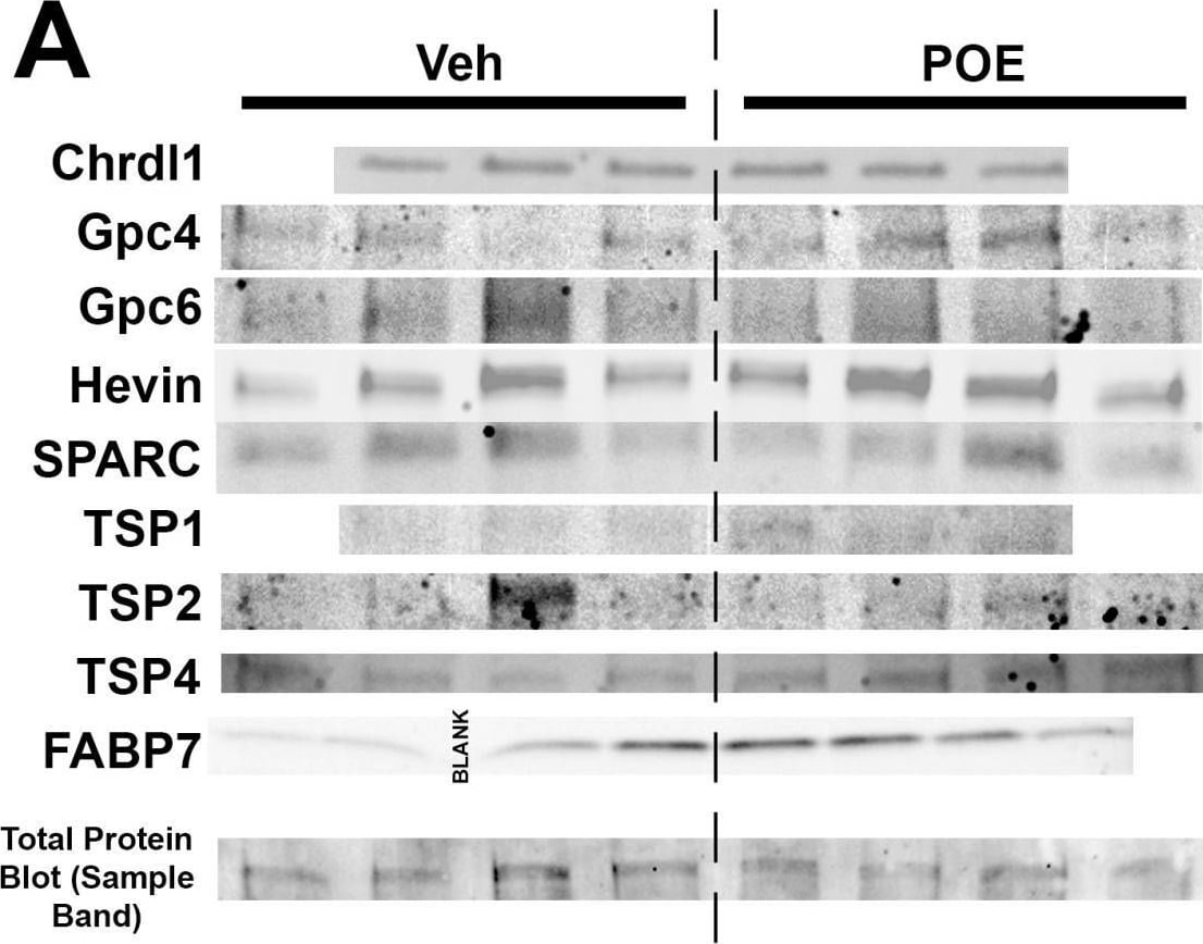
Detection of Glypican 6 by Western Blot
Changes in synapse-related protein expression in POE astrocytes. (A) Western blot images reveal expression patterns of prominent synapse-associated proteins from the DIV10 vehicle and POE astrocyte lysates. Bottom row shows a representative band from one of the total protein blot images that was used for normalization purposes (full image shown in Figure S1). (B) Quantification of Western blots for proteins indicated in (A). Data shown as fold-change in band intensity values for POE astrocyte lysates compared to vehicle (n = 3–4 experimental replicates; unpaired t-test (unlabeled p values were not significant)). Image collected and cropped by CiteAb from the following open publication (https://pubmed.ncbi.nlm.nih.gov/38786059), licensed under a CC-BY license. Not internally tested by R&D Systems.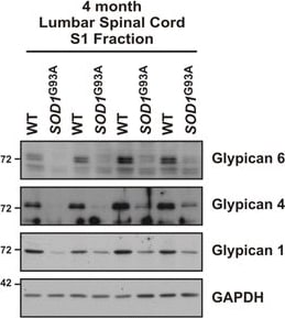
Detection of Glypican 6 by Western Blot
GDE2 substrates show impaired release in SOD1G93A mice. a Schematic of the sequential solubilization protocol used to separate hydrophilic non-membrane proteins (S1) from hydrophobic membrane proteins (S2). b Western blot demonstrating the successful enrichment of non-membrane protein (GAPDH) in the S1 fraction and membrane protein (Na/K ATPase) in the S2 fraction for WT and SOD1G93A transgenic mice. c Western blots visualizing the impaired production of non-membrane bound (S1) Glypican 6, Glypican 4, and Glypican 1 in the lumbar spinal cord of 4 month SOD1G93A mice. d Quantification of the reduced membrane release of Glypican 6, *p = 0.038; Glypican 4, *p = 0.016; and Glypican 1, *p = 0.030 relative to GAPDH. Graph represents mean ± SEM. Student’s t test, n = 4 WT, 4 SOD1G93A. Image collected and cropped by CiteAb from the following open publication (https://pubmed.ncbi.nlm.nih.gov/28103900), licensed under a CC-BY license. Not internally tested by R&D Systems.Applications for Mouse Glypican 6 Antibody
Application
Recommended Usage
Immunohistochemistry
5-15 µg/mL
Sample: Immersion fixed frozen sections of mouse embryo (E13)
Sample: Immersion fixed frozen sections of mouse embryo (E13)
Western Blot
0.1 µg/mL
Sample: Recombinant Mouse Glypican 6
Sample: Recombinant Mouse Glypican 6
Formulation, Preparation, and Storage
Purification
Antigen Affinity-purified
Reconstitution
Reconstitute at 0.2 mg/mL in sterile PBS. For liquid material, refer to CoA for concentration.
Loading...
Formulation
Lyophilized from a 0.2 μm filtered solution in PBS with Trehalose. *Small pack size (SP) is supplied either lyophilized or as a 0.2 µm filtered solution in PBS.
Shipping
Lyophilized product is shipped at ambient temperature. Liquid small pack size (-SP) is shipped with polar packs. Upon receipt, store immediately at the temperature recommended below.
Stability & Storage
Use a manual defrost freezer and avoid repeated freeze-thaw cycles.
- 12 months from date of receipt, -20 to -70 °C as supplied.
- 1 month, 2 to 8 °C under sterile conditions after reconstitution.
- 6 months, -20 to -70 °C under sterile conditions after reconstitution.
Calculators
Background: Glypican 6
Additional Glypican 6 Products
Product Documents for Mouse Glypican 6 Antibody
Certificate of Analysis
To download a Certificate of Analysis, please enter a lot or batch number in the search box below.
Note: Certificate of Analysis not available for kit components.
Product Specific Notices for Mouse Glypican 6 Antibody
For research use only
Related Research Areas
Citations for Mouse Glypican 6 Antibody
 Powered by Bioz
Customer Reviews for Mouse Glypican 6 Antibody
There are currently no reviews for this product. Be the first to review Mouse Glypican 6 Antibody and earn rewards!
Have you used Mouse Glypican 6 Antibody?
Submit a review and receive an Amazon gift card!
$25/€18/£15/$25CAN/¥2500 for a review with an image
$10/€7/£6/$10CAN/¥1110 for a review without an image
Submit a review
Protocols
Find general support by application which include: protocols, troubleshooting, illustrated assays, videos and webinars.
- Antigen Retrieval Protocol (PIER)
- Antigen Retrieval for Frozen Sections Protocol
- Appropriate Fixation of IHC/ICC Samples
- Cellular Response to Hypoxia Protocols
- Chromogenic IHC Staining of Formalin-Fixed Paraffin-Embedded (FFPE) Tissue Protocol
- Chromogenic Immunohistochemistry Staining of Frozen Tissue
- ClariTSA™ Fluorophore Kits
- Detection & Visualization of Antibody Binding
- Fluorescent IHC Staining of Frozen Tissue Protocol
- Graphic Protocol for Heat-induced Epitope Retrieval
- Graphic Protocol for the Preparation and Fluorescent IHC Staining of Frozen Tissue Sections
- Graphic Protocol for the Preparation and Fluorescent IHC Staining of Paraffin-embedded Tissue Sections
- Graphic Protocol for the Preparation of Gelatin-coated Slides for Histological Tissue Sections
- IHC Sample Preparation (Frozen sections vs Paraffin)
- Immunofluorescent IHC Staining of Formalin-Fixed Paraffin-Embedded (FFPE) Tissue Protocol
- Immunohistochemistry (IHC) and Immunocytochemistry (ICC) Protocols
- Immunohistochemistry Frozen Troubleshooting
- Immunohistochemistry Paraffin Troubleshooting
- Preparing Samples for IHC/ICC Experiments
- Preventing Non-Specific Staining (Non-Specific Binding)
- Primary Antibody Selection & Optimization
- Protocol for Heat-Induced Epitope Retrieval (HIER)
- Protocol for Making a 4% Formaldehyde Solution in PBS
- Protocol for VisUCyte™ HRP Polymer Detection Reagent
- Protocol for the Preparation & Fixation of Cells on Coverslips
- Protocol for the Preparation and Chromogenic IHC Staining of Frozen Tissue Sections
- Protocol for the Preparation and Chromogenic IHC Staining of Frozen Tissue Sections - Graphic
- Protocol for the Preparation and Chromogenic IHC Staining of Paraffin-embedded Tissue Sections
- Protocol for the Preparation and Chromogenic IHC Staining of Paraffin-embedded Tissue Sections - Graphic
- Protocol for the Preparation and Fluorescent IHC Staining of Frozen Tissue Sections
- Protocol for the Preparation and Fluorescent IHC Staining of Paraffin-embedded Tissue Sections
- Protocol for the Preparation of Gelatin-coated Slides for Histological Tissue Sections
- R&D Systems Quality Control Western Blot Protocol
- TUNEL and Active Caspase-3 Detection by IHC/ICC Protocol
- The Importance of IHC/ICC Controls
- Troubleshooting Guide: Immunohistochemistry
- Troubleshooting Guide: Western Blot Figures
- Western Blot Conditions
- Western Blot Protocol
- Western Blot Protocol for Cell Lysates
- Western Blot Troubleshooting
- Western Blot Troubleshooting Guide
- View all Protocols, Troubleshooting, Illustrated assays and Webinars
Loading...
Associated Pathways