
IGF-II (Insulin-like growth factor II; also multiplication-stimulating polypeptide/MSP and somatomedin-A) is a secreted 8 kDa polypeptide that belongs to the insulin family of peptide growth factors (1‑3). It is part of a complex system of growth and metabolic-regulating proteins that is particularly important during development. It has been associated with nervous system proliferation and differentiation, myelination, adrenal cortical proliferation, and skeletal growth and differentiation (4). In humans, IGF-II is primarily synthesized by the liver and circulates at high levels in both fetus and adult. In rodents, however, IGF-II levels drop after the perinatal period, an effect attributed to the lack of a key gene promoter (2, 5). This may indicate that postnatally, IGF-II has either a limited or local effect only in rodents. For example, evidence suggests IGF-II may be the intermediary for SHH induction of VEGF attendant with local neovascularization (6). Rodent cells known to express IGF-II include astrocytes (7), hepatocytes (8), osteoblasts (9), embryonic striated muscle cells (10, 11), plus Kupffer cells and Ito cells (12). Mouse IGF-II is synthesized as a 180 amino acid (aa) preproprecursor (13). It contains a 24 aa signal sequence, a 67 aa mature region, and an 89 aa C-terminal prodomain that is alternatively referred to as the E-peptide. Mature IGF-II is 91% and 97% aa identical to human and rat IGF-II, respectively. Proper processing of IGF-II requires the chaperone activity of GRP94 (14). This generates an 8 kDa mature form, an 18 kDa, 156 aa proform, and a potential 11 kDa, 88 aa “Big” form (aa 25-112). This 11 kDa ”Big” form would be equivalent to human 15-16 kDa IGF-II, with the 5 kDa difference attributable to the presence of O-linked glycosylation (15). There is an additional 34 aa proteolytic fragment that is termed preptin and contains aa 93-126 of the preproprecursor. This is distinct from IGF-II, is secreted by pancreatic B cells, and facilitates insulin secretion (16, 17). IGF-II has multiple binding partners. It binds to IGF-I R, the Insulin receptor (IR)-type A and IGF-IR:IR-A hybrids, the type II IGF receptor (IGF-II R), and IGF binding proteins 1-6 (18, 19). The first three receptors initiate downstream signaling events, the IGF-II R sequesters local IGF‑II, and the six IGFBPs regulate IGF-II activity in various tissues.
Mouse IGF-II/IGF2 Antibody
R&D Systems | Catalog # AF792

Scientific images may be resized, cropped, or adjusted for brightness or contrast for presentation purposes. These updates are limited to presentation and do not affect the underlying scientific interpretation of the data. Where available, additional source data may be provided upon request.
Key Product Details
Species Reactivity
Validated:
Cited:
Applications
Validated:
Cited:
Label
Antibody Source
Product Specifications
Immunogen
Ala25-Glu91
Accession # P09535
Specificity
Clonality
Host
Isotype
Endotoxin Level
Scientific Data Images for Mouse IGF-II/IGF2 Antibody
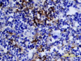
IGF-II/IGF2 in Mouse Embryo.
IGF-II/IGF2 was detected in immersion fixed frozen sections of mouse embryo (E13-17) using Mouse IGF-II/IGF2 Antigen Affinity-purified Polyclonal Antibody (Catalog # AF792) at 15 µg/mL overnight at 4 °C. Tissue was stained using the Anti-Goat HRP-DAB Cell & Tissue Staining Kit (brown; Catalog # CTS008) and counterstained with hematoxylin (blue). View our protocol for Chromogenic IHC Staining of Frozen Tissue Sections.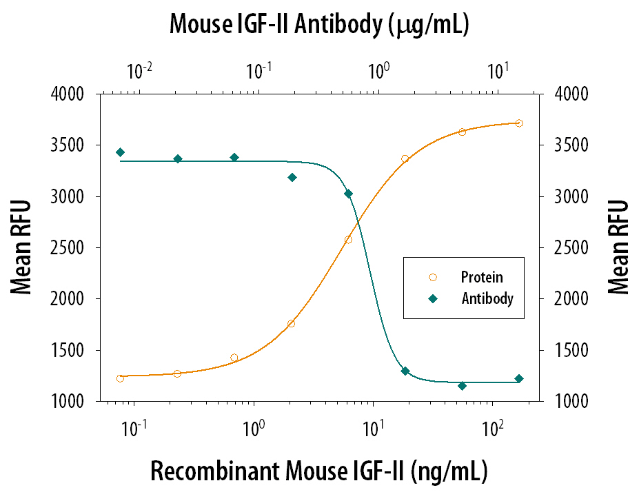
Cell Proliferation Induced by IGF-II/IGF2 and Neutralization by Mouse IGF-II/IGF2 Antibody.
Recombinant Mouse IGF-II/IGF2 (Catalog # 792-MG) stimulates proliferation in the MCF-7 human breast cancer cell line in a dose-dependent manner (orange line). Proliferation elicited by Recombinant Mouse IGF-II/IGF2 (30 ng/mL) is neutralized (green line) by increasing concentrations of Mouse IGF-II/IGF2 Antigen Affinity-purified Polyclonal Antibody (Catalog # AF792). The ND50 is typically 0.5-2.5 µg/mL.
Mouse IGF-II ELISA Standard Curve
Recombinant Mouse IGF-II/IGF2 (Catalog # 792-MG) was serially diluted and captured by Rat Anti-Mouse IGF-II/IGF2 Monoclonal Antibody (Catalog # MAB792) coated on a Clear Polystyrene Microplate (Catalog # DY990). Goat Anti-Mouse IGF-II/IGF2 Antigen Affinity-purified Polyclonal Antibody (Catalog # AF792) was biotinylated and incubated with the protein captured on the plate. Detection of the standard curve was achieved by incubating Streptavidin-HRP (Catalog # DY998)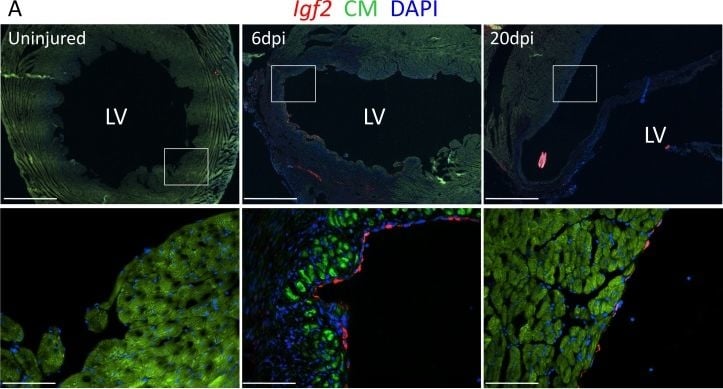
Detection of IGF-II/IGF2 by Immunohistochemistry
Adult heart injury induces Igf2 expression.(A) Sections of adult hearts that were uninjured or at 6 days and 20 days after coronary artery ligation. In the injured hearts, the infarction region is evident by absence of cardiomyocyte autofluorescence (green). Igf2 is not expressed in the uninjured heart, but is expressed after infarction in the endocardium. (B) RT-PCR analysis of Igf2 expression normalized to 18S rRNA, showing minimal expression in uninjured heart and induction after injury. The first lane is a size marker. (C) No activation of INSR phosphorylation after adult heart injury. Image collected and cropped by CiteAb from the following open publication (https://pubmed.ncbi.nlm.nih.gov/32167474), licensed under a CC-BY license. Not internally tested by R&D Systems.Applications for Mouse IGF-II/IGF2 Antibody
Immunohistochemistry
Sample: Immersion fixed frozen sections of mouse embryo (E13-17)
Western Blot
Sample: Recombinant Mouse IGF-II/IGF2 (Catalog # 792-MG)
Neutralization
Formulation, Preparation, and Storage
Purification
Reconstitution
Reconstitute at 0.2 mg/mL in sterile PBS. For liquid material, refer to CoA for concentration.
Formulation
Shipping
Stability & Storage
- 12 months from date of receipt, -20 to -70 °C as supplied.
- 1 month, 2 to 8 °C under sterile conditions after reconstitution.
- 6 months, -20 to -70 °C under sterile conditions after reconstitution.
Calculators
Background: IGF-II/IGF2
References
- LeRoith, D. and C.T. Roberts Jr. (2003) Cancer Lett. 195:127.
- Werner, H. and D. LeRoith (2000) Cell. Mol. Life Sci. 57:932.
- Pavelic, J. et al. (2007) Indian J. Med. Res. 125:511.
- Varela-Nieto, I. et al. (2007) Curr. Pharm. Des. 13:687.
- Rotwein, P. and L.J. Hall (1990) DNA Cell Biol. 10:725.
- Chao, W. and P.A. D-Amore (2008) Cytokine Growth Factor Rev. 19:111.
- Rotwein, P. et al. (1988) Proc. Natl. Acad. Sci. USA 85:265.
- Goya, L. et al. (1999) J. Biol. Chem. 274:24633.
- McCarthy, T.L. et al. (1992) Endocrinology 130:1303.
- Zindy, F. et al. (1992) J. Hepatol. 14:30.
- Holthuizen, P.E. et al. (1993) Regul. Pept. 48:77.
- Merrick, D. et al. (2007) BMC Dev. Biol. 7:65.
- Stempien, M.M. et al. (1986) DNA 5:357.
- Ostrovsky, O. et al. (2009) Mol. Biol. Cell 20:1855.
- Daughaday, W.H. et al. (1993) Proc. Natl. Acad. Sci. USA 90:5823.
- Buchanan, C.M. et al. (2001) Biochem. J. 360:431.
- Cornish, J. et al. (2007) Am. J. Physiol. Endocrinol. Metab. 292:E117.
- Denley, A. et al. (2005) Cytokine Growth Factor Rev. 16:421.
- Belfiore, A. (2007) Curr. Pharm. Des. 13:671.
Long Name
Alternate Names
Gene Symbol
UniProt
Additional IGF-II/IGF2 Products
Product Documents for Mouse IGF-II/IGF2 Antibody
Certificate of Analysis
To download a Certificate of Analysis, please enter a lot or batch number in the search box below.
Note: Certificate of Analysis not available for kit components.
Product Specific Notices for Mouse IGF-II/IGF2 Antibody
For research use only
Citations for Mouse IGF-II/IGF2 Antibody
 Powered by Bioz
Customer Reviews for Mouse IGF-II/IGF2 Antibody
There are currently no reviews for this product. Be the first to review Mouse IGF-II/IGF2 Antibody and earn rewards!
Have you used Mouse IGF-II/IGF2 Antibody?
Submit a review and receive an Amazon gift card!
$25/€18/£15/$25CAN/¥2500 for a review with an image
$10/€7/£6/$10CAN/¥1110 for a review without an image
Submit a review
Protocols
Find general support by application which include: protocols, troubleshooting, illustrated assays, videos and webinars.
- Antigen Retrieval Protocol (PIER)
- Antigen Retrieval for Frozen Sections Protocol
- Appropriate Fixation of IHC/ICC Samples
- Cellular Response to Hypoxia Protocols
- Chromogenic IHC Staining of Formalin-Fixed Paraffin-Embedded (FFPE) Tissue Protocol
- Chromogenic Immunohistochemistry Staining of Frozen Tissue
- ClariTSA™ Fluorophore Kits
- Detection & Visualization of Antibody Binding
- Fluorescent IHC Staining of Frozen Tissue Protocol
- Graphic Protocol for Heat-induced Epitope Retrieval
- Graphic Protocol for the Preparation and Fluorescent IHC Staining of Frozen Tissue Sections
- Graphic Protocol for the Preparation and Fluorescent IHC Staining of Paraffin-embedded Tissue Sections
- Graphic Protocol for the Preparation of Gelatin-coated Slides for Histological Tissue Sections
- IHC Sample Preparation (Frozen sections vs Paraffin)
- Immunofluorescent IHC Staining of Formalin-Fixed Paraffin-Embedded (FFPE) Tissue Protocol
- Immunohistochemistry (IHC) and Immunocytochemistry (ICC) Protocols
- Immunohistochemistry Frozen Troubleshooting
- Immunohistochemistry Paraffin Troubleshooting
- Preparing Samples for IHC/ICC Experiments
- Preventing Non-Specific Staining (Non-Specific Binding)
- Primary Antibody Selection & Optimization
- Protocol for Heat-Induced Epitope Retrieval (HIER)
- Protocol for Making a 4% Formaldehyde Solution in PBS
- Protocol for VisUCyte™ HRP Polymer Detection Reagent
- Protocol for the Preparation & Fixation of Cells on Coverslips
- Protocol for the Preparation and Chromogenic IHC Staining of Frozen Tissue Sections
- Protocol for the Preparation and Chromogenic IHC Staining of Frozen Tissue Sections - Graphic
- Protocol for the Preparation and Chromogenic IHC Staining of Paraffin-embedded Tissue Sections
- Protocol for the Preparation and Chromogenic IHC Staining of Paraffin-embedded Tissue Sections - Graphic
- Protocol for the Preparation and Fluorescent IHC Staining of Frozen Tissue Sections
- Protocol for the Preparation and Fluorescent IHC Staining of Paraffin-embedded Tissue Sections
- Protocol for the Preparation of Gelatin-coated Slides for Histological Tissue Sections
- R&D Systems Quality Control Western Blot Protocol
- TUNEL and Active Caspase-3 Detection by IHC/ICC Protocol
- The Importance of IHC/ICC Controls
- Troubleshooting Guide: Immunohistochemistry
- Troubleshooting Guide: Western Blot Figures
- Western Blot Conditions
- Western Blot Protocol
- Western Blot Protocol for Cell Lysates
- Western Blot Troubleshooting
- Western Blot Troubleshooting Guide
- View all Protocols, Troubleshooting, Illustrated assays and Webinars