
Legumain is a lysosomal cysteine protease found in all mouse tissues examined, but was particularly abundant in kidney and placenta (1). Legumain plays a pivotal role in the endosomal/lysosomal degradation system because the Legumain deficiency causes the accumulation of pro cathepsins B, H and L, another group of lysosomal cysteine proteases (2). Over-expression of Legumain in tumors is significant for invasion/metastasis (3). Also known as asparaginyl endopeptidase, it specifically cleaves peptide bonds with Asn at the P1 position. Nevertheless, it also cleaves peptide bonds with Asp at the P1 position. Auto-activation of pro Legumain involves both types of cleavage, which results in the removal of the pro peptides in both C- and N-termini (4). In addition, Legumain activates pro MMP-2 and processes bacterial antigens for MHC class II presentation and pro thymosin alpha to thymosin alpha 1 and thymosin alpha 11, two acidic peptides with immunoregulatory properties (5-7).
Mouse Legumain/Asparaginyl Endopeptidase Antibody
R&D Systems | Catalog # AF2058

Scientific images may be resized, cropped, or adjusted for brightness or contrast for presentation purposes. These updates are limited to presentation and do not affect the underlying scientific interpretation of the data. Where available, additional source data may be provided upon request.
Key Product Details
Species Reactivity
Validated:
Cited:
Applications
Validated:
Cited:
Label
Antibody Source
Product Specifications
Immunogen
Val18-Tyr435
Accession # O89017
Specificity
Clonality
Host
Isotype
Scientific Data Images for Mouse Legumain/Asparaginyl Endopeptidase Antibody
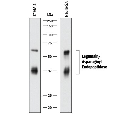
Detection of Mouse Legumain/Asparaginyl Endopeptidase by Western Blot.
Western blot shows lysates of J774A.1 mouse reticulum cell sarcoma macrophage cell line and Neuro-2A mouse neuroblastoma cell line. PVDF membrane was probed with 0.2 µg/mL of Sheep Anti-Mouse Legumain/Asparaginyl Endopeptidase Antigen Affinity-purified Polyclonal Antibody (Catalog # AF2058) followed by HRP-conjugated Anti-Sheep IgG Secondary Antibody (Catalog # HAF016). Specific bands were detected for Legumain/Asparaginyl Endopeptidase at approximately 37 and 56 kDa (as indicated). This experiment was conducted under reducing conditions and using Immunoblot Buffer Group 1.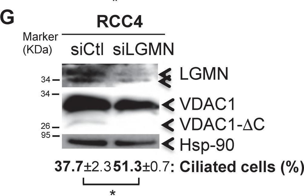
Detection of Legumain/Asparaginyl Endopeptidase by Western Blot
The presence of VDAC1-DC in ccRCC cells decreases or abolishes ciliation. A, Triple immunofluorescence labeling and merged images with acetylated alpha -tubulin (Acet. alpha -tubulin in red), Arl13b (in green) and DAPI (in blue). B, Electron microscopy of RCC4+pVHL cells. C, Quantitative analysis of the ciliation percentage was assessed by confocal fluorescence microscopy (n=100-300 cells). D, Both cell lines were seeded at the same density and incubated in Nx for 48h with or without serum. Percentage of ciliated cells, proliferation and FACS analysis were measured. The mean ± SEM is representative of three independent experiments carried out in duplicate. E, F and G, RCC4 cells were transfected with control siRNA (siCtl), (E) siHIF-1 alpha, siHIF-2 alpha and siHIF-1/2 alpha, (F) siVDAC1 and (G) siLGMN. Cell lysates were analyzed by immunoblotting for HIF-1 alpha, HIF-2 alpha, VDAC1, LGMN and beta - tubulin/Actin or HSP90 were used as a loading control. Quantitative analysis of the ciliation percentage was assessed by confocal fluorescence microscopy (n=100-300 cells). A * p<0.05 shows significant differences. Quantification of VDAC1 and VDAC1-delta C protein levels (E). Experiments have been proceeded without serum. Image collected and cropped by CiteAb from the following open publication (https://pubmed.ncbi.nlm.nih.gov/32194829), licensed under a CC-BY license. Not internally tested by R&D Systems.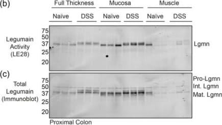
Detection of Legumain/Asparaginyl Endopeptidase by Western Blot
Mucosal legumain is increased during acute colitis. (a) Legumain activity in colons harvested from naïve mice or mice treated with DSS for 6 days, as shown by ex vivo imaging of LE28 fluorescence. Three biological replicates per group are shown. Color scale represents radiant efficiency/109 (p/sec/cm2/sr)/(μW/cm2). (b) Legumain activity in proximal colon tissues shown in A, either full thickness, or dissected into mucosal and muscle layers, as shown by in‐gel fluorescence of LE28. (c) Immunoblot of gel shown in B with a legumain‐specific antibody. (d) Ex vivo labeling of lysates prepared from naïve and DSS‐treated colon tissue with LE28, as shown by in‐gel fluorescence. Three biological replicates per group are shown. (e) Immunoprecipitation of LE28‐labeled lysates prepared from full thickness colons in shown in A with a legumain‐specific antibody. I, input; P, pulldown. (f) In‐gel fluorescence showing LE28 labeling in luminal fluids and fecal samples from mice imaged in A. *Designates LE28‐labeled species of unknown identity. (g) LE28 fluorescence in naïve mice compared to those treated with TNBS for 3 days or DSS for 6 days. Three biological replicates are shown. (h) In‐gel fluorescence of the LE28‐labeled proximal and distal colons shown in H. Note the x designates a lost distal colon sample. Color scale represents radiant efficiency/108 (p/sec/cm2/sr)/(μW/cm2). Image collected and cropped by CiteAb from the following open publication (https://pubmed.ncbi.nlm.nih.gov/39392222), licensed under a CC-BY license. Not internally tested by R&D Systems.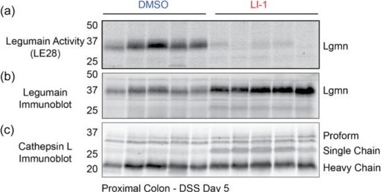
Detection of Legumain/Asparaginyl Endopeptidase by Western Blot
Legumain inhibition does not prevent DSS‐induced acute colitis. (a) Assessment of legumain activity in DSS‐treated proximal colons after daily treatment with DMSO or LI‐1 (25 mg/kg), as shown by in‐gel fluorescence of LE28. (b, c) Immunoblot with antibodies specific to legumain or cathepsin L on the samples shown in A. Five biological replicates are shown. (d) Body weight, shown as a percentage of starting weight, over the course of six days in naïve mice or those treated with DSS and DMSO or LI‐1). (e) Disease activity index (DAI) of mice over time. (f) Colon length at endpoint (Day 6). (g–j) Histological scoring of colon sections, including immune infiltration, crypt disorganization, goblet cell cavitation and total histology score. Error bars represent mean ± SEM, and 5–6 mice/group are reported. No significant differences between DMSO‐ and LI‐1‐treated mice were observed. Image collected and cropped by CiteAb from the following open publication (https://pubmed.ncbi.nlm.nih.gov/39392222), licensed under a CC-BY license. Not internally tested by R&D Systems.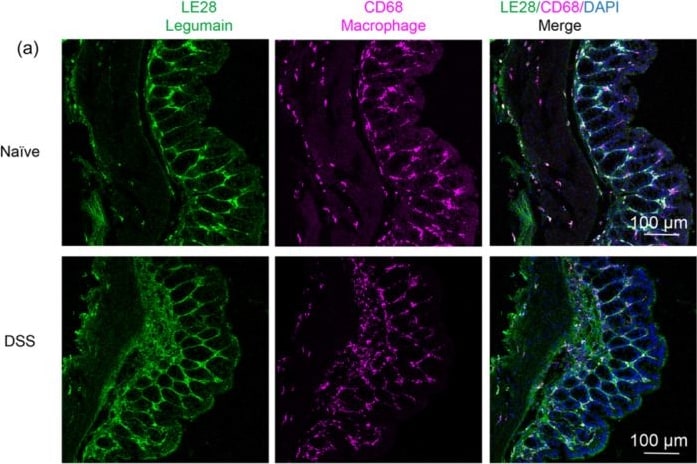
Detection of Legumain/Asparaginyl Endopeptidase by Immunohistochemistry
Legumain is present in macrophages and colonocytes of the colon. (a) LE28 fluorescence showing legumain activity (green), CD68 immunoreactivity (magenta), or the two merged with DAPI (blue) in colon tissue sections from naïve mice or those treated with DSS for 6 days. (b) Legumain immunoreactivity in colon tissue sections from naïve or DSS‐treated mice. Black arrowheads denote colonocytes while red show macrophages. Image collected and cropped by CiteAb from the following open publication (https://pubmed.ncbi.nlm.nih.gov/39392222), licensed under a CC-BY license. Not internally tested by R&D Systems.Applications for Mouse Legumain/Asparaginyl Endopeptidase Antibody
Western Blot
Sample: J774A.1 mouse reticulum cell sarcoma macrophage cell line and Neuro‑2A mouse neuroblastoma cell line
Formulation, Preparation, and Storage
Purification
Reconstitution
Reconstitute at 0.2 mg/mL in sterile PBS. For liquid material, refer to CoA for concentration.
Formulation
Shipping
Stability & Storage
- 12 months from date of receipt, -20 to -70 °C as supplied.
- 1 month, 2 to 8 °C under sterile conditions after reconstitution.
- 6 months, -20 to -70 °C under sterile conditions after reconstitution.
Calculators
Background: Legumain/Asparaginyl Endopeptidase
References
- Chen, J.M. et al. (1998) Biochem. J. 335:111.
- Shirahama-Noda, K. et al. (2003) J. Biol. Chem. 278:33194.
- Liu, C. et al. (2003) Cancer Res. 63: 2957.
- Li D.N. et al. (2003) J. Biol. Chem. 278:38980.
- Chen, J.M. et al. (2001) Biol. Chem. 382:777.
- Schwarz, G. et al. (2002) Biol. Chem. 383:1813.
- Sarndeses, C.S. et al. (2003) J. Biol. Chem. 278:13286.
Alternate Names
Gene Symbol
UniProt
Additional Legumain/Asparaginyl Endopeptidase Products
Product Documents for Mouse Legumain/Asparaginyl Endopeptidase Antibody
Certificate of Analysis
To download a Certificate of Analysis, please enter a lot or batch number in the search box below.
Note: Certificate of Analysis not available for kit components.
Product Specific Notices for Mouse Legumain/Asparaginyl Endopeptidase Antibody
For research use only
Related Research Areas
Citations for Mouse Legumain/Asparaginyl Endopeptidase Antibody
 Powered by Bioz
Customer Reviews for Mouse Legumain/Asparaginyl Endopeptidase Antibody
There are currently no reviews for this product. Be the first to review Mouse Legumain/Asparaginyl Endopeptidase Antibody and earn rewards!
Have you used Mouse Legumain/Asparaginyl Endopeptidase Antibody?
Submit a review and receive an Amazon gift card!
$25/€18/£15/$25CAN/¥2500 for a review with an image
$10/€7/£6/$10CAN/¥1110 for a review without an image
Submit a review
Protocols
Find general support by application which include: protocols, troubleshooting, illustrated assays, videos and webinars.
- Cellular Response to Hypoxia Protocols
- R&D Systems Quality Control Western Blot Protocol
- Troubleshooting Guide: Western Blot Figures
- Western Blot Conditions
- Western Blot Protocol
- Western Blot Protocol for Cell Lysates
- Western Blot Troubleshooting
- Western Blot Troubleshooting Guide
- View all Protocols, Troubleshooting, Illustrated assays and Webinars