
Oncostatin M (OSM) is a member of a cytokine subfamily that includes IL-6, IL-11, LIF, CNTF, and Cardiotrophin-1. These cytokines have overlapping biological functions and shared receptor components. Mouse OSM was cloned and identified as an immediate early gene induced in various myeloid and lymphoid cell lines by a subset of cytokines including IL-2, IL-3, GM-CSF, and Erythropoietin. The mouse OSM cDNA encodes a 263 amino acid residue precursor protein that shows 48% identity with human OSM. Similar to human OSM, the C-terminal region of mouse OSM contains a highly charged region. Deletion of this C-terminal region appears to be essential for the formation of biologically active mouse OSM. The biological activity of human OSM has been shown to be mediated either by the LIF/OSM receptor complex composed of gp130 and LIF R alpha or by a human OSM specific receptor composed of gp130 and OSM R alpha. It remains to be determined if the biological activities of mouse OSM can also be mediated by both receptor complexes in mouse cells.
Mouse Oncostatin M/OSM Antibody (824157)
R&D Systems | Catalog # MAB4951

Scientific images may be resized, cropped, or adjusted for brightness or contrast for presentation purposes. These updates are limited to presentation and do not affect the underlying scientific interpretation of the data. Where available, additional source data may be provided upon request.
Key Product Details
Species Reactivity
Validated:
Cited:
Applications
Validated:
Cited:
Label
Antibody Source
Product Specifications
Immunogen
Ala24-Arg206
Accession # P53347
Specificity
Clonality
Host
Isotype
Endotoxin Level
Scientific Data Images for Mouse Oncostatin M/OSM Antibody (824157)
Cell Proliferation Induced by Oncostatin M/OSM and Neutralization by Mouse Oncostatin M/OSM Antibody
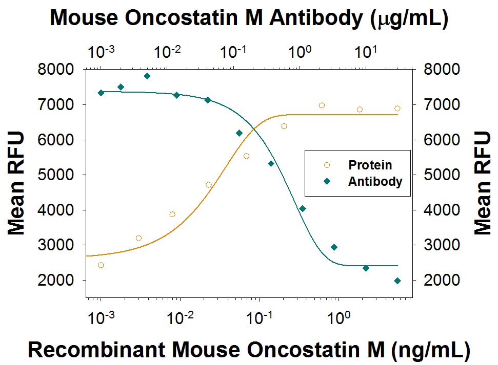
Recombinant Mouse Oncostatin M/OSM (Catalog # 495-MO) stimulates proliferation in the NIH-3T3 mouse embryonic fibroblast cell line in a dose-dependent manner (orange line). Proliferation elicited by Recombinant Mouse Oncostatin M/OSM (1 ng/mL) is neutralized (green line) by increasing concentrations of Rat Anti-Mouse Oncostatin M/OSM Monoclonal Antibody (Catalog # MAB4951). The ND50 is typically 0.2-1.2 µg/mL.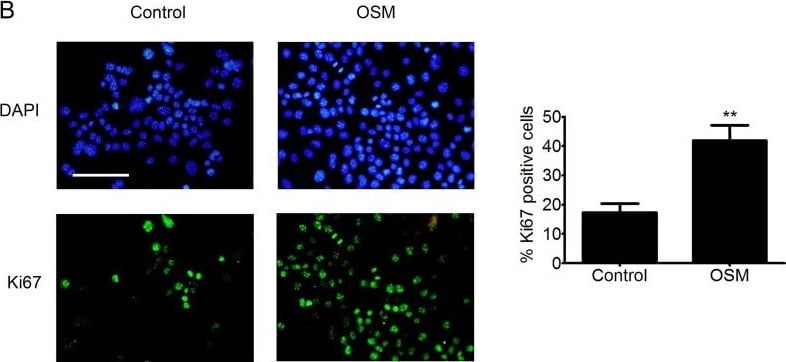
Detection of Oncostatin M (OSM) by Immunocytochemistry/ Immunofluorescence
OSM increases the migration and proliferation o PDVC57 cells(A)In vitro wounds were made in cultures of mitomycin-treated PDVC57 cells and the cells cultured for a further 48 h, with or without OSM, (10 ng/mL). Cell migration into the wounded area was assessed as described in Materials & Methods. Scale bar: 100 μm. Data are presented as the mean ± SEM of the migration area (mm2) from three independent experiments. Mann-Whitney test, *p < 0.05. (B) PDVC57 cells were cultured, with or without OSM (10 ng/mL), for 48,h. Cells were fixed and stained with an anti-Ki67 antibody (green) and the cell nuclei stained with DAPI (blue). Scale bar: 100 μm. Data are represented as the mean ± SEM of the percentage of Ki67+ cells from three independent experiments. Mann-Whitney test, *p < 0.05, **p < 0.01. Image collected and cropped by CiteAb from the following open publication (https://pubmed.ncbi.nlm.nih.gov/30559930), licensed under a CC-BY license. Not internally tested by R&D Systems.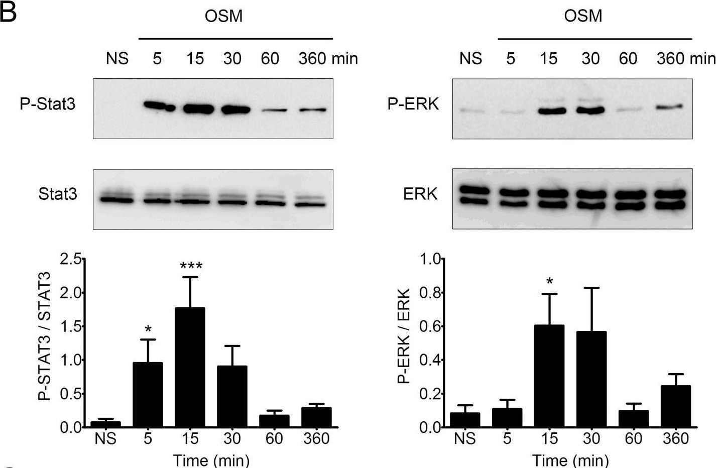
Detection of Oncostatin M (OSM) by Western Blot
PDVC57 cells express functional OSMR type II involved in ERK1/2 and STAT3 signaling and modulation of gene expression(A) Relative mRNA expression of normal murine epidermal keratinocytes (NMEK) and PDVC57 cells cultured under normal conditions for 6 h was measured by real-time qPCR. Data are presented as the mean ± SEM of relative mRNA expression (A.U.). Two-way ANOVA test with Bonferroni post-tests, *p < 0.05, **p < 0.01. (B) PDVC57 cells were cultured with or without OSM (10 ng/mL) for 5 min, 15 min, 30 min, 1 h, or 6 h. Protein levels of P-STAT3, total STAT3, P-ERK 1/2 and total ERK were measured by western blotting. Data are presented as the mean ± SEM of phosphorylated to-total protein ratio for STAT3 or ERK from three independent experiments. Kruskall-Wallis test with Dunn's Multiple Comparison Test, *p < 0.05, **p < 0.01, ***p < 0.001. (C) Scatter-plot graphs representing the relative mRNA expression of PDVC57 cells stimulated, or not, with OSM (10 ng/mL) for 6 or 24 h. The X axis indicates the relative expression in the control and the Y axis the relative expression of OSM stimulated PDVC57 cells. The red dots reflect up-regulated genes and the green dots, down-regulated genes. Total RNA was isolated and labelled according to the Affymetrix protocol. Transcriptional profiles were obtained using Affymetrix GeneChip™ Mouse Gene 2.1 ST Array Plate. Image collected and cropped by CiteAb from the following open publication (https://pubmed.ncbi.nlm.nih.gov/30559930), licensed under a CC-BY license. Not internally tested by R&D Systems.Applications for Mouse Oncostatin M/OSM Antibody (824157)
Neutralization
Reviewed Applications
Read 1 review rated 4 using MAB4951 in the following applications:
Formulation, Preparation, and Storage
Purification
Reconstitution
Reconstitute at 0.5 mg/mL in sterile PBS. For liquid material, refer to CoA for concentration.
Formulation
Shipping
Stability & Storage
- 12 months from date of receipt, -20 to -70 °C as supplied.
- 1 month, 2 to 8 °C under sterile conditions after reconstitution.
- 6 months, -20 to -70 °C under sterile conditions after reconstitution.
Calculators
Background: Oncostatin M/OSM
References
- Yoshimura, A. et al. (1996) EMBO J. 15:1055.
- Ray, P. et al. (1996) Endocrinology 137:1151.
- Rose, T.M. and A.G. Bruce (1994) in Guidebook to Cytokines and Their Receptors, N.A. Nicola, editor, Oxford University Press, New York, p. 127.
Alternate Names
Gene Symbol
UniProt
Additional Oncostatin M/OSM Products
Product Documents for Mouse Oncostatin M/OSM Antibody (824157)
Certificate of Analysis
To download a Certificate of Analysis, please enter a lot or batch number in the search box below.
Note: Certificate of Analysis not available for kit components.
Product Specific Notices for Mouse Oncostatin M/OSM Antibody (824157)
For research use only
Citations for Mouse Oncostatin M/OSM Antibody (824157)
 Powered by Bioz
Customer Reviews for Mouse Oncostatin M/OSM Antibody (824157) (1)
Have you used Mouse Oncostatin M/OSM Antibody (824157)?
Submit a review and receive an Amazon gift card!
$25/€18/£15/$25CAN/¥2500 for a review with an image
$10/€7/£6/$10CAN/¥1110 for a review without an image
Submit a review
Customer Images
-
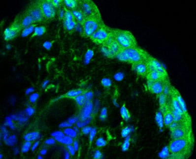
Application: ImmunocytochemistrySample Tested: Mouse Ear Skin (frozen section)Species: MouseVerified Customer | Posted 03/30/2017Frozen slide Fixation: faraformaldhehyde 4% mouse ear skin injected with OSM adenovirus

There are no reviews that match your criteria.