
Mouse Peptidase Inhibitor 16/PI16 Antibody
R&D Systems | Catalog # AF4929


Scientific images may be resized, cropped, or adjusted for brightness or contrast for presentation purposes. These updates are limited to presentation and do not affect the underlying scientific interpretation of the data. Where available, additional source data may be provided upon request.
Key Product Details
Species Reactivity
Validated:
Cited:
Applications
Validated:
Cited:
Label
Antibody Source
Product Specifications
Immunogen
Leu21-Glu464
Accession # Q9ET66
Specificity
Clonality
Host
Isotype
Scientific Data Images for Mouse Peptidase Inhibitor 16/PI16 Antibody
Peptidase Inhibitor 16/PI16 in Mouse Pancreas.
Mouse pancreas cryosections stained with CD324 (green) and PI16 (white). Image from a verified customer review.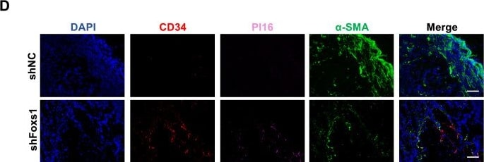
Detection of Peptidase Inhibitor 16/PI16 by Immunohistochemistry
Foxs1 knockdown in fibroblasts inhibits tumor growth.(A) Photographs of excised tumors from shNC and shFoxs1 mice. (B) Tumor volume over time. Data represent mean ± SD; n = 8. Statistical significance was assessed at each time point using an unpaired two-tailed Student’s t test. Significant exact P values were: Day 10: shNC vs. shFoxs1, P = 0.0008; Day 12: shNC vs. shFoxs1, P = 1.49 × 10⁻⁴; Day 14: shNC vs. shFoxs1, P = 5.66 × 10⁻⁶; Day 16: shNC vs. shFoxs1, P = 5.13 × 10⁻⁶. *P < 0.05. (C) Quantification of tumor weight. Data represent mean ± SD; n = 8. Statistical significance was assessed using an unpaired two-tailed Student’s t test; shNC vs. shFoxs1, P = 6.82 × 10⁻⁷; *P < 0.05. (D, E) IF staining for CD34, PI16, and alpha -SMA/COL1A1 in tumor tissues from the two mouse groups (D), with quantification (E). Scale Bars:100 μm. Data are mean ± SD; n = 5. Statistical significance was assessed using an unpaired two-tailed Student’s t test; Significant exact P values were: CD34, shNC vs. shFoxs1, P = 0.0002; PI16, shNC vs. shFoxs1, P = 0.0011; alpha -SMA, shNC vs. shFoxs1, P = 0.0001; *P < 0.05. Image collected and cropped by CiteAb from the following open publication (https://link.springer.com/10.1038/s44321-026-00476-8), licensed under a CC-BY license. Not internally tested by R&D Systems.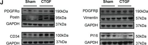
Detection of Peptidase Inhibitor 16/PI16 by Western Blot
CD34+ PI16+ SPC subpopulation and the pro-inflammatory effect of CD34+ PI16- cell subpopulation. (A) UMAP plots showing the major cell clusters of CD34+ cells in the normal thoracic aortas and grafted arteries (n = 43178 cells). (B) Bar chart showing the proportion of identified cell clusters in the normal thoracic aortas and grafted arteries. (C) Volcano plots showing the up-regulated gene for clusters 0 and 3. (D) Feature plots showing CD34 and PI16 expression in different clusters. (E) Gene Ontology analysis of top enriched functions in cluster 0. (F) Gene Ontology analysis of top enriched functions in cluster 3. (G) Analysis of the correlation between each cluster and the chemokine family. (H) Pseudotime trajectories showing CD34high and PI16high cells differentiated into endothelial cells and fibroblasts in the normal thoracic aortas and grafted arteries. (I) Trajectory plot showing the pseudotime representing trajectories of Mef2c+ and PDGFR-alpha + cells differentiation and the expression of 2 markers (CD34/Ly6a) in the endothelial cells. (J) Representative western blot images of these markers (PDGFR-alpha /Postn/CD34/PDGFR-beta /Vimentin/PI16) from CD34+ PI16+ cells treated with PBS and CTGF 50ug/L for 48 h. n = 6 per group. (K) Quantification analysis of these markers in (J). Data were presented as the mean ± SEM and analyzed by using an unpaired two-tailed Student's t-test. * p < 0.05, ** p < 0.01, *** p < 0.001, **** p < 0.0001. (L) Cartoon showing that CD34+ PI16- cells, derived from CD34+ PI16+ cells, have a chemotactic effect on SMCs. Image collected and cropped by CiteAb from the following open publication (https://www.thno.org/v15p2523.htm), licensed under a CC-BY license. Not internally tested by R&D Systems.Applications for Mouse Peptidase Inhibitor 16/PI16 Antibody
Western Blot
Sample: Recombinant Mouse Peptidase Inhibitor 16/PI16
Reviewed Applications
Read 1 review rated 4 using AF4929 in the following applications:
Formulation, Preparation, and Storage
Purification
Reconstitution
Reconstitute at 0.2 mg/mL in sterile PBS. For liquid material, refer to CoA for concentration.
Formulation
Shipping
Stability & Storage
- 12 months from date of receipt, -20 to -70 °C as supplied.
- 1 month, 2 to 8 °C under sterile conditions after reconstitution.
- 6 months, -20 to -70 °C under sterile conditions after reconstitution.
Calculators
Background: Peptidase Inhibitor 16/PI16
Alternate Names
Gene Symbol
UniProt
Additional Peptidase Inhibitor 16/PI16 Products
Product Documents for Mouse Peptidase Inhibitor 16/PI16 Antibody
Certificate of Analysis
To download a Certificate of Analysis, please enter a lot or batch number in the search box below.
Note: Certificate of Analysis not available for kit components.
Product Specific Notices for Mouse Peptidase Inhibitor 16/PI16 Antibody
For research use only
Related Research Areas
Citations for Mouse Peptidase Inhibitor 16/PI16 Antibody
 Powered by Bioz
Customer Reviews for Mouse Peptidase Inhibitor 16/PI16 Antibody (1)
Have you used Mouse Peptidase Inhibitor 16/PI16 Antibody?
Submit a review and receive an Amazon gift card!
$25/€18/£15/$25CAN/¥2500 for a review with an image
$10/€7/£6/$10CAN/¥1110 for a review without an image
Submit a review
Customer Images

-
Application: ImmunohistochemistrySample Tested: Adult pancreasSpecies: MouseVerified Customer | Posted 10/14/2025Mouse pancreas cryosections stained with CD324 (green) and PI16 (white)
 Bio-Techne ResponseThis review reflects a new species or application tested on a primary antibody.
There are no reviews that match your criteria.
Protocols
Find general support by application which include: protocols, troubleshooting, illustrated assays, videos and webinars.
- Cellular Response to Hypoxia Protocols
- R&D Systems Quality Control Western Blot Protocol
- Troubleshooting Guide: Western Blot Figures
- Western Blot Conditions
- Western Blot Protocol
- Western Blot Protocol for Cell Lysates
- Western Blot Troubleshooting
- Western Blot Troubleshooting Guide
- View all Protocols, Troubleshooting, Illustrated assays and Webinars