
Butyrylcholinesterase (BCHE) is a major acetylcholine hydrolyzing enzyme in the circulation (1). Although it is present in significant amounts (~3 mg/L) in human plasma, no endogenous physiological substrate has been described for this enzyme. It can degrade a large number of ester-containing compounds in addition to acylcholines. Thus, it is likely to play significant pharmacological and toxicological roles. It is thought to be involved in the pathological process of Alzheimer's disease (AD) by depleting acetylcholine. In contrast to ACHE, it attenuates amyloid fibril formation in vitro (2). BCHE inhibitors have been used to delay symptoms of AD patients by virtue of their ability to enhance acetylcholine availability (3). Its involvement in a cholinergic anti-inflammatory pathway connect BCHE and ACHE with a possible marker of low-grade systemic inflammation observed in Type-2 diabetes, obesity, hypertension, coronary heart disease, and AD (4). BCHE can exist in monomeric and multimeric forms (1).
Butyrylcholinesterase/BCHE Antibody
R&D Systems | Catalog # AF9024

Scientific images may be resized, cropped, or adjusted for brightness or contrast for presentation purposes. These updates are limited to presentation and do not affect the underlying scientific interpretation of the data. Where available, additional source data may be provided upon request.
Key Product Details
Species Reactivity
Validated:
Cited:
Applications
Validated:
Cited:
Label
Antibody Source
Product Specifications
Immunogen
His28-Leu603
Accession # AAH99977
Specificity
Clonality
Host
Isotype
Scientific Data Images for Butyrylcholinesterase/BCHE Antibody
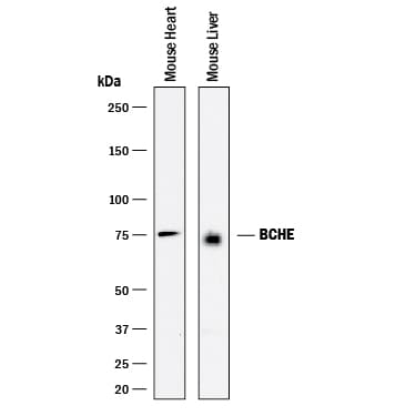
Detection of Mouse Butyrylcholinesterase/BCHE by Western Blot.
Western blot shows lysates of mouse heart tissue and mouse liver tissue. PVDF membrane was probed with 1 µg/mL of Goat Anti-Mouse/Rat Butyrylcholinesterase/BCHE Polyclonal Antibody (Catalog # AF9024) followed by HRP-conjugated Anti-Goat IgG Secondary Antibody (Catalog # HAF017). A specific band was detected for Butyrylcholinesterase/BCHE at approximately 75 kDa (as indicated). This experiment was conducted under reducing conditions and using Immunoblot Buffer Group 1.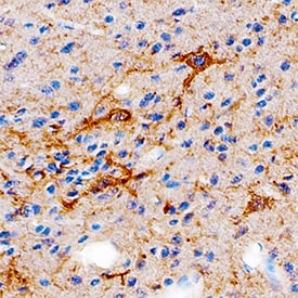
Butyrylcholinesterase/BCHE in Mouse Brain.
Butyrylcholinesterase/BCHE was detected in perfusion fixed frozen sections of mouse brain using Goat Anti-Mouse/Rat Butyrylcholinesterase/BCHE Antigen Affinity-purified Polyclonal Antibody (Catalog # AF9024) at 5 µg/mL overnight at 4 °C. Tissue was stained using the Anti-Goat HRP-DAB Cell & Tissue Staining Kit (brown; Catalog # CTS008) and counterstained with hematoxylin (blue). Specific staining was localized to glial cells. View our protocol for Chromogenic IHC Staining of Frozen Tissue Sections.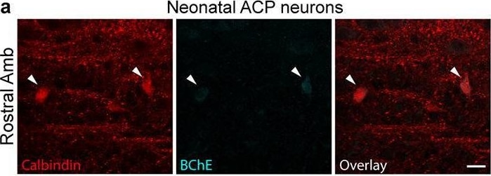
Detection of Butyrylcholinesterase/BCHE by Immunohistochemistry
Conservation of ACP and ACV markers across postnatal development.a, Immunostaining of ACP neurons in the rostral Amb of a neonatal (postnatal day 2) mouse. ACP neurons stained positive for calbindin (red) and stained weakly positive for BChE (cyan). b, Immunostaining of neonatal ACV neurons in the caudal Amb of a postnatal day 2 mouse. ACV neurons stained positive for BChE and negative for calbindin. Bars, 20 μm. c, Map of ACP and ACV neurons in neonatal Amb. Sagittal schematic view showing overlay of soma of all neurons (circles) across all sections spanning a single neonatal (postnatal day 2) Amb that was stained for ACP marker calbindin (dark blue circles) and ACV marker BChE (light blue circles). d, Quantification of absolute numbers of ACP neurons (calbindin+, dark blue) and ACV neurons (BChE+, light blue) per Amb in neonatal mice (mean ± s.d., n = 3 mice). e, Immunostaining of ACP neurons in the rostral Amb of an adult (postnatal day 60) mouse. ACP neurons stained positive for calbindin (red) and stained weakly positive for BChE (cyan). f, Immunostaining of adult ACV neurons in the caudal Amb of a postnatal day 60 mouse. ACV neurons stained positive for BChE and negative for calbindin. Bars, 20 μm. g, Map of ACP and ACV neurons in adult Amb. Sagittal schematic view showing overlay of soma of all neurons (circles) across all sections spanning a single adult (postnatal day 60) Amb that was stained for calbindin (dark blue) and BChE (light blue). Note similarity in marker expression and cell type distribution in neonatal and adult Amb. h, Quantification of absolute numbers of ACP neurons (calbindin+, dark blue) and ACV neurons (BChE+, light blue) per Amb in adult mice (mean ± s.d., n = 3 mice). Image collected and cropped by CiteAb from the following open publication (https://www.nature.com/articles/s41586-022-04760-8), licensed under a CC-BY license. Not internally tested by R&D Systems.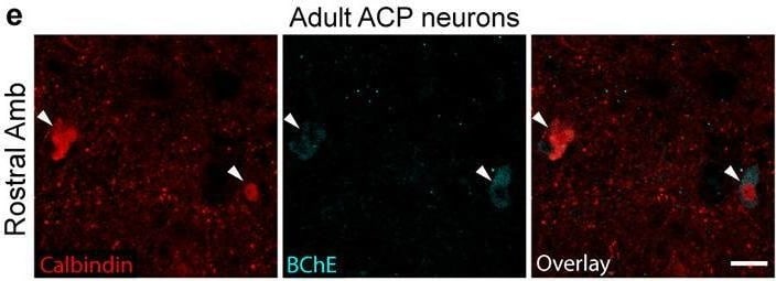
Detection of Butyrylcholinesterase/BCHE by Immunohistochemistry
Conservation of ACP and ACV markers across postnatal development.a, Immunostaining of ACP neurons in the rostral Amb of a neonatal (postnatal day 2) mouse. ACP neurons stained positive for calbindin (red) and stained weakly positive for BChE (cyan). b, Immunostaining of neonatal ACV neurons in the caudal Amb of a postnatal day 2 mouse. ACV neurons stained positive for BChE and negative for calbindin. Bars, 20 μm. c, Map of ACP and ACV neurons in neonatal Amb. Sagittal schematic view showing overlay of soma of all neurons (circles) across all sections spanning a single neonatal (postnatal day 2) Amb that was stained for ACP marker calbindin (dark blue circles) and ACV marker BChE (light blue circles). d, Quantification of absolute numbers of ACP neurons (calbindin+, dark blue) and ACV neurons (BChE+, light blue) per Amb in neonatal mice (mean ± s.d., n = 3 mice). e, Immunostaining of ACP neurons in the rostral Amb of an adult (postnatal day 60) mouse. ACP neurons stained positive for calbindin (red) and stained weakly positive for BChE (cyan). f, Immunostaining of adult ACV neurons in the caudal Amb of a postnatal day 60 mouse. ACV neurons stained positive for BChE and negative for calbindin. Bars, 20 μm. g, Map of ACP and ACV neurons in adult Amb. Sagittal schematic view showing overlay of soma of all neurons (circles) across all sections spanning a single adult (postnatal day 60) Amb that was stained for calbindin (dark blue) and BChE (light blue). Note similarity in marker expression and cell type distribution in neonatal and adult Amb. h, Quantification of absolute numbers of ACP neurons (calbindin+, dark blue) and ACV neurons (BChE+, light blue) per Amb in adult mice (mean ± s.d., n = 3 mice). Image collected and cropped by CiteAb from the following open publication (https://www.nature.com/articles/s41586-022-04760-8), licensed under a CC-BY license. Not internally tested by R&D Systems.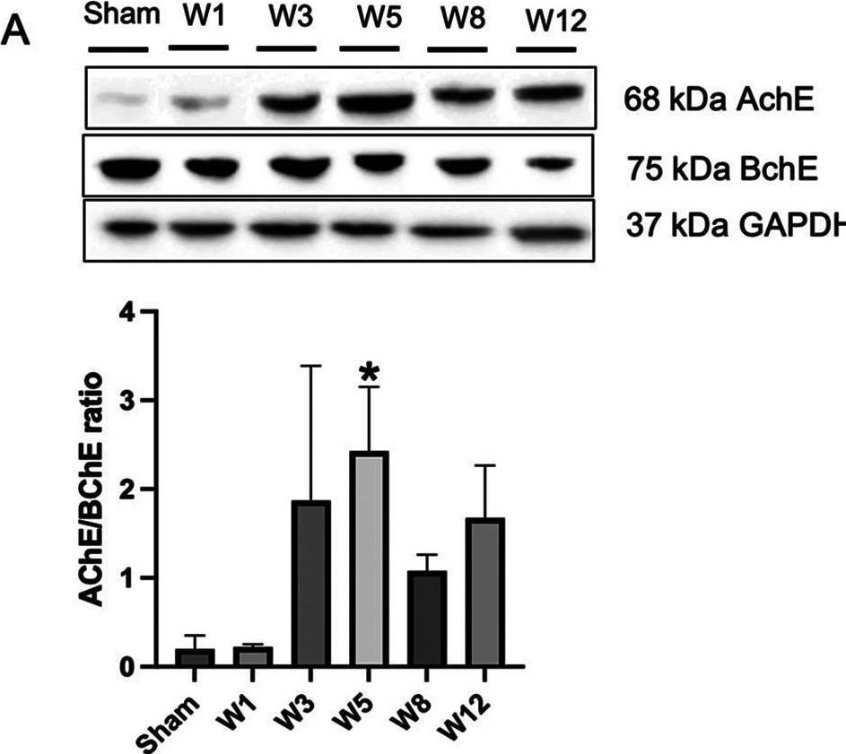
Detection of Butyrylcholinesterase/BCHE by Western Blot
Benzalkonium chloride-induced hypoganglionosis results in hypertrophy of cholinergic nerve fiber, loss of intestinal tight junctions, and inflammation. (A) Western blotting revealed an increased AChE/BChE ratio in the rectosigmoid colon of BAC-treated groups (n = 6) compared to the sham group (n = 6) according to the time of termination. (B) Western blotting revealed decreased occludin tight junction in the rectosigmoid colon of BAC-treated groups (n = 6) compared to the sham group (n = 6) according to the time of termination. (C) mRNA expression of IL-1 beta on rectosigmoid colon tissue between groups. Results are presented as mean ± SE. A one-way ANOVA test followed by Tukey's test was performed to compare the data. *p ≤ 0.05 vs. sham; **p ≤ 0.01 vs. sham. Image collected and cropped by CiteAb from the following open publication (https://www.frontiersin.org/articles/10.3389/fsurg.2024.1407948/full), licensed under a CC-BY license. Not internally tested by R&D Systems.Applications for Butyrylcholinesterase/BCHE Antibody
Immunohistochemistry
Sample: Perfusion fixed frozen sections of mouse brain
Western Blot
Sample: Mouse heart tissue and mouse liver tissue
Formulation, Preparation, and Storage
Purification
Reconstitution
Reconstitute at 0.5 mg/mL in sterile PBS. For liquid material, refer to CoA for concentration.
Formulation
Shipping
Stability & Storage
- 12 months from date of receipt, -20 to -70 °C as supplied.
- 1 month, 2 to 8 °C under sterile conditions after reconstitution.
- 6 months, -20 to -70 °C under sterile conditions after reconstitution.
Calculators
Background: Butyrylcholinesterase/BCHE
References
- Darvesh, S. et al. (2003) Nat. Rev. Neuroscience 4:131.
- Diamant, S. et al. (2006) Proc. Natl. Acad. Sci. USA 103:8628.
- Campbell, V. A. and Gowran, A. (2007) Br. J. Pharm. 152:655.
- Das, U. N. (2007) Med Sci Monit. 13:RA214.
Alternate Names
Gene Symbol
UniProt
Additional Butyrylcholinesterase/BCHE Products
Product Documents for Butyrylcholinesterase/BCHE Antibody
Certificate of Analysis
To download a Certificate of Analysis, please enter a lot or batch number in the search box below.
Note: Certificate of Analysis not available for kit components.
Product Specific Notices for Butyrylcholinesterase/BCHE Antibody
For research use only
Related Research Areas
Citations for Butyrylcholinesterase/BCHE Antibody
 Powered by Bioz
Customer Reviews for Butyrylcholinesterase/BCHE Antibody
There are currently no reviews for this product. Be the first to review Butyrylcholinesterase/BCHE Antibody and earn rewards!
Have you used Butyrylcholinesterase/BCHE Antibody?
Submit a review and receive an Amazon gift card!
$25/€18/£15/$25CAN/¥2500 for a review with an image
$10/€7/£6/$10CAN/¥1110 for a review without an image
Submit a review
Protocols
Find general support by application which include: protocols, troubleshooting, illustrated assays, videos and webinars.
- Antigen Retrieval Protocol (PIER)
- Antigen Retrieval for Frozen Sections Protocol
- Appropriate Fixation of IHC/ICC Samples
- Cellular Response to Hypoxia Protocols
- Chromogenic IHC Staining of Formalin-Fixed Paraffin-Embedded (FFPE) Tissue Protocol
- Chromogenic Immunohistochemistry Staining of Frozen Tissue
- ClariTSA™ Fluorophore Kits
- Detection & Visualization of Antibody Binding
- Fluorescent IHC Staining of Frozen Tissue Protocol
- Graphic Protocol for Heat-induced Epitope Retrieval
- Graphic Protocol for the Preparation and Fluorescent IHC Staining of Frozen Tissue Sections
- Graphic Protocol for the Preparation and Fluorescent IHC Staining of Paraffin-embedded Tissue Sections
- Graphic Protocol for the Preparation of Gelatin-coated Slides for Histological Tissue Sections
- IHC Sample Preparation (Frozen sections vs Paraffin)
- Immunofluorescent IHC Staining of Formalin-Fixed Paraffin-Embedded (FFPE) Tissue Protocol
- Immunohistochemistry (IHC) and Immunocytochemistry (ICC) Protocols
- Immunohistochemistry Frozen Troubleshooting
- Immunohistochemistry Paraffin Troubleshooting
- Preparing Samples for IHC/ICC Experiments
- Preventing Non-Specific Staining (Non-Specific Binding)
- Primary Antibody Selection & Optimization
- Protocol for Heat-Induced Epitope Retrieval (HIER)
- Protocol for Making a 4% Formaldehyde Solution in PBS
- Protocol for VisUCyte™ HRP Polymer Detection Reagent
- Protocol for the Preparation & Fixation of Cells on Coverslips
- Protocol for the Preparation and Chromogenic IHC Staining of Frozen Tissue Sections
- Protocol for the Preparation and Chromogenic IHC Staining of Frozen Tissue Sections - Graphic
- Protocol for the Preparation and Chromogenic IHC Staining of Paraffin-embedded Tissue Sections
- Protocol for the Preparation and Chromogenic IHC Staining of Paraffin-embedded Tissue Sections - Graphic
- Protocol for the Preparation and Fluorescent IHC Staining of Frozen Tissue Sections
- Protocol for the Preparation and Fluorescent IHC Staining of Paraffin-embedded Tissue Sections
- Protocol for the Preparation of Gelatin-coated Slides for Histological Tissue Sections
- R&D Systems Quality Control Western Blot Protocol
- TUNEL and Active Caspase-3 Detection by IHC/ICC Protocol
- The Importance of IHC/ICC Controls
- Troubleshooting Guide: Immunohistochemistry
- Troubleshooting Guide: Western Blot Figures
- Western Blot Conditions
- Western Blot Protocol
- Western Blot Protocol for Cell Lysates
- Western Blot Troubleshooting
- Western Blot Troubleshooting Guide
- View all Protocols, Troubleshooting, Illustrated assays and Webinars