Matrix metalloproteinases are a family of zinc and calcium dependent endopeptidases with the combined ability to degrade all the components of the extracellular matrix. MMP-2 (gelatinase A), a type IV collagenase, can degrade a broad range of substrates including type IV, V, VII and X collagens as well as elastin and fibronectin. It is believed to act synergistically with interstitial collagenase (MMP-1) in the degradation of fibrillar collagens as it degrades their denatured gelatin forms. MMP-2 has been shown to be associated with many connective tissue cells as well as neutrophils, macrophages and monocytes. Structurally, MMP-2 may be divided into several distinct domains: a pro-domain which is cleaved upon activation; a catalytic domain containing the zinc binding site; a fibronectin-like domain thought to play a role in substrate targeting; and a carboxyl terminal (hemopexin-like) domain containing 2 N-linked glycosylation sites. The amino acid sequences of the proenzymes are identical between mouse and rat.


Scientific images may be resized, cropped, or adjusted for brightness or contrast for presentation purposes. These updates are limited to presentation and do not affect the underlying scientific interpretation of the data. Where available, additional source data may be provided upon request.
Key Product Details
Validated by
Species Reactivity
Validated:
Cited:
Applications
Validated:
Cited:
Label
Antibody Source
Product Specifications
Immunogen
Ile34-Cys662
Accession # P33434
Specificity
Clonality
Host
Isotype
Scientific Data Images for MMP-2 Antibody
MMP‑2 in Mouse Thymus.
MMP-2 was detected in perfusion fixed frozen sections of mouse thymus using Goat Anti-Mouse/ Rat MMP-2 Antigen Affinity-purified Polyclonal Antibody (Catalog # AF1488) at 15 µg/mL overnight at 4 °C. Tissue was stained using the Anti-Goat HRP-DAB Cell & Tissue Staining Kit (brown; Catalog # CTS008) and counterstained with hematoxylin (blue). View our protocol for Chromogenic IHC Staining of Frozen Tissue Sections.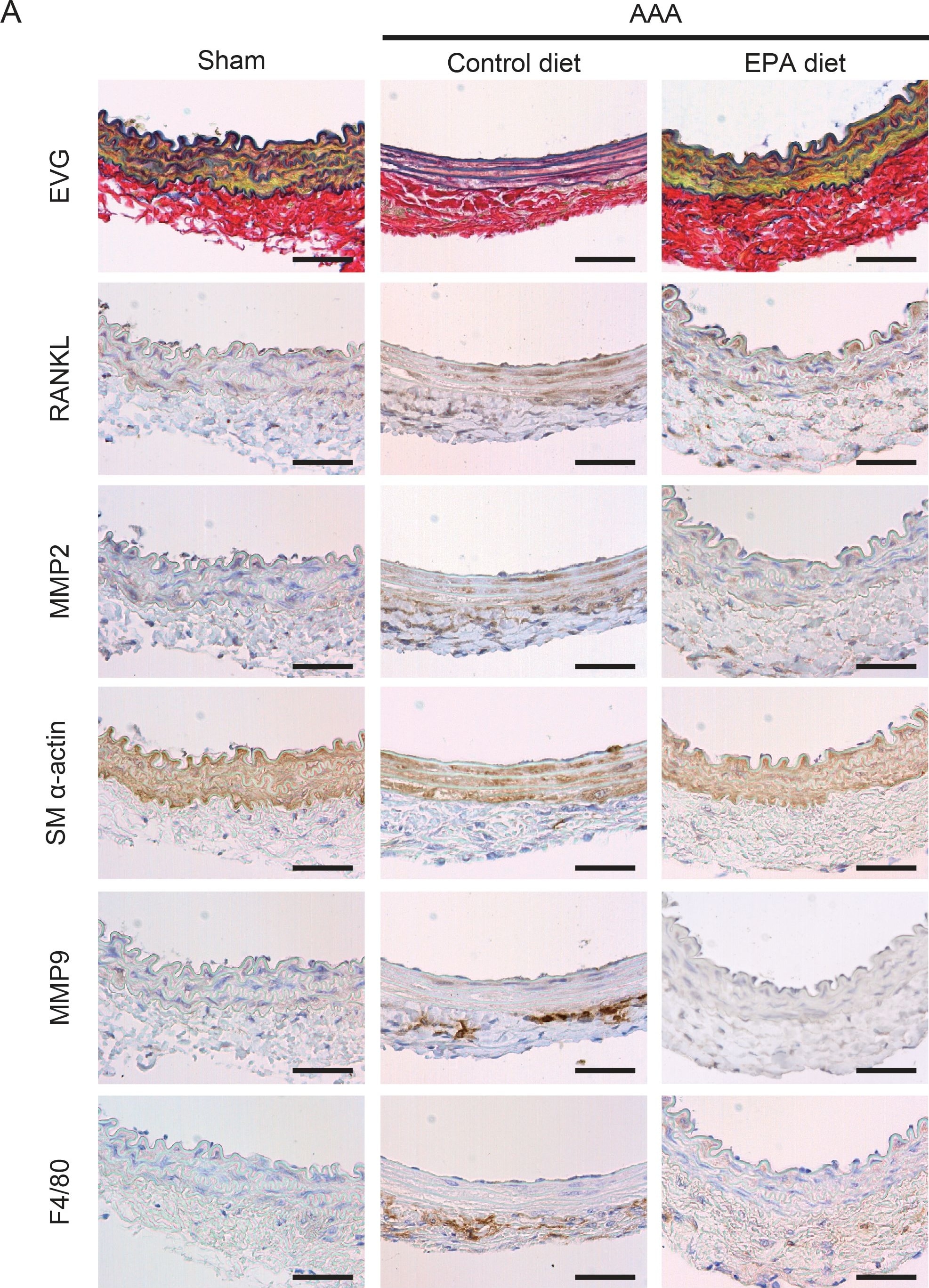
Detection of Mouse MMP-2 by Immunohistochemistry-Paraffin
MMP2, MMP9, and RANKL expression in AAAs.A. Immunohistochemical staining for indicated proteins of serial sections of aortas one week after CaCl2 treatment. Elastic van Gieson staining is also shown. SM alpha -actin and F4/80 were stained to locate SMCs and macrophages, respectively. Shown are representative images of 4 or more samples in each group. Scale bars, 50 µm. B. Relative positive staining area of MMP2, MMP9, and RANKL in sections from control diet and EPA diet groups. n = 4–5. *P<0.05. Image collected and cropped by CiteAb from the following open publication (https://dx.plos.org/10.1371/journal.pone.0096286), licensed under a CC-BY license. Not internally tested by R&D Systems.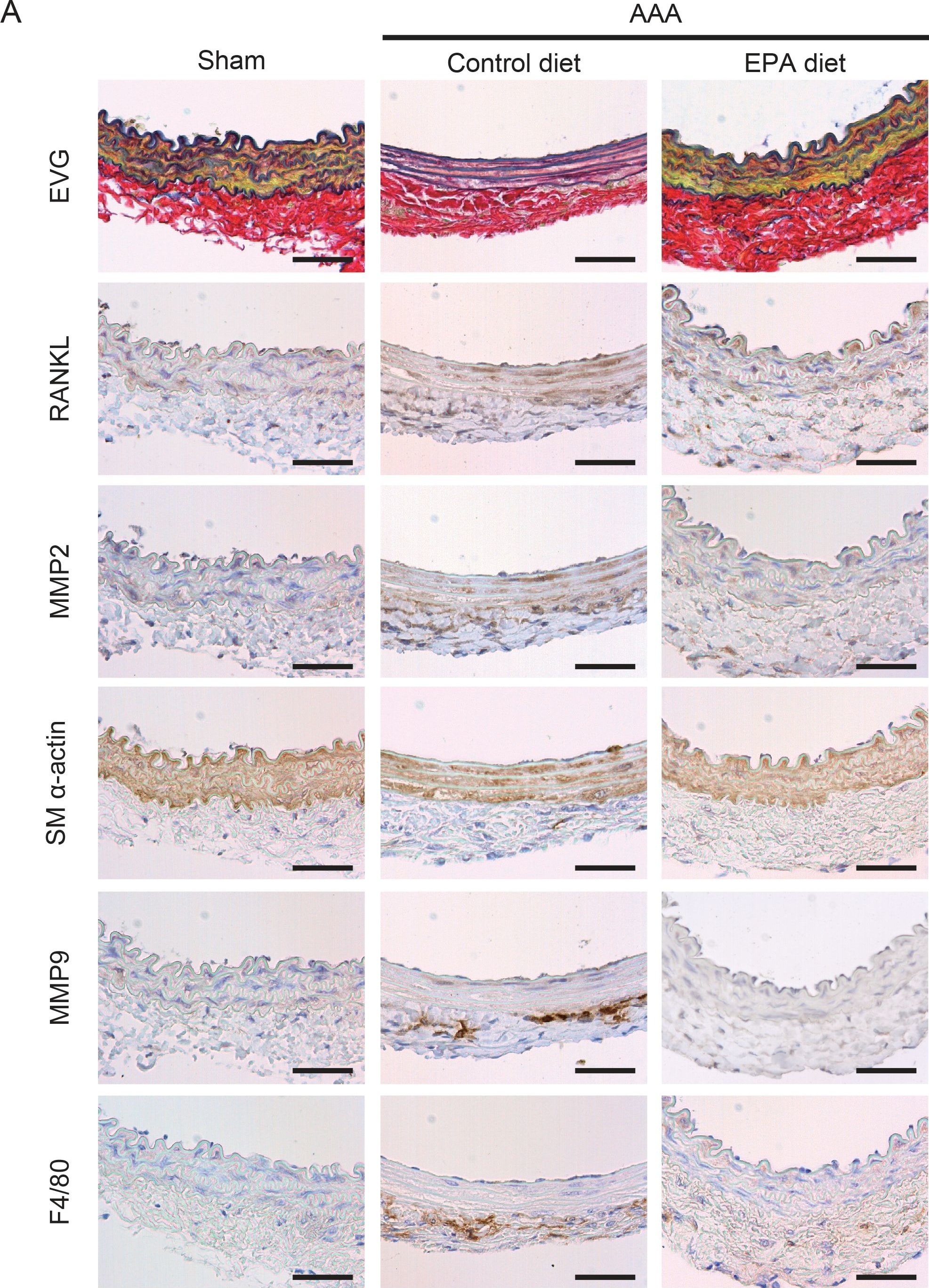
Detection of MMP-2 by Immunohistochemistry-Paraffin
MMP2, MMP9, and RANKL expression in AAAs.A. Immunohistochemical staining for indicated proteins of serial sections of aortas one week after CaCl2 treatment. Elastic van Gieson staining is also shown. SM alpha -actin and F4/80 were stained to locate SMCs and macrophages, respectively. Shown are representative images of 4 or more samples in each group. Scale bars, 50 µm. B. Relative positive staining area of MMP2, MMP9, and RANKL in sections from control diet and EPA diet groups. n = 4–5. *P<0.05. Image collected and cropped by CiteAb from the following open publication (https://pubmed.ncbi.nlm.nih.gov/24798452), licensed under a CC-BY license. Not internally tested by R&D Systems.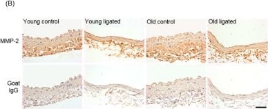
Detection of MMP-2 by Immunohistochemistry
Quantification of MMP-2 and AXiN-2 in the right carotid artery before and 21 days after ligation of the left carotid artery in young and old mice. Young and old mice were subjected to ligation of the left carotid artery for 2 days, Animals with unligated arteries served as controls. (A) MMP-2 protein detected by immunohistochemistry in the media of right carotid arteries was quantified by pixel analysis on sections and expressed as a percentage of medial area. * indicates p < 0.05 vs. young contol, Kruskal-Walls test and Dunn’s multiple comparisons test, n = 4 young controls, n = 12 young ligated, n = 3 old controls, n = 13 old ligated. (B) Representative images of MMP-2 immunohistochemistry, MMP-2 protein is brown and haematoxylin-stained nuclei are blue. (C) Medial cells positive for AXiN-2 (detected by immunohistochemistry) were counted on sections of right carotid arteries and expressed as a percentage of the total medial cell number viewed, * indicates p < 0 05 vs. young control, ANOVA and Student Newman-Keuls post hoc test, n = 4 young controls, n = 11 young ligated, n = 4 old controls, n = 12 old ligated. (D) Representative images ofAXN-2immunohistochemistry with some positive cells with brown cytoplasm are indicated by red arrows. Haematoxylin-stained nuclei are blue. (E,F). The percentage of AXIN-2 positive cells and MMP-2 protein in the media of right carotid arteries were plotted and correlation was assessed for young (E) and old (F) vessels (n = 15 young, n = 16 old. A linear trendline is shown on each graph. Significant correlation was detected in young arteries (r2 = 0.4891) but not old arteries (r2 = 0.07847). The scale bars represent 50 μm. Image collected and cropped by CiteAb from the following open publication (https://www.mdpi.com/1422-0067/26/21/10274), licensed under a CC-BY license. Not internally tested by R&D Systems.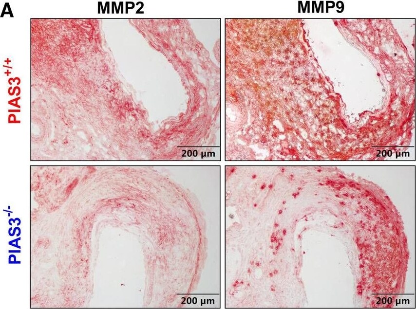
Detection of MMP-2 by Immunohistochemistry
PIAS3 deficiency decreases the expression levels of matrix metalloproteinases 2 and 9. (A): Representative immunohistochemical staining images of MMP2 and MMP9 in aneurysmal aortas from mild type (PIAS3+/+) and PIAS3 deficient (PIAS3−/−) mice. (B): Semi-quantification of MMP2 and MMP9 expression levels in aneurysmal lesions quantitated as the positively stained area per aortic cross-section. N = 10 for each group, *p < 0.05 and **p < 0.01 as compared to PIAS3+/+ mice. Image collected and cropped by CiteAb from the following open publication (https://pubmed.ncbi.nlm.nih.gov/37008329), licensed under a CC-BY license. Not internally tested by R&D Systems.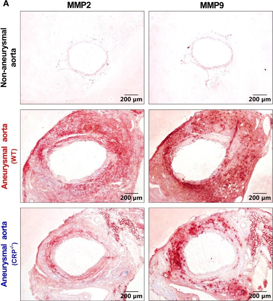
Detection of MMP-2 by Immunohistochemistry
CRP deficiency reduces aneurysmal aortic MMP2 expression. (A): Representative immunostaining images for MMP2 and MMP9 in non-aneurysmal normal or aneurysmal (WT and CRP-/-) aortas. (B, C): Quantification of MMP2 and MMP9 expression (mean ± standard for the percentage of positively stained area in total aortic cross section area) in wild type and CRP-/- aneurysmal aortas. Student’s t tests, n=10-11 mice per group, **p<0.01 compared to wild type mice. Image collected and cropped by CiteAb from the following open publication (https://pubmed.ncbi.nlm.nih.gov/37753091), licensed under a CC-BY license. Not internally tested by R&D Systems.Applications for MMP-2 Antibody
Immunohistochemistry
Sample: Perfusion fixed frozen sections of mouse thymus
Immunoprecipitation
Sample: Conditioned cell culture medium spiked with Recombinant Mouse/Rat MMP‑2 (Catalog # 924‑MP), see our available Western blot detection antibodies
Western Blot
Sample: Recombinant Mouse/Rat MMP‑2 (Catalog # 924-MP)
Formulation, Preparation, and Storage
Purification
Reconstitution
Reconstitute at 0.2 mg/mL in sterile PBS. For liquid material, refer to CoA for concentration.
Formulation
Shipping
Stability & Storage
- 12 months from date of receipt, -20 to -70 °C as supplied.
- 1 month, 2 to 8 °C under sterile conditions after reconstitution.
- 6 months, -20 to -70 °C under sterile conditions after reconstitution.
Calculators
Background: MMP-2
Long Name
Alternate Names
Gene Symbol
UniProt
Additional MMP-2 Products
Product Documents for MMP-2 Antibody
Certificate of Analysis
To download a Certificate of Analysis, please enter a lot or batch number in the search box below.
Note: Certificate of Analysis not available for kit components.
Product Specific Notices for MMP-2 Antibody
For research use only
Citations for MMP-2 Antibody
 Powered by Bioz
Customer Reviews for MMP-2 Antibody
There are currently no reviews for this product. Be the first to review MMP-2 Antibody and earn rewards!
Have you used MMP-2 Antibody?
Submit a review and receive an Amazon gift card!
$25/€18/£15/$25CAN/¥2500 for a review with an image
$10/€7/£6/$10CAN/¥1110 for a review without an image
Submit a review
Protocols
Find general support by application which include: protocols, troubleshooting, illustrated assays, videos and webinars.
- Antigen Retrieval Protocol (PIER)
- Antigen Retrieval for Frozen Sections Protocol
- Appropriate Fixation of IHC/ICC Samples
- Cellular Response to Hypoxia Protocols
- Chromogenic IHC Staining of Formalin-Fixed Paraffin-Embedded (FFPE) Tissue Protocol
- Chromogenic Immunohistochemistry Staining of Frozen Tissue
- ClariTSA™ Fluorophore Kits
- Detection & Visualization of Antibody Binding
- Fluorescent IHC Staining of Frozen Tissue Protocol
- Graphic Protocol for Heat-induced Epitope Retrieval
- Graphic Protocol for the Preparation and Fluorescent IHC Staining of Frozen Tissue Sections
- Graphic Protocol for the Preparation and Fluorescent IHC Staining of Paraffin-embedded Tissue Sections
- Graphic Protocol for the Preparation of Gelatin-coated Slides for Histological Tissue Sections
- IHC Sample Preparation (Frozen sections vs Paraffin)
- Immunofluorescent IHC Staining of Formalin-Fixed Paraffin-Embedded (FFPE) Tissue Protocol
- Immunohistochemistry (IHC) and Immunocytochemistry (ICC) Protocols
- Immunohistochemistry Frozen Troubleshooting
- Immunohistochemistry Paraffin Troubleshooting
- Immunoprecipitation Protocol
- Preparing Samples for IHC/ICC Experiments
- Preventing Non-Specific Staining (Non-Specific Binding)
- Primary Antibody Selection & Optimization
- Protocol for Heat-Induced Epitope Retrieval (HIER)
- Protocol for Making a 4% Formaldehyde Solution in PBS
- Protocol for VisUCyte™ HRP Polymer Detection Reagent
- Protocol for the Preparation & Fixation of Cells on Coverslips
- Protocol for the Preparation and Chromogenic IHC Staining of Frozen Tissue Sections
- Protocol for the Preparation and Chromogenic IHC Staining of Frozen Tissue Sections - Graphic
- Protocol for the Preparation and Chromogenic IHC Staining of Paraffin-embedded Tissue Sections
- Protocol for the Preparation and Chromogenic IHC Staining of Paraffin-embedded Tissue Sections - Graphic
- Protocol for the Preparation and Fluorescent IHC Staining of Frozen Tissue Sections
- Protocol for the Preparation and Fluorescent IHC Staining of Paraffin-embedded Tissue Sections
- Protocol for the Preparation of Gelatin-coated Slides for Histological Tissue Sections
- R&D Systems Quality Control Western Blot Protocol
- TUNEL and Active Caspase-3 Detection by IHC/ICC Protocol
- The Importance of IHC/ICC Controls
- Troubleshooting Guide: Immunohistochemistry
- Troubleshooting Guide: Western Blot Figures
- Western Blot Conditions
- Western Blot Protocol
- Western Blot Protocol for Cell Lysates
- Western Blot Troubleshooting
- Western Blot Troubleshooting Guide
- View all Protocols, Troubleshooting, Illustrated assays and Webinars