
Mouse repulsive guidance molecule (RGM-A) is a 33 kDa GPI-linked member of an expanding RGM-related family of neuronal and muscle-expressed membrane proteins (1). It is synthesized as a 454 amino acid (aa) preproprotein that contains a 47 aa signal sequence, a 122 aa N-terminal prosegment, a 258 mature region and a 27 aa C-terminal prosegment (2, 3). The N-terminal prosegment contains an RGD tripeptide and the molecule’s only two potential N-linked glycosylation sites. The mature segment shows an abbreviated von Willebrand factor domain. Proteolytic processing occurs at an aspartic acid-proline bond, creating a predicted 32 kDa mature region (2). The mature region of mouse RGM-A is 93% and 87% aa identical to human and chick mature region RGM-A, respectively. When compared to mouse RGMb and c, the mature region of mouse RGM-A shows 77% and 76% aa identity, respectively (2, 3). Recombinant chick RGM has been reported to induce collapse of temporal but not nasal growth cones, and to repel temporal retinal axons in vitro. This suggests a role in the development of the retina-superior colliculus connection. In mice, however, this activity is not so obvious, and thus its function in this system is uncertain (3). Alternatively, in mice, RGM-A is said to be needed for neural tube closure (3). And in a mouse culture system, chick RGM-A is reported to be responsible for the layered segmentation of of entorhinal cortical projections to the hippocampus (4). The receptor for RGM is reported to be neogenin (5, 6).

Scientific images may be resized, cropped, or adjusted for brightness or contrast for presentation purposes. These updates are limited to presentation and do not affect the underlying scientific interpretation of the data. Where available, additional source data may be provided upon request.
Key Product Details
Species Reactivity
Validated:
Cited:
Applications
Validated:
Cited:
Label
Antibody Source
Product Specifications
Immunogen
Cys48-Gly421
Accession # Q6PCX7
Specificity
Clonality
Host
Isotype
Scientific Data Images for Mouse RGM-A Antibody
RGM‑A in Mouse Embryo.
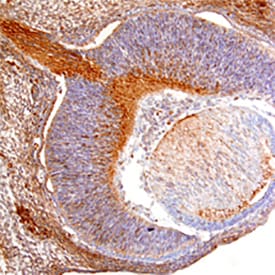
RGM-A was detected in immersion fixed frozen sections of mouse embryo (13 d.p.c) using Goat Anti-Mouse RGM-A Antigen Affinity-purified Polyclonal Antibody (Catalog # AF2458) at 15 µg/mL overnight at 4 °C. Tissue was stained using the Anti-Goat HRP-DAB Cell & Tissue Staining Kit (brown; Catalog # CTS008) and counterstained with hematoxylin (blue). Specific staining was localized to retina and developing neurons in cross-section of the developing eye. View our protocol for Chromogenic IHC Staining of Frozen Tissue Sections.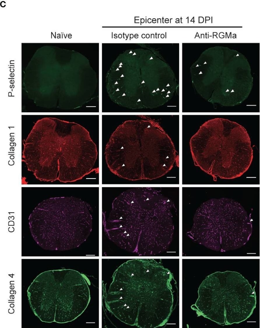
Detection of RGM-A by Immunohistochemistry
Immunohistochemical analysis of the spinal cord in targeted EAE mice treated by control/anti-RGMa antibody at 14 DPI. (A) Graphical representation of IHC experiments. (B) Representative images of vascular abnormalities at 14 DPI obtained from the isotype control group. (C) Representative images of the spinal cord stained for P-selectin, type I collagen, CD31, and type IV collagen in the naïve (left), isotype control (middle), and anti-RGMa antibody groups (right) at 14 DPI. Arrows indicate abnormal vessels. Scale bar; 50 μm (b, magnified image), 250 μm (c, whole spinal cord). (D) Quantification of P-selectin, CD31, perivascular type I and IV collagen-positive areas within white matter in each group (naïve; n=8, isotype control; n=4, anti-RGMa; n=4. Error bars represent the mean ± SEM. *p < 0.05 vs naïve, #p <0.05 vs isotype control, assessed via one-way ANOVA, followed by the Tukey-Kramer test). Image collected and cropped by CiteAb from the following open publication (https://www.frontiersin.org/articles/10.3389/fimmu.2022.870126/full), licensed under a CC-BY license. Not internally tested by R&D Systems.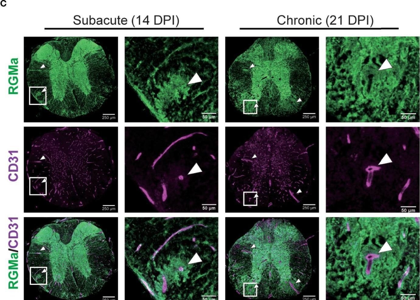
Detection of RGM-A by Immunohistochemistry
Immunohistochemical analysis of the spinal cord in targeted EAE mice. (A) Experimental time course of IHC analysis. (B) Representative image of the whole spinal cord stained with anti-CD31 and anti-type IV collagen antibodies at each disease phase in naïve mice and targeted EAE mice at 8, 14, 21 DPI. (C) Representative image of anti-RGMa and anti-CD31 antibodies staining in targeted EAE mouse spinal cords at 14 and 21 DPI. White and yellow arrows indicate abnormal vessels and collagen mesh formation, respectively. Scale bar; 50 μm (magnified), 250 μm (whole spinal cord). Image collected and cropped by CiteAb from the following open publication (https://www.frontiersin.org/articles/10.3389/fimmu.2022.870126/full), licensed under a CC-BY license. Not internally tested by R&D Systems.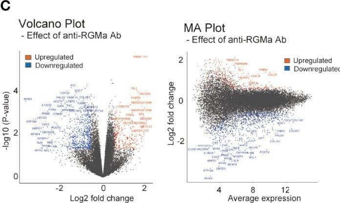
Detection of RGM-A by Flow Cytometry
Transcriptional analysis of targeted EAE mice treated with control/anti-RGMa antibody. (A) Graphical representation of transcriptional analysis experiments. (B) Principal component analysis plot of each group. (C) Volcano plot (left) and MA plot (right) of the comparison between the isotype control group and the anti-RGMa antibody group. Differentially expressed genes (fold change>1.5, p-value < 0.05) were represented by color dots. (D) GSEA plot of CNS cell markers with positive enrichment in anti-RGMa antibody group compared to isotype control group (p-adjust < 0.05). (E) GSEA plot of CNS cell markers with negative enrichment in anti-RGMa antibody group compared to isotype control group (p-adjust < 0.05). (F–H) Top 5 GSEA plots with negative enrichment gene sets (p-adjust < 0.05) of GO biological process (F), KEGG pathway (G), and MsigDB hall mark (H). (I) GSEA plot of BBB related genes sets (p-adjust < 0.05, n.s.; not significant). (J) Key features related to BBB disruption and corresponding genes (left) and heatmap of representative genes in BBB dysfunction modules, ECM production, and EndMT (right). Image collected and cropped by CiteAb from the following open publication (https://www.frontiersin.org/articles/10.3389/fimmu.2022.870126/full), licensed under a CC-BY license. Not internally tested by R&D Systems.Applications for Mouse RGM-A Antibody
Immunohistochemistry
Sample: Immersion fixed frozen sections of mouse embryo (13 d.p.c.)
Western Blot
Sample: Recombinant Mouse Repulsive Guidance Molecule A/RGM-A (Catalog # 2458-RG)
Formulation, Preparation, and Storage
Purification
Reconstitution
Reconstitute at 0.2 mg/mL in sterile PBS. For liquid material, refer to CoA for concentration.
Formulation
Shipping
Stability & Storage
- 12 months from date of receipt, -20 to -70 °C as supplied.
- 1 month, 2 to 8 °C under sterile conditions after reconstitution.
- 6 months, -20 to -70 °C under sterile conditions after reconstitution.
Calculators
Background: RGM-A
References
- Samad, T.A. et al. (2004) J. Neurosci. 24:2027.
- Schmidtmer, J. et al. (2004) Gene Expr. Patterns 4:105.
- Niederkofler V. et al. (2004) J. Neurosci. 24:808.
- Brinks, H. et al. (2004) J. Neurosci. 24:3862.
- Rajagopalan S. et al. (2004) Nat. Cell Biol. 6:756.
- Matsunaga E. et al. (2004) Nat. Cell Biol. 6:749.
Long Name
Alternate Names
Gene Symbol
UniProt
Additional RGM-A Products
Product Documents for Mouse RGM-A Antibody
Certificate of Analysis
To download a Certificate of Analysis, please enter a lot or batch number in the search box below.
Note: Certificate of Analysis not available for kit components.
Product Specific Notices for Mouse RGM-A Antibody
For research use only
Related Research Areas
Citations for Mouse RGM-A Antibody
 Powered by Bioz
Customer Reviews for Mouse RGM-A Antibody
There are currently no reviews for this product. Be the first to review Mouse RGM-A Antibody and earn rewards!
Have you used Mouse RGM-A Antibody?
Submit a review and receive an Amazon gift card!
$25/€18/£15/$25CAN/¥2500 for a review with an image
$10/€7/£6/$10CAN/¥1110 for a review without an image
Submit a review
Protocols
Find general support by application which include: protocols, troubleshooting, illustrated assays, videos and webinars.
- Antigen Retrieval Protocol (PIER)
- Antigen Retrieval for Frozen Sections Protocol
- Appropriate Fixation of IHC/ICC Samples
- Cellular Response to Hypoxia Protocols
- Chromogenic IHC Staining of Formalin-Fixed Paraffin-Embedded (FFPE) Tissue Protocol
- Chromogenic Immunohistochemistry Staining of Frozen Tissue
- ClariTSA™ Fluorophore Kits
- Detection & Visualization of Antibody Binding
- Fluorescent IHC Staining of Frozen Tissue Protocol
- Graphic Protocol for Heat-induced Epitope Retrieval
- Graphic Protocol for the Preparation and Fluorescent IHC Staining of Frozen Tissue Sections
- Graphic Protocol for the Preparation and Fluorescent IHC Staining of Paraffin-embedded Tissue Sections
- Graphic Protocol for the Preparation of Gelatin-coated Slides for Histological Tissue Sections
- IHC Sample Preparation (Frozen sections vs Paraffin)
- Immunofluorescent IHC Staining of Formalin-Fixed Paraffin-Embedded (FFPE) Tissue Protocol
- Immunohistochemistry (IHC) and Immunocytochemistry (ICC) Protocols
- Immunohistochemistry Frozen Troubleshooting
- Immunohistochemistry Paraffin Troubleshooting
- Preparing Samples for IHC/ICC Experiments
- Preventing Non-Specific Staining (Non-Specific Binding)
- Primary Antibody Selection & Optimization
- Protocol for Heat-Induced Epitope Retrieval (HIER)
- Protocol for Making a 4% Formaldehyde Solution in PBS
- Protocol for VisUCyte™ HRP Polymer Detection Reagent
- Protocol for the Preparation & Fixation of Cells on Coverslips
- Protocol for the Preparation and Chromogenic IHC Staining of Frozen Tissue Sections
- Protocol for the Preparation and Chromogenic IHC Staining of Frozen Tissue Sections - Graphic
- Protocol for the Preparation and Chromogenic IHC Staining of Paraffin-embedded Tissue Sections
- Protocol for the Preparation and Chromogenic IHC Staining of Paraffin-embedded Tissue Sections - Graphic
- Protocol for the Preparation and Fluorescent IHC Staining of Frozen Tissue Sections
- Protocol for the Preparation and Fluorescent IHC Staining of Paraffin-embedded Tissue Sections
- Protocol for the Preparation of Gelatin-coated Slides for Histological Tissue Sections
- R&D Systems Quality Control Western Blot Protocol
- TUNEL and Active Caspase-3 Detection by IHC/ICC Protocol
- The Importance of IHC/ICC Controls
- Troubleshooting Guide: Immunohistochemistry
- Troubleshooting Guide: Western Blot Figures
- Western Blot Conditions
- Western Blot Protocol
- Western Blot Protocol for Cell Lysates
- Western Blot Troubleshooting
- Western Blot Troubleshooting Guide
- View all Protocols, Troubleshooting, Illustrated assays and Webinars