
Mouse S100A8 (also CP-10, calgranulin A and MRP8) is a 10 kDa member of the S100 family of calcium-binding proteins. The S100A8 protein is 89 amino acids (aa) in length and contains short, sequential modules. There is an N-terminal alpha -helix, followed by a calcium-binding EF-hand segment, and a C-terminal alpha -helix. S100A8 will noncovalently heterodimerize with S100A9. In the presence of calcium, this heterodimer will form a heterotetramer. The dimeric complex is both intracellular and extracellular. It binds heparan sulfate and is chemotactic for PMNs and macrophages. Mouse S100A8 shares 80% and 57% aa sequence identity with rat and human S100A8, respectively.


Scientific images may be resized, cropped, or adjusted for brightness or contrast for presentation purposes. These updates are limited to presentation and do not affect the underlying scientific interpretation of the data. Where available, additional source data may be provided upon request.
Key Product Details
Species Reactivity
Validated:
Mouse
Cited:
Human, Mouse, Transgenic Mouse
Applications
Validated:
Western Blot, Immunocytochemistry
Cited:
Immunohistochemistry, Immunohistochemistry-Paraffin, Immunohistochemistry-Frozen, Western Blot, Immunocytochemistry, ELISA Development
Label
Unconjugated
Antibody Source
Monoclonal Rat IgG2B Clone # 335806
Loading...
Product Specifications
Immunogen
E. coli-derived recombinant mouse S100A8
Met1-Glu89 (predicted)
Accession # P27005
Met1-Glu89 (predicted)
Accession # P27005
Specificity
Detects mouse S100A8 in direct ELISAs and Western blots. In direct ELISAs and Western blots, 40% cross-reactivity with recombinant human (rh) S100A8 is observed and no cross-reactivity with recombinant mouse (rm) S100A4, rmS100A9, rmS100A10, rmS100A11, rhS100B, or rhS100P is observed.
Clonality
Monoclonal
Host
Rat
Isotype
IgG2B
Scientific Data Images for Mouse S100A8 Antibody (335806)
Detection of S100A8 Rat by Western Blot.
Western blot shows lysates of mouse leukocyte. PVDF membrane was probed with 2 µg/mL of Rat Anti-Mouse S100A8 Monoclonal Antibody (Catalog # MAB3059) followed by HRP-conjugated Anti-Rat IgG Secondary Antibody (Catalog # HAF005). A specific band was detected for S100A8 at approximately 10 kDa (as indicated). This experiment was conducted under reducing conditions and using Immunoblot Buffer Group 1.
S100A8 in NIH-3T3 Mouse Cell Line.
S100A8 was detected in immersion fixed NIH-3T3 mouse embryonic fibroblast cell line using 10 µg/mL Rat Anti-Mouse S100A8 Monoclonal Antibody (Catalog # MAB3059) for 3 hours at room temperature. Cells were stained (red) and counterstained with DAPI (blue). View our protocol for Fluorescent ICC Staining of Cells on Coverslips.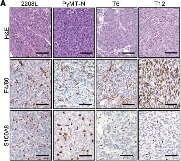
Detection of S100A8 by Immunohistochemistry
IACS-70654 suppressed the growth of neutrophil-enriched preclinical mouse models of TNBC.(A) Representative images of H&E and immunostaining of all TNBC preclinical mouse models used. F4/80 is a macrophage marker, and S100A8 is a neutrophil marker. Scale bars: 50 μm. (B) Flow cytometry analysis of TANs and TAMs in all TNBC preclinical mouse models used (T6, n = 6; all other models, n = 5). (C) The treatment scheme of IACS-70654 in a 7-day experiment. When the average tumor volume reached 80–250 mm3, the mice were randomized to initiate treatment. Vehicle or IACS-70654 at 3.75 mg/kg was administered orally on a 3-on/2-off schedule. On day 7, mice were euthanized 2 hours after receiving the last treatment. (D) Tumor growth curves of all TNBC models treated with IACS-70654 for 7 days (2208L, n = 6; all other models, n = 5). Two-way ANOVA and Šidák’s multiple-comparison test were used. (E) Left: Representative images of immunostaining of BrdU in 2208L tumor sections treated with vehicle or IACS-70654 for 7 days. Scale bar: 50 μm. Right: Quantification of BrdU staining (n = 15). A 2-tailed, unpaired Student’s t test was used. For B, D, and E, data are presented as mean ± SD. *P < 0.05; ****P < 0.0001. The experiments were conducted 3 times for 2208L and twice for other models. Data from 1 representative experiment are shown. (F) Tumor growth curves (from 1 representative experiment) of 2208L tumors treated with IACS-70654 for 27 days. The experiment was conducted twice. Image collected and cropped by CiteAb from the following open publication (https://pubmed.ncbi.nlm.nih.gov/39287984), licensed under a CC-BY license. Not internally tested by R&D Systems.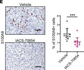
Detection of S100A8 by Immunohistochemistry
IACS-70654 reduced the growth of established 2208L lung metastases.(A) Violin plots showing RNA expression of representative genes that might be involved in migration and metastasis in 2208L tumor cells treated in vivo. Adjusted P < 0.01 for all genes. (B) Experimental design for studying the effects of IACS-70654 on established lung metastases. Dissociated 2208L tumor cells (100,000) were injected into the tail vein of each mouse. The mice were treated for 23 days. One mouse from the vehicle group had to be euthanized on day 22 due to poor body condition. (C) Representative images from H&E staining of lungs with 2208L metastases from mice after treatment. Scale bars: 5 mm. (D) Quantifications of lung metastases after treatment. Serial sectioning was performed to collect a total of 10 sections for each sample (vehicle, n = 40; IACS-70654, n = 50). Metastases with sizes of <1 mm, 1–3 mm, 3–5 mm, and >5 mm were assigned scores of 1, 2, 3, and 4, respectively. A 2-tailed, unpaired Student’s t test was used. Data are presented as mean ± SD. (E) Left: Representative images of S100A8 immunostaining on 2208L lung metastasis sections. Scale bar: 50 μm. Right: Quantification of S100A8 staining (n = 12). A 2-tailed, unpaired Student’s t test was used. ***P < 0.001; ****P < 0.0001. Data are presented as mean ± SD. Image collected and cropped by CiteAb from the following open publication (https://pubmed.ncbi.nlm.nih.gov/39287984), licensed under a CC-BY license. Not internally tested by R&D Systems.Applications for Mouse S100A8 Antibody (335806)
Application
Recommended Usage
Immunocytochemistry
8-25 µg/mL
Sample: Immersion fixed NIH-3T3 mouse embryonic fibroblast cell line
Sample: Immersion fixed NIH-3T3 mouse embryonic fibroblast cell line
Western Blot
2 µg/mL
Sample: Mouse leukocyte
Sample: Mouse leukocyte
Reviewed Applications
Read 1 review rated 5 using MAB3059 in the following applications:
Formulation, Preparation, and Storage
Purification
Protein A or G purified from hybridoma culture supernatant
Reconstitution
Reconstitute at 0.5 mg/mL in sterile PBS. For liquid material, refer to CoA for concentration.
Loading...
Formulation
Lyophilized from a 0.2 μm filtered solution in PBS with Trehalose. *Small pack size (SP) is supplied either lyophilized or as a 0.2 µm filtered solution in PBS.
Shipping
Lyophilized product is shipped at ambient temperature. Liquid small pack size (-SP) is shipped with polar packs. Upon receipt, store immediately at the temperature recommended below.
Stability & Storage
Use a manual defrost freezer and avoid repeated freeze-thaw cycles.
- 12 months from date of receipt, -20 to -70 °C as supplied.
- 1 month, 2 to 8 °C under sterile conditions after reconstitution.
- 6 months, -20 to -70 °C under sterile conditions after reconstitution.
Calculators
Background: S100A8
Long Name
S100 Calcium Binding Protein A8
Alternate Names
Calgranulin A, CFAG, MRP8
Gene Symbol
S100A8
UniProt
Additional S100A8 Products
Product Documents for Mouse S100A8 Antibody (335806)
Certificate of Analysis
To download a Certificate of Analysis, please enter a lot or batch number in the search box below.
Note: Certificate of Analysis not available for kit components.
Product Specific Notices for Mouse S100A8 Antibody (335806)
For research use only
Citations for Mouse S100A8 Antibody (335806)
 Powered by Bioz
Customer Reviews for Mouse S100A8 Antibody (335806) (1)
5 out of 5
1 Customer Rating
Have you used Mouse S100A8 Antibody (335806)?
Submit a review and receive an Amazon gift card!
$25/€18/£15/$25CAN/¥2500 for a review with an image
$10/€7/£6/$10CAN/¥1110 for a review without an image
Submit a review
Customer Images
Showing
1
-
1 of
1 review
Showing All
Filter By:
-
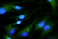
Application: Immunocytochemistry/ImmunofluorescenceSample Tested: Pulmonary Artery Smooth Muscle Cells and Artery Smooth Muscle CellsSpecies: MouseVerified Customer | Posted 11/02/2021

There are no reviews that match your criteria.
Protocols
Find general support by application which include: protocols, troubleshooting, illustrated assays, videos and webinars.
- Appropriate Fixation of IHC/ICC Samples
- Cellular Response to Hypoxia Protocols
- ClariTSA™ Fluorophore Kits
- Detection & Visualization of Antibody Binding
- ICC Cell Smear Protocol for Suspension Cells
- ICC Immunocytochemistry Protocol Videos
- ICC for Adherent Cells
- Immunocytochemistry (ICC) Protocol
- Immunocytochemistry Troubleshooting
- Immunofluorescence of Organoids Embedded in Cultrex Basement Membrane Extract
- Immunohistochemistry (IHC) and Immunocytochemistry (ICC) Protocols
- Preparing Samples for IHC/ICC Experiments
- Preventing Non-Specific Staining (Non-Specific Binding)
- Primary Antibody Selection & Optimization
- Protocol for VisUCyte™ HRP Polymer Detection Reagent
- Protocol for the Fluorescent ICC Staining of Cell Smears - Graphic
- Protocol for the Fluorescent ICC Staining of Cultured Cells on Coverslips - Graphic
- Protocol for the Preparation and Fluorescent ICC Staining of Cells on Coverslips
- Protocol for the Preparation and Fluorescent ICC Staining of Non-adherent Cells
- Protocol for the Preparation and Fluorescent ICC Staining of Stem Cells on Coverslips
- Protocol for the Preparation of a Cell Smear for Non-adherent Cell ICC - Graphic
- R&D Systems Quality Control Western Blot Protocol
- TUNEL and Active Caspase-3 Detection by IHC/ICC Protocol
- The Importance of IHC/ICC Controls
- Troubleshooting Guide: Western Blot Figures
- Western Blot Conditions
- Western Blot Protocol
- Western Blot Protocol for Cell Lysates
- Western Blot Troubleshooting
- Western Blot Troubleshooting Guide
- View all Protocols, Troubleshooting, Illustrated assays and Webinars
Loading...