
TNF receptor 1 (TNF RI; also called TNF R-p55/p60 and TNFRSF1A) is a type I transmembrane protein member of the TNF receptor superfamily, designated TNFRSF1A (1, 2). Both TNF RI and TNF RII (TNFRSF1B) are widely expressed and contain four TNF-alpha trimer-binding cysteine-rich domains (CRD) in their extracellular domains (ECD). However, TNF RI is thought to mediate most of the cellular effects of TNF-alpha (3). It is essential for proper development of lymph node germinal centers and Peyer’s patches, and for combating intracellular pathogens such as Listeria (1‑3). TNF RI is also a receptor for TNF-beta /TNFSF1B (lymphotoxin-alpha ) (4). TNF RI is present on the cell surface as a trimer of 55 kDa subunits (4, 5). TNF-alpha induces sequestering of TNF RI in lipid rafts, where it activates NF kappa B and is cleaved by ADAM-17/TACE (9, 10). Release of the 28 - 34 kDa TNF RI ECD also occurs constitutively and in response to products of pathogens such as LPS, CpG DNA or S. aureus protein A (1, 6‑8). Full-length TNF RI may also be released in exosome-like vesicles (11). Release helps to resolve inflammatory reactions, since it down-regulates cell surface TNF RI and provides soluble TNF RI to bind TNF-alpha (6, 12, 13). Exclusion from lipid rafts causes endocytosis of TNF RI complexes and induces apoptosis (1). Mouse TNF RI is a 454 amino acid (aa) protein that contains a 21 aa signal sequence, a 191 aa ECD with a PLAD domain (5) that mediates constitutive trimer formation, followed by the four CRD, a 23 aa transmembrane domain, and a 219 aa cytoplasmic sequence that contains a neutral sphingomyelinase activation domain and a death domain (15). The ECD of mouse TNF RI shows 67%, 70%, 64%, 70% and 88% aa identity with canine, feline, porcine, human, and rat TNF RI, respectively; it shows 23% aa identity with the ECD of TNF RII.
Mouse TNF RI/TNFRSF1A Antibody (55R170)
R&D Systems | Catalog # MAB430

Scientific images may be resized, cropped, or adjusted for brightness or contrast for presentation purposes. These updates are limited to presentation and do not affect the underlying scientific interpretation of the data. Where available, additional source data may be provided upon request.
Key Product Details
Species Reactivity
Validated:
Cited:
Applications
Validated:
Cited:
Label
Antibody Source
Product Specifications
Immunogen
Extracellular domain
Specificity
Clonality
Host
Isotype
Endotoxin Level
Scientific Data Images for Mouse TNF RI/TNFRSF1A Antibody (55R170)
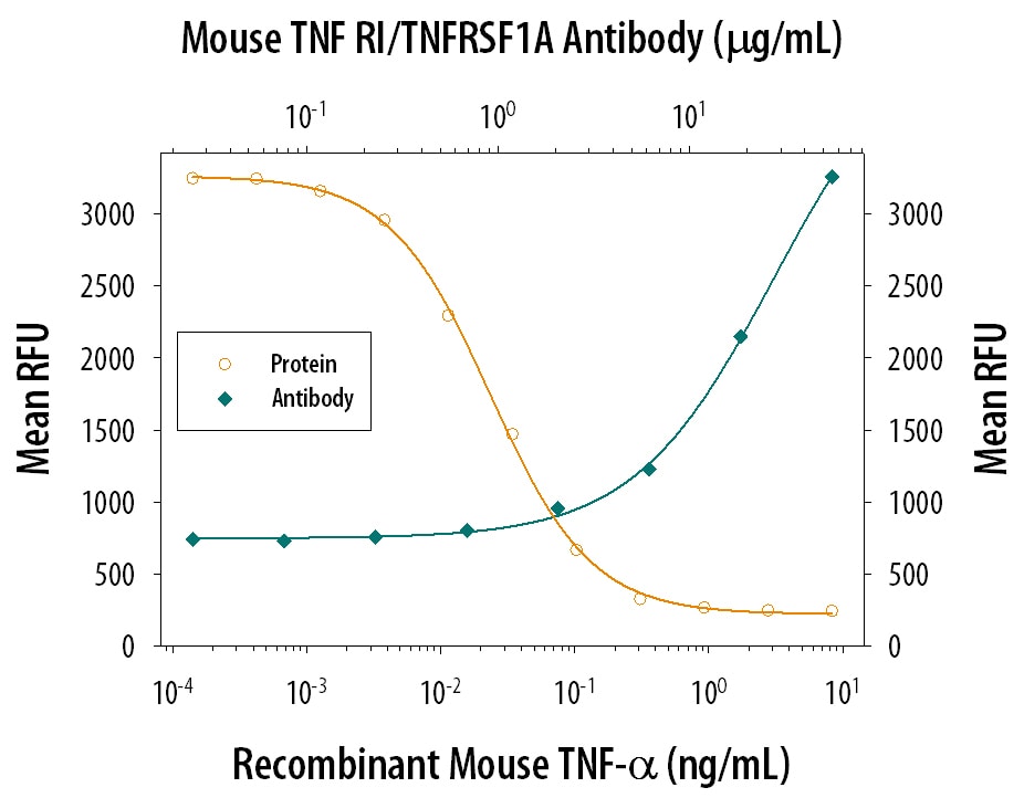
Cytotoxicity Induced by TNF‑ alpha and Neutralization by Mouse TNF RI/TNFRSF1A Antibody.
Recombinant Mouse TNF-a (Catalog # 410-MT) induces cytotoxicity in the the L-929 mouse fibroblast cell line in a dose-dependent manner (orange line). Cytotoxicity elicited by Recombinant Mouse TNF-a (0.1 ng/mL) is neutralized (green line) by increasing concentrations of Hamster Anti-Mouse TNF RI/TNFRSF1A Monoclonal Antibody (Catalog # MAB430). The ND50 is typically 5-20 µg/mL in the presence of the metabolic inhibitor actinomycin D (1 µg/mL).Applications for Mouse TNF RI/TNFRSF1A Antibody (55R170)
Immunoprecipitation
Western Blot
Sample: Recombinant Mouse TNF RI/TNFRSF1A (Catalog # 425-R1)
under non-reducing conditions only
Neutralization
Reviewed Applications
Read 2 reviews rated 4.5 using MAB430 in the following applications:
Formulation, Preparation, and Storage
Purification
Reconstitution
Reconstitute at 0.5 mg/mL in sterile PBS. For liquid material, refer to CoA for concentration.
Formulation
Shipping
Stability & Storage
- 12 months from date of receipt, -20 to -70 °C as supplied.
- 1 month, 2 to 8 °C under sterile conditions after reconstitution.
- 6 months, -20 to -70 °C under sterile conditions after reconstitution.
Calculators
Background: TNF RI/TNFRSF1A
References
- Pfeffer, K. (2003) Cytokine Growth Factor Rev. 14:185.
- Hehlgans, T. and K. Pfeffer (2005) Immunology 115:1.
- Peschon, J.J. et al. (1998) J. Immunol. 160:943.
- Banner, D.W et al. (1993) Cell 73: 431.
- Chan, F.K. et al. (2000) Science 288:2351.
- Xanthoulea, S. et al. (2004) J. Exp. Med. 200:367.
- Jin, L. et al. (2000) J. Immunol. 165:5153.
- Gomez, M.I. et al. (2006) J. Biol. Chem. 281:20190.
- Legler, D.F. et al. (2003) Immunity 18:655.
- Tellier, E. et al. (2006) Exp. Cell Res. 312:3969.
- Islam, A. et al. (2006) J. Biol. Chem. 281:6860.
- Garton, K.J. et al. (2006) J. Leukoc. Biol. 79:1105.
- McDermott, M.F. et al. (1999) Cell 97:133.
- Schneider-Brachert, W. et al. (2004) Immunity 21:415.
- Lewis, M. et al. (1991) Proc. Natl. Acad. Sci. USA 88:2830.
Long Name
Alternate Names
Gene Symbol
Additional TNF RI/TNFRSF1A Products
Product Documents for Mouse TNF RI/TNFRSF1A Antibody (55R170)
Certificate of Analysis
To download a Certificate of Analysis, please enter a lot or batch number in the search box below.
Note: Certificate of Analysis not available for kit components.
Product Specific Notices for Mouse TNF RI/TNFRSF1A Antibody (55R170)
For research use only
Citations for Mouse TNF RI/TNFRSF1A Antibody (55R170)
 Powered by Bioz
Customer Reviews for Mouse TNF RI/TNFRSF1A Antibody (55R170) (2)
Have you used Mouse TNF RI/TNFRSF1A Antibody (55R170)?
Submit a review and receive an Amazon gift card!
$25/€18/£15/$25CAN/¥2500 for a review with an image
$10/€7/£6/$10CAN/¥1110 for a review without an image
Submit a review
Customer Images
-
Application: Western BlotSample Tested: MacrophagesSpecies: MouseVerified Customer | Posted 10/20/2021

-
Application: ELISASample Tested: purified IgGSpecies: HumanVerified Customer | Posted 06/28/2016murine cross-reactivity studies
There are no reviews that match your criteria.
Protocols
Find general support by application which include: protocols, troubleshooting, illustrated assays, videos and webinars.
- Cellular Response to Hypoxia Protocols
- Immunoprecipitation Protocol
- R&D Systems Quality Control Western Blot Protocol
- Troubleshooting Guide: Western Blot Figures
- Western Blot Conditions
- Western Blot Protocol
- Western Blot Protocol for Cell Lysates
- Western Blot Troubleshooting
- Western Blot Troubleshooting Guide
- View all Protocols, Troubleshooting, Illustrated assays and Webinars
Associated Pathways


