
Vascular endothelial growth factor B (VEGF-B; also known as VFR) is a member of the VEGF-PDGF supergene family of growth factor molecules (1 - 4). Five mouse members have been identified, including VEGF-A, -B, -C, -D, and PlGF(-2) (1, 5). VEGF family members are disulfide-linked homo- and heterodimeric proteins that are important regulators of vasculogenesis and lymphangiogenesis. Mouse VEGF-B has two isoforms, a 32 kDa single chain and a 21 kDa single chain form (6, 7). The long form (VEGF-B186) is 207 amino acids (aa) in length, with a 21 aa signal sequence and a 186 aa mature region. The short form (VEGF-B167) is 188 aa in length, with a 21 aa signal sequence and a 167 aa mature segment. Each mature isoform shows the same N-terminal 94 aa that contains a cysteine knot VEGF homology domain (6 - 8). VEGF-B186 is O-glycosylated; VEGF-B167 is not. VEGF-B167 binds heparin; VEGF-B186 does not. Thus, VEGF-B186 is secreted and freely diffusible in tissues (7). However, the VEGF-B167 isoform is the predominant form in tissue (9). Mouse VEGF-B186 is 93% and 87% aa identical to bovine and human VEGF-B186, respectively; mouse VEGF-B167 is 90% and 88% aa identical to bovine and human VEGF-B167, respectively. The mouse VEGF-B167 homodimer is 42 kDa in size, while the VEGF-B186 homodimer is 62 kDa in size. Unlike VEGF167, VEGF-B186 undergoes proteolytic processing that creates a partially processed 48 kDa homodimer and a fully processed 32 kDa homodimer. Processing appears to occur at Arg127 of the mature form (10). Both forms of VEGF-B can heterodimerize with VEGF (7). Both VEGF-B isoforms bind to VEGF receptor 1 (VEGF R1), but not VEGF R2 or VEGF R3 (11). VEGF-B167 also binds neuropilin-1, but only the 127 aa processed form of VEGF-B186 binds neuropilin-1 (10). As a dimer, full length VEGF-B186 does not interact with neuropilin-1, while any dimer that contains the processed VEGF-B127 subunit will interact with neuropilin-1 (10). The importance of differential neuropilin binding is unclear. VEGF-B deficient mice display an atrial conduction deficit (12). On endothelial cells, ligation of VEGF R1 by VEGF-B has been shown to regulate the expression and activity of urokinase type plasminogen activator and plasminogen activator inhibitor 1 (11).
Mouse VEGF-B 167/186 Antibody
R&D Systems | Catalog # AF590

Scientific images may be resized, cropped, or adjusted for brightness or contrast for presentation purposes. These updates are limited to presentation and do not affect the underlying scientific interpretation of the data. Where available, additional source data may be provided upon request.
Key Product Details
Species Reactivity
Validated:
Cited:
Applications
Validated:
Cited:
Label
Antibody Source
Product Specifications
Immunogen
Pro22-Ala207
Accession # P49766
Specificity
Clonality
Host
Isotype
Endotoxin Level
Scientific Data Images for Mouse VEGF-B 167/186 Antibody
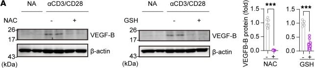
Detection of VEGF-B 167/186 by Western Blot
ROS-triggered VEGF-B protein acts on the VEGFR-1 expressed on activated T cells.(A) VEGF-B protein levels in activated CD8+ T cells treated with N-acetyl-L-cysteine (NAC) (5 μM) or glutathione (GSH) (5 μM); n = 6. (B) VEGF-B protein levels in activated CD8+ T cells treated with H2O2 at a gradient concentration; n = 3. (C) Alkaline phosphatase–labeled VEGF-B protein (VEGF-B-AP) binding in NA or activated CD4+ or CD8+ T cells. Scale bar: 2 μm. (D and E) The phosphorylation levels of VEGFR-1 (phospho-tyrosine), PI3K (P85), or ERK1/2 in activated CD8+ T cells treated by anti–VEGF-B ( alpha VB) or anti–VEGFR-1 ( alpha VEGFR-1) antibodies; n = 3. Data are shown as mean ± SEM. P values were calculated using 2-tailed unpaired t test in A and 1-way ANOVA with Holm-Šídák’s post hoc test in B. *P < 0.05, ***P < 0.001. Image collected and cropped by CiteAb from the following open publication (https://pubmed.ncbi.nlm.nih.gov/39145452), licensed under a CC-BY license. Not internally tested by R&D Systems.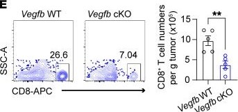
Detection of VEGF-B 167/186 by Flow Cytometry
Autocrine VEGF-B signaling is essential for the antitumor immunity of CD8+ T cells.(A–D) The tumor growth curve of MC38 cells or B16 cells on Vegfb WT or -cKO or Flt1 WT or -cKO mice by subcutaneous injection; n = 5. (E and F) Flow cytometry (FCM) analysis for the ratio of tumor-infiltrating CD8+ T cells in MC38 tumors on Vegfb WT or -cKO mice. (E) Cell numbers and (F) annexin V+ ratio of tumor-infiltrating CD8+ T cells were calculated; n = 5. (G and H) FCM analysis for the ratio of tumor-infiltrating CD8+ T cells in MC38 tumors on Flt1 WT or -cKO mice. (G) Cell numbers and (H) annexin V+ ratio of tumor-infiltrating CD8+ T cells were calculated; n = 5. (I) CD8+ Tn cells from Vegfb WT or -cKO OT-1 cells were activated, followed by Senp2 overexpression, and then these OT-1 T cells were transferred (i.v.) to recipient mice inoculated with MC38-OVA cells. (J) The tumor growth curves were recorded; n = 5. (K) The H-2Kb+ ratio in tumor-infiltrating CD8+ T cells and (L) the annexin V+ ratio of H-2Kb+ CD8+ T cells were calculated; n = 5. Data are shown as mean ± SEM. P values were calculated using 2-way ANOVA with Bonferroni’s post hoc test in A–D and J–L and 2-tailed unpaired t test in E–H. **P < 0.01, ***P < 0.001. Image collected and cropped by CiteAb from the following open publication (https://pubmed.ncbi.nlm.nih.gov/39145452), licensed under a CC-BY license. Not internally tested by R&D Systems.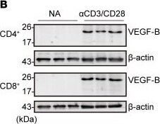
Detection of VEGF-B 167/186 by Western Blot
TCR activation induces VEGF-B protein expression in T cells.(A) Relative mRNA levels of the secreted factors in CD8+ Tn cells treated with or without (NA) alpha CD3/CD28 for 3 days; n = 3. Prf1, Ifng, Gzmb, and Tnf were positive indicators for T cell activation. (B) VEGF-B protein levels in NA or activated CD4+ and CD8+ T cells; n = 3. (C) Immunofluorescence images of VEGF-B in NA or activated CD4+ or CD8+ T cells. DAPI staining is in blue. Scale bar: 2 μm. (D) The mean fluorescent intensity (MFI) of VEGF-B in NA or activated CD4+ or CD8+ T cells; n = 3. (E) The population of VEGF-B+ and IFN-gamma + in NA or activated CD8+ T cells; n = 3. (F) VEGF-B protein levels in culture supernatants of NA or activated CD4+ or CD8+ T cells; n = 3. (G) The population of VEGF-B+ and H-2Kb+ cells in CD8+ T cells from the spleen of OT-1 mice infected by LM-OVA for 3 days; n = 5. (H) The population of VEGF-B+ cells in CD4+ or CD8+ T cells from colon tissue with DSS-induced colitis; n = 6. Data are shown as mean ± SEM. P values were calculated using 2-tailed unpaired t test in F. *P < 0.05, **P < 0.01. Image collected and cropped by CiteAb from the following open publication (https://pubmed.ncbi.nlm.nih.gov/39145452), licensed under a CC-BY license. Not internally tested by R&D Systems.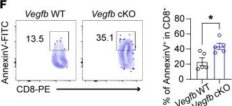
Detection of VEGF-B 167/186 by Flow Cytometry
Autocrine VEGF-B signaling is essential for the antitumor immunity of CD8+ T cells.(A–D) The tumor growth curve of MC38 cells or B16 cells on Vegfb WT or -cKO or Flt1 WT or -cKO mice by subcutaneous injection; n = 5. (E and F) Flow cytometry (FCM) analysis for the ratio of tumor-infiltrating CD8+ T cells in MC38 tumors on Vegfb WT or -cKO mice. (E) Cell numbers and (F) annexin V+ ratio of tumor-infiltrating CD8+ T cells were calculated; n = 5. (G and H) FCM analysis for the ratio of tumor-infiltrating CD8+ T cells in MC38 tumors on Flt1 WT or -cKO mice. (G) Cell numbers and (H) annexin V+ ratio of tumor-infiltrating CD8+ T cells were calculated; n = 5. (I) CD8+ Tn cells from Vegfb WT or -cKO OT-1 cells were activated, followed by Senp2 overexpression, and then these OT-1 T cells were transferred (i.v.) to recipient mice inoculated with MC38-OVA cells. (J) The tumor growth curves were recorded; n = 5. (K) The H-2Kb+ ratio in tumor-infiltrating CD8+ T cells and (L) the annexin V+ ratio of H-2Kb+ CD8+ T cells were calculated; n = 5. Data are shown as mean ± SEM. P values were calculated using 2-way ANOVA with Bonferroni’s post hoc test in A–D and J–L and 2-tailed unpaired t test in E–H. **P < 0.01, ***P < 0.001. Image collected and cropped by CiteAb from the following open publication (https://pubmed.ncbi.nlm.nih.gov/39145452), licensed under a CC-BY license. Not internally tested by R&D Systems.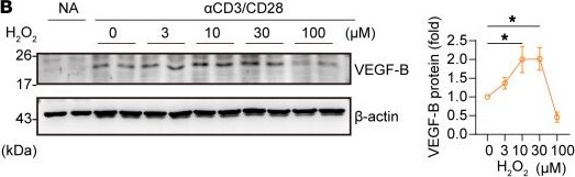
Detection of VEGF-B 167/186 by Western Blot
ROS-triggered VEGF-B protein acts on the VEGFR-1 expressed on activated T cells.(A) VEGF-B protein levels in activated CD8+ T cells treated with N-acetyl-L-cysteine (NAC) (5 μM) or glutathione (GSH) (5 μM); n = 6. (B) VEGF-B protein levels in activated CD8+ T cells treated with H2O2 at a gradient concentration; n = 3. (C) Alkaline phosphatase–labeled VEGF-B protein (VEGF-B-AP) binding in NA or activated CD4+ or CD8+ T cells. Scale bar: 2 μm. (D and E) The phosphorylation levels of VEGFR-1 (phospho-tyrosine), PI3K (P85), or ERK1/2 in activated CD8+ T cells treated by anti–VEGF-B ( alpha VB) or anti–VEGFR-1 ( alpha VEGFR-1) antibodies; n = 3. Data are shown as mean ± SEM. P values were calculated using 2-tailed unpaired t test in A and 1-way ANOVA with Holm-Šídák’s post hoc test in B. *P < 0.05, ***P < 0.001. Image collected and cropped by CiteAb from the following open publication (https://pubmed.ncbi.nlm.nih.gov/39145452), licensed under a CC-BY license. Not internally tested by R&D Systems.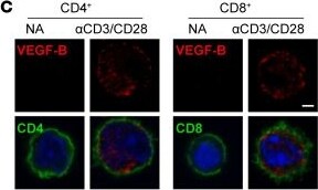
Detection of VEGF-B 167/186 by Immunocytochemistry/ Immunofluorescence
TCR activation induces VEGF-B protein expression in T cells.(A) Relative mRNA levels of the secreted factors in CD8+ Tn cells treated with or without (NA) alpha CD3/CD28 for 3 days; n = 3. Prf1, Ifng, Gzmb, and Tnf were positive indicators for T cell activation. (B) VEGF-B protein levels in NA or activated CD4+ and CD8+ T cells; n = 3. (C) Immunofluorescence images of VEGF-B in NA or activated CD4+ or CD8+ T cells. DAPI staining is in blue. Scale bar: 2 μm. (D) The mean fluorescent intensity (MFI) of VEGF-B in NA or activated CD4+ or CD8+ T cells; n = 3. (E) The population of VEGF-B+ and IFN-gamma + in NA or activated CD8+ T cells; n = 3. (F) VEGF-B protein levels in culture supernatants of NA or activated CD4+ or CD8+ T cells; n = 3. (G) The population of VEGF-B+ and H-2Kb+ cells in CD8+ T cells from the spleen of OT-1 mice infected by LM-OVA for 3 days; n = 5. (H) The population of VEGF-B+ cells in CD4+ or CD8+ T cells from colon tissue with DSS-induced colitis; n = 6. Data are shown as mean ± SEM. P values were calculated using 2-tailed unpaired t test in F. *P < 0.05, **P < 0.01. Image collected and cropped by CiteAb from the following open publication (https://pubmed.ncbi.nlm.nih.gov/39145452), licensed under a CC-BY license. Not internally tested by R&D Systems.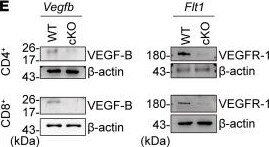
Detection of VEGF-B 167/186 by Western Blot
Autocrine VEGF-B is required for the survival of activated T cells.(A–D) sh-Vegfb or negative control (sh-NC) CD8+ OT-1 Tn cells were transferred into recipients infected with LM-OVA. The H-2Kb+ ratio in CD8+ T cells from the blood (B) or spleen (C) of recipients; n = 5. (D) The LM CFUs in the spleen and liver of the infected recipients; n = 5. (E) VEGF-B and VEGFR-1 protein levels in WT and cKO T cells; n = 3. (F) The MFI of CD69, CD25, and CD44 in Vegfb or Flt1 WT and cKO CD8+ Tn cells with TCR activation; n = 3. (G) The annexin V+ and PI+ ratio in VEGF-B–treated (VB-treated) Vegfb or Flt1 WT and cKO CD8+ T cells; n = 3. (H) CD8+ Tn cells were activated to induce memory T cells (Tm) by IL-15. The numbers and ratios of Tm (CD44+CD62L+) or TE (CD44+CD62L–) cells with IgG or alpha VB treatment were calculated; n = 8. (I–M) Vegfb or Flt1 WT and cKO CD8+ OT-1 T cells were transferred to recipients, followed by LM-OVA infection. (J and K) H-2Kb+ ratio in CD8+ T cells from the blood of recipients; n = 5. (L and M) The memory precursor (KLRG1–CD127A+) ratio in H-2Kb+ CD8+ T cells from the spleen of recipients on day 7 of prime infection; n = 5. Data are shown as mean ± SEM. P values were calculated using 2-tailed unpaired t test in C, D, H, L, and M and 2-way ANOVA with Holm-Šídák’s post-hoc test in B, G, J, and K. *P < 0.05, **P < 0.01, ***P < 0.001. Image collected and cropped by CiteAb from the following open publication (https://pubmed.ncbi.nlm.nih.gov/39145452), licensed under a CC-BY license. Not internally tested by R&D Systems.Applications for Mouse VEGF-B 167/186 Antibody
Blockade of Receptor-ligand Interaction
Western Blot
Sample: Recombinant Mouse VEGF-B167 (Catalog # 2595-VE)
Recombinant Mouse VEGF-B186 (Catalog # 767-VE)
Formulation, Preparation, and Storage
Purification
Reconstitution
Reconstitute at 0.2 mg/mL in sterile PBS. For liquid material, refer to CoA for concentration.
Formulation
Shipping
Stability & Storage
- 12 months from date of receipt, -20 to -70 °C as supplied.
- 1 month, 2 to 8 °C under sterile conditions after reconstitution.
- 6 months, -20 to -70 °C under sterile conditions after reconstitution.
Calculators
Background: VEGF-B
References
- Li, X. and U. Eriksson (2001) Int. J. Biochem Cell Biol. 33:421.
- Olofsson, B. et al. (1999) Curr. Opin. Biotechnol. 10:528.
- Clauss, M. (2000) Semin. Thromb. Hemost. 26:561.
- Matsumoto, T. and L. Claesson-Welsh (2001) Sci STKE Dec. 11(112):RE21.
- DiPalma, T. et al. (1996) Mamm. Genome 7:6.
- Olofsson, B. et al. (1996) Proc. Natl. Acad. Sci. USA 93:2576.
- Olofsson, B. et al. (1996) J. Biol. Chem. 271:19310.
- Twonson, S. et al. (1996) Biochem. Biophys. Res. Commun. 220:922.
- Li, X. et al. (2001) Growth Factors 19:49.
- Makinen, T. et al. (1999) J. Biol. Chem. 274:21217.
- Olofsson, B. et al. (1998) Proc. Nat. Acad. Sci. USA 95:11709.
- Aase, K. et al. (2001) Circulation 104:358.
Long Name
Alternate Names
Gene Symbol
UniProt
Additional VEGF-B Products
Product Documents for Mouse VEGF-B 167/186 Antibody
Certificate of Analysis
To download a Certificate of Analysis, please enter a lot or batch number in the search box below.
Note: Certificate of Analysis not available for kit components.
Product Specific Notices for Mouse VEGF-B 167/186 Antibody
For research use only
Citations for Mouse VEGF-B 167/186 Antibody
 Powered by Bioz
Customer Reviews for Mouse VEGF-B 167/186 Antibody
There are currently no reviews for this product. Be the first to review Mouse VEGF-B 167/186 Antibody and earn rewards!
Have you used Mouse VEGF-B 167/186 Antibody?
Submit a review and receive an Amazon gift card!
$25/€18/£15/$25CAN/¥2500 for a review with an image
$10/€7/£6/$10CAN/¥1110 for a review without an image
Submit a review
Protocols
Find general support by application which include: protocols, troubleshooting, illustrated assays, videos and webinars.
- Cellular Response to Hypoxia Protocols
- R&D Systems Quality Control Western Blot Protocol
- Troubleshooting Guide: Western Blot Figures
- Western Blot Conditions
- Western Blot Protocol
- Western Blot Protocol for Cell Lysates
- Western Blot Troubleshooting
- Western Blot Troubleshooting Guide
- View all Protocols, Troubleshooting, Illustrated assays and Webinars