
WARP (von Willebrand factor A [vWFA] domain-related protein) is a 50 kDa glycoprotein member of the vWFA domain superfamily of extracellular matrix proteins. It is expressed in embryonic articular cartilage, skeletal muscle and basement membranes in the PNS. WARP forms disulfide-linked homodimers and multimers, and complexes with perlecan. Secreted mouse WARP is 397 amino acids (aa) in length. It contains a vWFA domain (aa 34‑209), and two fibronectin type III domains (aa 211‑394) that likely bind to the GAG modification of perlecan. Cys369 and Cys393 contribute to intermolecular bond formation. There is one alternate start site at Met213. Mature mouse WARP (aa 19‑415) is 93% and 78% aa identical to rat and human WARP, respectively.

Scientific images may be resized, cropped, or adjusted for brightness or contrast for presentation purposes. These updates are limited to presentation and do not affect the underlying scientific interpretation of the data. Where available, additional source data may be provided upon request.
Key Product Details
Species Reactivity
Validated:
Mouse
Cited:
Mouse
Applications
Validated:
Western Blot
Cited:
Immunohistochemistry, Western Blot
Label
Unconjugated
Antibody Source
Polyclonal Goat IgG
Loading...
Product Specifications
Immunogen
Mouse myeloma cell line NS0-derived recombinant mouse WARP isoform 1
Arg19-Pro415
Accession # Q8R2Z5
Arg19-Pro415
Accession # Q8R2Z5
Specificity
Detects mouse WARP in direct ELISAs and Western blots. In direct ELISAs, approximately 20% cross‑reactivity with recombinant human WARP is observed.
Clonality
Polyclonal
Host
Goat
Isotype
IgG
Scientific Data Images for Mouse WARP Antibody
Detection of Mouse WARP/VWA1 by Immunocytochemistry/Immunofluorescence
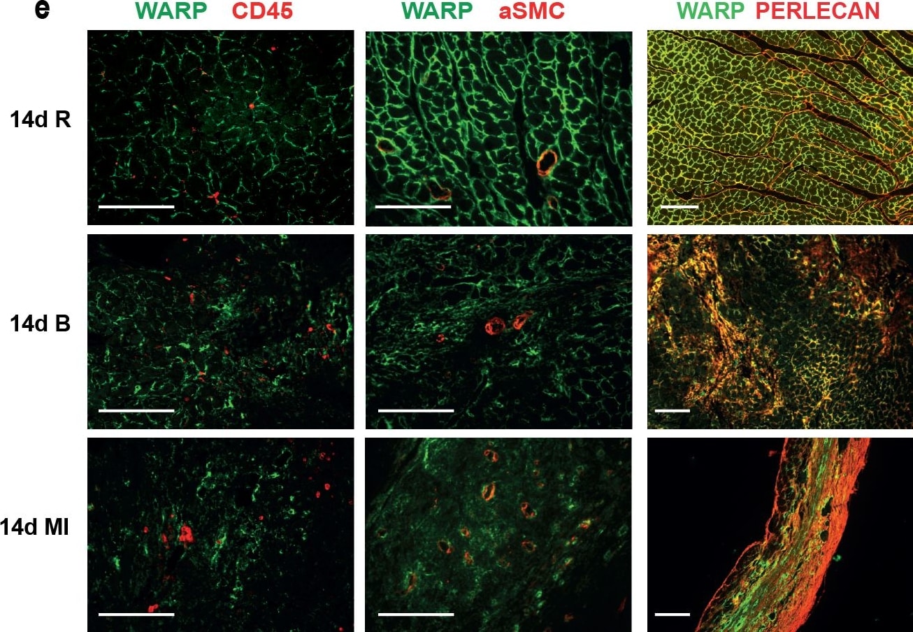
WARP expression pattern in the heart.(a) WARP mRNA levels were induced 3 and 7 days after MI. WARP mRNA levels peaked at 7 days after MI and then decreased again at 14 days. (b) Western blot analysis and (c) immunohistochemistry also showed an induction of WARP protein levels in the infarcted LV at 3, 7, and 14 days after MI. Because of different sample-loading, the blots of the 14 days MI were cut and pasted in the same order as the blots of the 3 and 7 days MI. Images of the original unadjusted blots are provided in S1 Fig (d and e) Confocal microscopy confirmed the co-localization of WARP with perlecan in the uninfarcted heart, showing a network of WARP and perlecan as a honeycomb-structure surrounding the cardiomyocytes. In the infarcted LV zones, the WARP-perlecan honeycomb-structure is reduced at 3 days after MI and completely absent at 7 and 14 days after MI and the interaction between WARP and perlecan is disrupted. (e) WARP did not localize on CD45 positive leukocytes or in alpha smooth muscle cells lining the vessels in the remote, border and infarcted zone of the heart 14 days after MI. n≥3; *p≤0.05; **p≤0.005; ***p≤0.001 versus sham, ##p≤0.005; ###p≤0.001 versus remote, bars 1000 μm for (c) and 100 μm for (d) and (e). Image collected and cropped by CiteAb from the following publication (https://dx.plos.org/10.1371/journal.pone.0139199), licensed under a CC-BY license. Not internally tested by R&D Systems.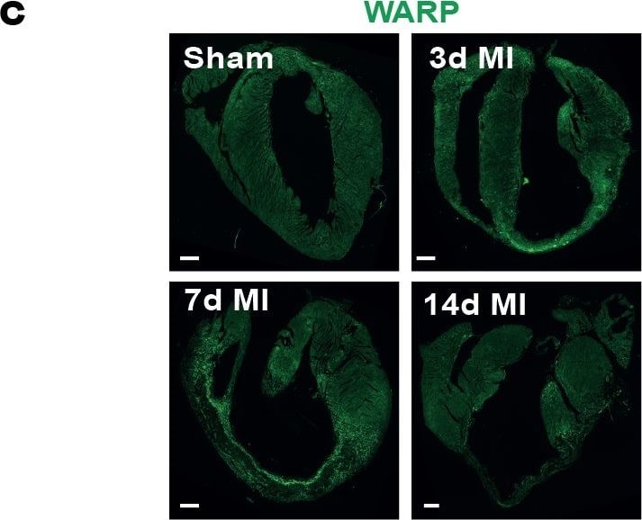
Detection of Mouse WARP/VWA1 by Immunocytochemistry/Immunofluorescence
WARP expression pattern in the heart.(a) WARP mRNA levels were induced 3 and 7 days after MI. WARP mRNA levels peaked at 7 days after MI and then decreased again at 14 days. (b) Western blot analysis and (c) immunohistochemistry also showed an induction of WARP protein levels in the infarcted LV at 3, 7, and 14 days after MI. Because of different sample-loading, the blots of the 14 days MI were cut and pasted in the same order as the blots of the 3 and 7 days MI. Images of the original unadjusted blots are provided in S1 Fig (d and e) Confocal microscopy confirmed the co-localization of WARP with perlecan in the uninfarcted heart, showing a network of WARP and perlecan as a honeycomb-structure surrounding the cardiomyocytes. In the infarcted LV zones, the WARP-perlecan honeycomb-structure is reduced at 3 days after MI and completely absent at 7 and 14 days after MI and the interaction between WARP and perlecan is disrupted. (e) WARP did not localize on CD45 positive leukocytes or in alpha smooth muscle cells lining the vessels in the remote, border and infarcted zone of the heart 14 days after MI. n≥3; *p≤0.05; **p≤0.005; ***p≤0.001 versus sham, ##p≤0.005; ###p≤0.001 versus remote, bars 1000 μm for (c) and 100 μm for (d) and (e). Image collected and cropped by CiteAb from the following publication (https://dx.plos.org/10.1371/journal.pone.0139199), licensed under a CC-BY license. Not internally tested by R&D Systems.
Detection of Mouse WARP/VWA1 by Immunocytochemistry/Immunofluorescence
WARP expression pattern in the heart.(a) WARP mRNA levels were induced 3 and 7 days after MI. WARP mRNA levels peaked at 7 days after MI and then decreased again at 14 days. (b) Western blot analysis and (c) immunohistochemistry also showed an induction of WARP protein levels in the infarcted LV at 3, 7, and 14 days after MI. Because of different sample-loading, the blots of the 14 days MI were cut and pasted in the same order as the blots of the 3 and 7 days MI. Images of the original unadjusted blots are provided in S1 Fig (d and e) Confocal microscopy confirmed the co-localization of WARP with perlecan in the uninfarcted heart, showing a network of WARP and perlecan as a honeycomb-structure surrounding the cardiomyocytes. In the infarcted LV zones, the WARP-perlecan honeycomb-structure is reduced at 3 days after MI and completely absent at 7 and 14 days after MI and the interaction between WARP and perlecan is disrupted. (e) WARP did not localize on CD45 positive leukocytes or in alpha smooth muscle cells lining the vessels in the remote, border and infarcted zone of the heart 14 days after MI. n≥3; *p≤0.05; **p≤0.005; ***p≤0.001 versus sham, ##p≤0.005; ###p≤0.001 versus remote, bars 1000 μm for (c) and 100 μm for (d) and (e). Image collected and cropped by CiteAb from the following publication (https://dx.plos.org/10.1371/journal.pone.0139199), licensed under a CC-BY license. Not internally tested by R&D Systems.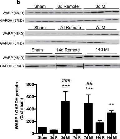
Detection of Mouse WARP/VWA1 by Western Blot
WARP expression pattern in the heart.(a) WARP mRNA levels were induced 3 and 7 days after MI. WARP mRNA levels peaked at 7 days after MI and then decreased again at 14 days. (b) Western blot analysis and (c) immunohistochemistry also showed an induction of WARP protein levels in the infarcted LV at 3, 7, and 14 days after MI. Because of different sample-loading, the blots of the 14 days MI were cut and pasted in the same order as the blots of the 3 and 7 days MI. Images of the original unadjusted blots are provided in S1 Fig (d and e) Confocal microscopy confirmed the co-localization of WARP with perlecan in the uninfarcted heart, showing a network of WARP and perlecan as a honeycomb-structure surrounding the cardiomyocytes. In the infarcted LV zones, the WARP-perlecan honeycomb-structure is reduced at 3 days after MI and completely absent at 7 and 14 days after MI and the interaction between WARP and perlecan is disrupted. (e) WARP did not localize on CD45 positive leukocytes or in alpha smooth muscle cells lining the vessels in the remote, border and infarcted zone of the heart 14 days after MI. n≥3; *p≤0.05; **p≤0.005; ***p≤0.001 versus sham, ##p≤0.005; ###p≤0.001 versus remote, bars 1000 μm for (c) and 100 μm for (d) and (e). Image collected and cropped by CiteAb from the following publication (https://dx.plos.org/10.1371/journal.pone.0139199), licensed under a CC-BY license. Not internally tested by R&D Systems.Applications for Mouse WARP Antibody
Application
Recommended Usage
Western Blot
0.1 µg/mL
Sample: Recombinant Mouse WARP
Sample: Recombinant Mouse WARP
Formulation, Preparation, and Storage
Purification
Antigen Affinity-purified
Reconstitution
Reconstitute at 0.2 mg/mL in sterile PBS. For liquid material, refer to CoA for concentration.
Loading...
Formulation
Lyophilized from a 0.2 μm filtered solution in PBS with Trehalose. *Small pack size (SP) is supplied either lyophilized or as a 0.2 µm filtered solution in PBS.
Shipping
Lyophilized product is shipped at ambient temperature. Liquid small pack size (-SP) is shipped with polar packs. Upon receipt, store immediately at the temperature recommended below.
Stability & Storage
Use a manual defrost freezer and avoid repeated freeze-thaw cycles.
- 12 months from date of receipt, -20 to -70 °C as supplied.
- 1 month, 2 to 8 °C under sterile conditions after reconstitution.
- 6 months, -20 to -70 °C under sterile conditions after reconstitution.
Calculators
Background: WARP
Long Name
von Willebrand factor A Domain-related Protein
Alternate Names
VWA1
Gene Symbol
VWA1
UniProt
Additional WARP Products
Product Documents for Mouse WARP Antibody
Certificate of Analysis
To download a Certificate of Analysis, please enter a lot or batch number in the search box below.
Note: Certificate of Analysis not available for kit components.
Product Specific Notices for Mouse WARP Antibody
For research use only
Related Research Areas
Citations for Mouse WARP Antibody
 Powered by Bioz
Customer Reviews for Mouse WARP Antibody
There are currently no reviews for this product. Be the first to review Mouse WARP Antibody and earn rewards!
Have you used Mouse WARP Antibody?
Submit a review and receive an Amazon gift card!
$25/€18/£15/$25CAN/¥2500 for a review with an image
$10/€7/£6/$10CAN/¥1110 for a review without an image
Submit a review
Protocols
Find general support by application which include: protocols, troubleshooting, illustrated assays, videos and webinars.
- Cellular Response to Hypoxia Protocols
- R&D Systems Quality Control Western Blot Protocol
- Troubleshooting Guide: Western Blot Figures
- Western Blot Conditions
- Western Blot Protocol
- Western Blot Protocol for Cell Lysates
- Western Blot Troubleshooting
- Western Blot Troubleshooting Guide
- View all Protocols, Troubleshooting, Illustrated assays and Webinars
Loading...