
PGAM1/2/4 Antibody
Novus Biologicals | Catalog # NB100-774

![Western Blot: PGAM1/2/4 Antibody [NB100-774]](https://resources.rndsystems.com/images/products/PGAM1-2-4-Antibody-Western-Blot-NB100-774-img0002.jpg "Western Blot: PGAM1/2/4 Antibody [NB100-774]")
Loading...
Key Product Details
Validated by
Biological Validation
Species Reactivity
Validated:
Human, Mouse, Rat, Porcine
Cited:
Human
Predicted:
Bovine (100%), Canine (100%). Backed by our 100% Guarantee.
Applications
Western Blot, Peptide ELISA, Immunocytochemistry/ Immunofluorescence
Label
Unconjugated
Antibody Source
Polyclonal Goat IgG
Loading...
Product Specifications
Immunogen
Peptide with sequence C-KAMEAVAAQGKAKK, from the C Terminus of the protein sequence according to NP_002620.1; NP_000281.2; NP_001025062.1.
Specificity
Please note this antibody is expected to recognize the products of 3 highly similar genes.
Clonality
Polyclonal
Host
Goat
Isotype
IgG
Theoretical MW
29 kDa.
Disclaimer note: The observed molecular weight of the protein may vary from the listed predicted molecular weight due to post translational modifications, post translation cleavages, relative charges, and other experimental factors.
Disclaimer note: The observed molecular weight of the protein may vary from the listed predicted molecular weight due to post translational modifications, post translation cleavages, relative charges, and other experimental factors.
Scientific Data Images for PGAM1/2/4 Antibody
Western Blot: PGAM1/2/4 Antibody [NB100-774]
Western Blot: PGAM1/2/4 Antibody [NB100-774] - staining of Mouse (A), Rat (B) and Pig (C) Liver lysate (35ug protein in RIPA buffer). Primary incubation was 1 hour. Detected by chemiluminescence.![Immunocytochemistry/ Immunofluorescence: PGAM1/2/4 Antibody [NB100-774]](https://resources.rndsystems.com/images/products/PGAM1-2-4-Antibody-Immunocytochemistry-Immunofluorescence-NB100-774-img0004.jpg "Immunocytochemistry/ Immunofluorescence: PGAM1/2/4 Antibody [NB100-774]")
Immunocytochemistry/ Immunofluorescence: PGAM1/2/4 Antibody [NB100-774]
PGAM1-2-4-Antibody-Immunocytochemistry-Immunofluorescence-NB100-774-img0004.jpg![Immunocytochemistry/ Immunofluorescence: PGAM1/2/4 Antibody [NB100-774]](https://resources.rndsystems.com/images/products/PGAM1-2-4-Antibody-Immunocytochemistry-Immunofluorescence-NB100-774-img0003.jpg "Immunocytochemistry/ Immunofluorescence: PGAM1/2/4 Antibody [NB100-774]")
Western Blot: PGAM1/2/4 Antibody [NB100-774] -
Validation of R-Ras2 as the defatty-acylation target of SIRT6.(A) mRNA and protein levels of R-Ras2 in Sirt6 WT and KO MEFs. (B) In-gel fluorescence (with NH2OH treatment) showing that R-Ras2 has higher lysine fatty acylation level in Sirt6 KO MEFs than in Sirt6 WT MEFs. Right histogram shows the quantification of bands on the fluorescence gel. Values with error bars indicate mean ± s.d. of three biological replicates. * indicates p<0.05. The full fluorescence gel is shown in Figure 2—figure supplement 2A. (C) Detection of R-Ras2 lysine fatty acylation levels in control and SIRT6 knockown HEK 293 T cells by in-gel fluorescence. Right histogram shows the quantification of bands on the fluorescence gel. Values with error bars indicate mean ± s.d. of three biological replicates. * indicates p<0.05. (D) Lysine fatty acylation levels of endogenous R-Ras2 in Sirt6 WT and KO MEFs. (E, F) SIRT6 defatty-acylated R-Ras2 in a NAD+-dependent manner in vitro. In-gel fluorescence was used to detect R-Ras2 lysine fatty acylation (E). A 32P-NAD+ assay was used to detect fatty acyl ADPR product from defatty-acylation reaction. (F). (G) In-gel fluorescence (with NH2OH treatment) showing that mutation of four lysine residues at the C-terminus of R-Ras2 significantly decreased lysine fatty acylation in Sirt6 KO MEFs. Right histogram shows the quantification of bands on the fluorescence gel. Values with error bars indicate mean ± s.d. of three biological replicates. * indicates p<0.05. The full fluorescence gel including R-Ras2 total fatty acylation levels (without NH2OH treatment) is shown in Figure 2—figure supplement 2C. (H) Tandem mass (MS/MS) spectrum of doubly charged Alk14 modified (on K194) R-Ras2 peptide. The b- and y- ions are shown along with the peptide sequence. (I) In-gel fluorescence (with NH2OH treatment) showing that single mutation of four lysine residues at the C-terminus of R-Ras2 did not affect R-Ras2 lysine fatty acylation. (J) Confocal imaging showed that R-Ras2 W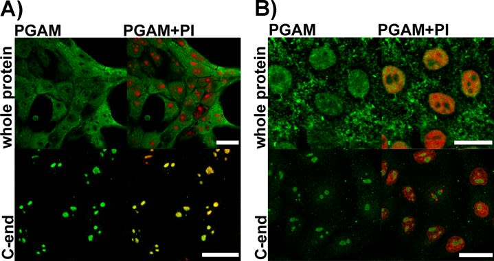
Immunocytochemistry/ Immunofluorescence: PGAM1/2/4 Antibody [NB100-774] -
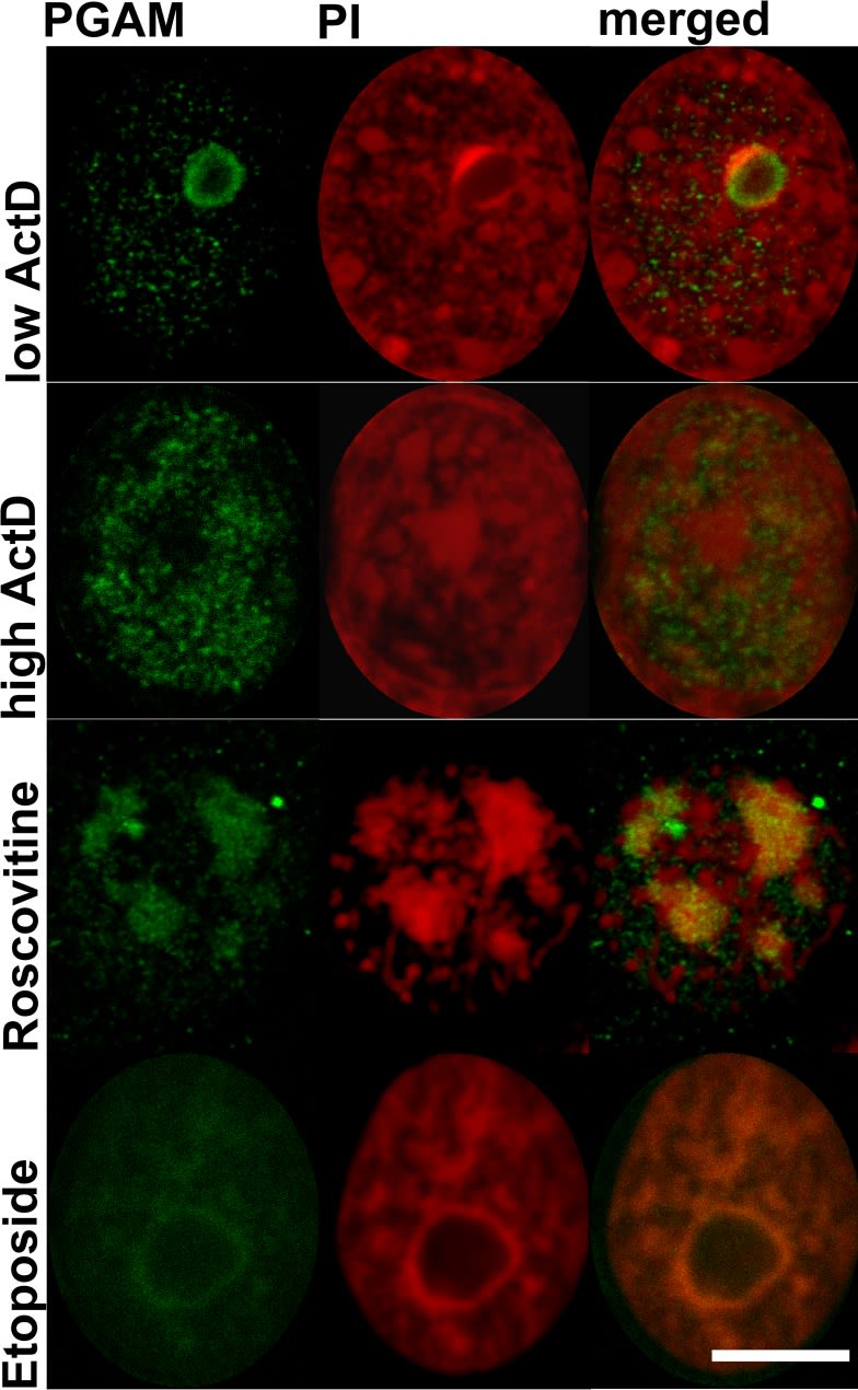
Immunocytochemistry/ Immunofluorescence: PGAM1/2/4 Antibody [NB100-774] - Detection of PGAM in KLN-205 cells with antibodies directed to whole PGAM protein or to C-terminal peptide of PGAMA. control conditions (scan parameters in red channel were set to emphasize nucleolar staining with propidium iodide – PI) B. RNase-treated cells. Bar=15 μm. Image collected & cropped by CiteAb from the following publication (https://www.oncotarget.com/lookup/doi/10.18632/oncotarget.4044), licensed under a CC-BY license. Not internally tested by Novus Biologicals.Applications for PGAM1/2/4 Antibody
Application
Recommended Usage
Peptide ELISA
Detection limit 1:28000
Western Blot
0.05-0.15 ug/ml
Application Notes
WB: Approx 27kDa band observed in Human Cerebellum and in Human and Mouse Liver lysates, and approx. 28kDa band observed in Rat and Pig Liver lysates (calculated MW of 28.8kDa according to Human NP_002620.1, NP_000281.2, and NP_001025062.1, Mouse NP_075907.2, Rat NP_445742.2 and Pig XP_003483583.1 ).
Formulation, Preparation, and Storage
Purification
Immunogen affinity purified
Formulation
Tris saline (20 mM Tris pH 7.3, 150 mM NaCl), 0.5% BSA
Preservative
0.02% Sodium Azide
Concentration
0.5 mg/ml
Shipping
The product is shipped with polar packs. Upon receipt, store it immediately at the temperature recommended below.
Stability & Storage
Store at -20C. Avoid freeze-thaw cycles.
Background: PGAM1/2/4
Alternate Names
PGAM1, PGAMA, PGAM-B, phosphoglycerate mutase 1
Entrez Gene IDs
5223 (Human)
Gene Symbol
PGAM1
UniProt
Additional PGAM1/2/4 Products
Product Documents for PGAM1/2/4 Antibody
Certificate of Analysis
To download a Certificate of Analysis, please enter a lot or batch number in the search box below.
Product Specific Notices for PGAM1/2/4 Antibody
This product is for research use only and is not approved for use in humans or in clinical diagnosis. Primary Antibodies are guaranteed for 1 year from date of receipt.
Citations for PGAM1/2/4 Antibody
 Powered by Bioz
Customer Reviews for PGAM1/2/4 Antibody
There are currently no reviews for this product. Be the first to review PGAM1/2/4 Antibody and earn rewards!
Have you used PGAM1/2/4 Antibody?
Submit a review and receive an Amazon gift card!
$25/€18/£15/$25CAN/¥2500 for a review with an image
$10/€7/£6/$10CAN/¥1110 for a review without an image
Submit a review
Protocols
Find general support by application which include: protocols, troubleshooting, illustrated assays, videos and webinars.
- Appropriate Fixation of IHC/ICC Samples
- Cellular Response to Hypoxia Protocols
- ClariTSA™ Fluorophore Kits
- Detection & Visualization of Antibody Binding
- ELISA Sample Preparation & Collection Guide
- ELISA Troubleshooting Guide
- How to Run an R&D Systems DuoSet ELISA
- How to Run an R&D Systems Quantikine ELISA
- How to Run an R&D Systems Quantikine™ QuicKit™ ELISA
- ICC Cell Smear Protocol for Suspension Cells
- ICC Immunocytochemistry Protocol Videos
- ICC for Adherent Cells
- Immunocytochemistry (ICC) Protocol
- Immunocytochemistry Troubleshooting
- Immunofluorescence of Organoids Embedded in Cultrex Basement Membrane Extract
- Immunohistochemistry (IHC) and Immunocytochemistry (ICC) Protocols
- Preparing Samples for IHC/ICC Experiments
- Preventing Non-Specific Staining (Non-Specific Binding)
- Primary Antibody Selection & Optimization
- Protocol for VisUCyte™ HRP Polymer Detection Reagent
- Protocol for the Fluorescent ICC Staining of Cell Smears - Graphic
- Protocol for the Fluorescent ICC Staining of Cultured Cells on Coverslips - Graphic
- Protocol for the Preparation and Fluorescent ICC Staining of Cells on Coverslips
- Protocol for the Preparation and Fluorescent ICC Staining of Non-adherent Cells
- Protocol for the Preparation and Fluorescent ICC Staining of Stem Cells on Coverslips
- Protocol for the Preparation of a Cell Smear for Non-adherent Cell ICC - Graphic
- Quantikine HS ELISA Kit Assay Principle, Alkaline Phosphatase
- Quantikine HS ELISA Kit Principle, Streptavidin-HRP Polymer
- R&D Systems Quality Control Western Blot Protocol
- Sandwich ELISA (Colorimetric) – Biotin/Streptavidin Detection Protocol
- Sandwich ELISA (Colorimetric) – Direct Detection Protocol
- TUNEL and Active Caspase-3 Detection by IHC/ICC Protocol
- The Importance of IHC/ICC Controls
- Troubleshooting Guide: ELISA
- Troubleshooting Guide: Western Blot Figures
- Western Blot Conditions
- Western Blot Protocol
- Western Blot Protocol for Cell Lysates
- Western Blot Troubleshooting
- Western Blot Troubleshooting Guide
- View all Protocols, Troubleshooting, Illustrated assays and Webinars
Loading...