
POT1 Antibody - BSA Free
Novus Biologicals | Catalog # NB500-176

![Western Blot: POT1 Antibody [NB500-176]](https://resources.rndsystems.com/images/products/POT1-Antibody-Western-Blot-NB500-176-img0004.jpg "Western Blot: POT1 Antibody [NB500-176]")
![Western Blot: POT1 Antibody [NB500-176]](https://resources.rndsystems.com/images/products/POT1-Antibody-Western-Blot-NB500-176-img0003.jpg "Western Blot: POT1 Antibody [NB500-176]")
Scientific images may be resized, cropped, or adjusted for brightness or contrast for presentation purposes. These updates are limited to presentation and do not affect the underlying scientific interpretation of the data. Where available, additional source data may be provided upon request.
Key Product Details
Validated by
Species Reactivity
Validated:
Cited:
Predicted:
Applications
Validated:
Cited:
Label
Antibody Source
Format
Product Specifications
Immunogen
Reactivity Notes
Localization
Clonality
Host
Isotype
Theoretical MW
Disclaimer note: The observed molecular weight of the protein may vary from the listed predicted molecular weight due to post translational modifications, post translation cleavages, relative charges, and other experimental factors.
Scientific Data Images for POT1 Antibody - BSA Free
Western Blot: POT1 Antibody [NB500-176]
Western Blot: POT1 Antibody [NB500-176] - Detection of POT1 in HeLa nuclear extracts (25 ug) using NB500-176(1:500). ECL detection 1 min.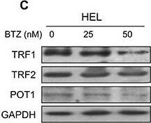
Western Blot: POT1 Antibody [NB500-176] -
Widespread dysregulation of shelterin protein expression in bortezomib-treated HEL & BGC-823 cellsA. & B. mRNA levels of shelterin factors TRF1, TRF2, TPP1, POT1, RAP1 & TIN2 in bortezomib-treated cells. Cells were treated with bortezomib for 24 hours & qPCR was used for quantitative assays. The levels of each target mRNA in bortezomib-treated cells were expressed as percentages of those in untreated cells. (A) HEL cells & (B) BGC-823 cells. C. & D. Immunoblotting assessment of TRF1, TRF2 & POT1 protein expression in bortezomib-treated cells. Same sets of cells above were analyzed for TRF1, TRF2 & POT1 protein levels & shown was representative of three independent experiments. (C) HEL cells & (D) BGC-823 cells. * & **: P < 0.05 & 0.01, respectively. BTZ, bortezomib. Image collected & cropped by CiteAb from the following publication (https://www.oncotarget.com/lookup/doi/10.18632/oncotarget.5752), licensed under a CC-BY license. Not internally tested by Novus Biologicals.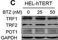
Western Blot: POT1 Antibody [NB500-176] -
Attenuation of bortezomib-induced shelterin protein dysregulation by hTERT over-expressionA. & B. Cells expressing ectopic hTERT were treated with bortezomib for 24 hours & mRNA levels of shelterin proteins then analyzed using qPCR. The levels of each target mRNA in bortezomib-treated cells were expressed as percentages of those in untreated cells. (A) HEL-hTERT cells & (B) BGC-823-hTERT cells. C. & D. Immunoblotting assessment of TRF1, TRF2 & POT1 protein expression in bortezomib-treated cells. Same sets of cells above were analyzed for TRF1, TRF2 & POT1 protein levels & shown was representative of three independent experiments. (C) HEL-hTERT cells & (D) BGC-823-hTERT cells.* & **: P < 0.05 & 0.01, respectively. BTZ, bortezomib. Image collected & cropped by CiteAb from the following publication (https://www.oncotarget.com/lookup/doi/10.18632/oncotarget.5752), licensed under a CC-BY license. Not internally tested by Novus Biologicals.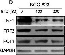
Western Blot: POT1 Antibody [NB500-176] -
Widespread dysregulation of shelterin protein expression in bortezomib-treated HEL & BGC-823 cellsA. & B. mRNA levels of shelterin factors TRF1, TRF2, TPP1, POT1, RAP1 & TIN2 in bortezomib-treated cells. Cells were treated with bortezomib for 24 hours & qPCR was used for quantitative assays. The levels of each target mRNA in bortezomib-treated cells were expressed as percentages of those in untreated cells. (A) HEL cells & (B) BGC-823 cells. C. & D. Immunoblotting assessment of TRF1, TRF2 & POT1 protein expression in bortezomib-treated cells. Same sets of cells above were analyzed for TRF1, TRF2 & POT1 protein levels & shown was representative of three independent experiments. (C) HEL cells & (D) BGC-823 cells. * & **: P < 0.05 & 0.01, respectively. BTZ, bortezomib. Image collected & cropped by CiteAb from the following publication (https://www.oncotarget.com/lookup/doi/10.18632/oncotarget.5752), licensed under a CC-BY license. Not internally tested by Novus Biologicals.Applications for POT1 Antibody - BSA Free
Western Blot
Reviewed Applications
Read 1 review rated 4 using NB500-176 in the following applications:
Formulation, Preparation, and Storage
Purification
Formulation
Format
Preservative
Concentration
Shipping
Stability & Storage
Background: POT1
Long Name
Alternate Names
Entrez Gene IDs
Gene Symbol
UniProt
Additional POT1 Products
Product Documents for POT1 Antibody - BSA Free
Certificate of Analysis
To download a Certificate of Analysis, please enter a lot or batch number in the search box below.
Product Specific Notices for POT1 Antibody - BSA Free
This product is for research use only and is not approved for use in humans or in clinical diagnosis. Primary Antibodies are guaranteed for 1 year from date of receipt.
Citations for POT1 Antibody - BSA Free
 Powered by Bioz
Customer Reviews for POT1 Antibody - BSA Free (1)
Have you used POT1 Antibody - BSA Free?
Submit a review and receive an Amazon gift card!
$25/€18/£15/$25CAN/¥2500 for a review with an image
$10/€7/£6/$10CAN/¥1110 for a review without an image
Submit a review
-
Application: Western BlotSample Tested: Human lympoblast cells, Sample Amount: 35 ugSpecies: HumanVerified Customer | Posted 09/23/2009
There are no reviews that match your criteria.
Protocols
View specific protocols for POT1 Antibody - BSA Free (NB500-176):
1. Perform SDS-PAGE on samples to be analyzed, loading 10-25 ug of total protein per lane.
2. Transfer proteins to PVDF membrane according to the instructions provided by the manufacturer of the membrane and transfer apparatus.
3. Stain the membrane with Ponceau S (or similar product) to assess transfer success, and mark molecular weight standards where appropriate.
4. Rinse the blot TBS -0.05% Tween 20 (TBST).
5. Block the membrane in 5% Non-fat milk in TBST (blocking buffer) for at least 1 hour.
6. Wash the membrane in TBST three times for 10 minutes each.
7. Dilute primary antibody in blocking buffer and incubate overnight at 4C with gentle rocking.
8. Wash the membrane in TBST three times for 10 minutes each.
9. Incubate the membrane in diluted HRP conjugated secondary antibody in blocking buffer (as per manufacturer's instructions) for 1 hour at room temperature.
10. Wash the blot in TBST three times for 10 minutes each (this step can be repeated as required to reduce background).
11. Apply the detection reagent of choice in accordance with the manufacturer's instructions.
Find general support by application which include: protocols, troubleshooting, illustrated assays, videos and webinars.
- Cellular Response to Hypoxia Protocols
- R&D Systems Quality Control Western Blot Protocol
- Troubleshooting Guide: Western Blot Figures
- Western Blot Conditions
- Western Blot Protocol
- Western Blot Protocol for Cell Lysates
- Western Blot Troubleshooting
- Western Blot Troubleshooting Guide
- View all Protocols, Troubleshooting, Illustrated assays and Webinars