
IL-13 is a 17 kDa immunoregulatory cytokine that plays a key role in the pathogenesis of allergic asthma and atopy. It is secreted by Th1 and Th2 CD4+ T cells, NK cells, visceral smooth muscle cells, eosinophils, mast cells, and basophils (1-3). IL-13 circulates as a monomer with two internal disulfide bonds that contribute to a bundled four alpha -helix configuration (4, 5). Mature rat IL-13 shares 59%, 75%, and 60% amino acid sequence identity with human, mouse, and rhesus IL-13, respectively. Despite the low homology, it exhibits cross-species activity between human, mouse, and rat (6, 7). IL-13 has diverse activities on numerous cell types (8). On macrophages, IL-13 suppresses the production of proinflammatory cytokines and other cytotoxic substances. On B cells, IL-13 induces immunoglobulin class switching to IgE, upregulates the expression of MHC class II, CD71, CD72, and CD23, and costimulates proliferation. IL-13 upregulates IL-6 while downregulating IL-1 and TNF-alpha production by fibroblasts and endothelial cells. IL-13 binds with low affinity to IL-13 R alpha 1, triggering IL-13 R alpha 1 association with IL-4 R alpha. This high affinity receptor complex also functions as the type 2 IL-4 receptor complex (9, 10). Additionally, IL-13 binds with high affinity to IL-13 R alpha 2 which is expressed intracellularly, on the cell surface, and as a soluble molecule (11-14). IL-13 R alpha 2 regulates the bioavailability of both IL-13 and IL-4 and is over-expressed in glioma and several bronchial pathologies (10, 15, 16). Compared to wild type IL-13, the atopy-associated R110Q variant of IL-13 elicits increased responsiveness from eosinophils that express low levels of IL-13 R alpha 2 (17).
Rat IL-13 Antibody (296202) [Biotin]
R&D Systems | Catalog # BAM1945

Scientific images may be resized, cropped, or adjusted for brightness or contrast for presentation purposes. These updates are limited to presentation and do not affect the underlying scientific interpretation of the data. Where available, additional source data may be provided upon request.
Key Product Details
Validated by
Species Reactivity
Applications
Label
Antibody Source
Product Specifications
Immunogen
Thr19-His131
Accession # P42203
Specificity
Clonality
Host
Isotype
Scientific Data Images for Rat IL-13 Antibody (296202) [Biotin]
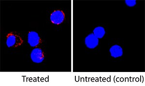
IL‑13 in Rat Splenocytes.
IL-13 was detected in immersion fixed rat splenocytes treated with PHA using Mouse Anti-Rat IL-13 Biotinylated Monoclonal Antibody (Catalog # BAM1945) at 25 µg/mL for 3 hours at room temperature. Cells were stained using the NorthernLights™ 557-conjugated Streptavidin (red; Catalog # NL999) and counterstained with DAPI (blue). Specific staining was localized to cytoplasm. View our protocol for Fluorescent ICC Staining of Non-adherent Cells.Applications for Rat IL-13 Antibody (296202) [Biotin]
Immunocytochemistry
Sample: Immersion fixed rat splenocytes treated with PHA
Western Blot
Sample: Recombinant Rat IL‑13 (Catalog # 1945-RL)
under non-reducing conditions only
Formulation, Preparation, and Storage
Purification
Reconstitution
Reconstitute at 0.5 mg/mL in sterile PBS.
Formulation
Shipping
Stability & Storage
- 12 months from date of receipt, -20 to -70 °C as supplied.
- 1 month, 2 to 8 °C under sterile conditions after reconstitution.
- 6 months, -20 to -70 °C under sterile conditions after reconstitution.
Calculators
Background: IL-13
References
- Wills-Karp, M. (2004) Immunol. Rev. 202:175.
- Nakajima H. and K. Takatsu (2007) Int. Arch. Allergy Immunol. 142:265.
- Lakkis, F.G and E.N. Cruet (1993) Biochem. Biophys. Res. Commun. 197:612.
- Moy, F.J. et al. (2001) J. Mol. Biol. 310:219.
- Eisenmesser, E.Z. et al. (2001) J. Mol. Biol. 310:231.
- Ruetten, H. and C. Thiemermann (1997) Shock 8:409.
- Lakkis, F.G. et al. (1997) Biochem. Biophys. Res. Commun. 235:529.
- Wynn, T.A. (2003) Annu. Rev. Immunol. 21:425.
- Andrews, A.L. et al. (2002) J. Biol. Chem. 277:46073.
- Tabata, Y. et al. (2007) Curr. Allergy Asthma Rep. 7:338.
- Chiaramonte, M.G. et al. (2003) J. Exp. Med. 197:687.
- Daines, M.O. and G.K. Hershey (2002) J. Biol. Chem. 227:10387
- Matsumura, M. et al. (2007) Biochem. Biophys. Res. Commun.360:464.
- Tabata, Y. et al. (2007) J. Immunol. 177:7905.
- Andrews, A.L. et al. (2006) J. Allergy Clin. Immunol. 118:858.
- Joshi, B.H. et al. (2006) Vitam. Horm. 74:479.
- Andrews, A-L. et al. (2007) J. Allergy Clin. Immunol. 120:91.
Long Name
Alternate Names
Gene Symbol
UniProt
Additional IL-13 Products
Product Documents for Rat IL-13 Antibody (296202) [Biotin]
Certificate of Analysis
To download a Certificate of Analysis, please enter a lot or batch number in the search box below.
Note: Certificate of Analysis not available for kit components.
Product Specific Notices for Rat IL-13 Antibody (296202) [Biotin]
For research use only
Customer Reviews for Rat IL-13 Antibody (296202) [Biotin]
There are currently no reviews for this product. Be the first to review Rat IL-13 Antibody (296202) [Biotin] and earn rewards!
Have you used Rat IL-13 Antibody (296202) [Biotin]?
Submit a review and receive an Amazon gift card!
$25/€18/£15/$25CAN/¥2500 for a review with an image
$10/€7/£6/$10CAN/¥1110 for a review without an image
Submit a review
Protocols
Find general support by application which include: protocols, troubleshooting, illustrated assays, videos and webinars.
- Appropriate Fixation of IHC/ICC Samples
- Cellular Response to Hypoxia Protocols
- ClariTSA™ Fluorophore Kits
- Detection & Visualization of Antibody Binding
- ICC Cell Smear Protocol for Suspension Cells
- ICC Immunocytochemistry Protocol Videos
- ICC for Adherent Cells
- Immunocytochemistry (ICC) Protocol
- Immunocytochemistry Troubleshooting
- Immunofluorescence of Organoids Embedded in Cultrex Basement Membrane Extract
- Immunohistochemistry (IHC) and Immunocytochemistry (ICC) Protocols
- Preparing Samples for IHC/ICC Experiments
- Preventing Non-Specific Staining (Non-Specific Binding)
- Primary Antibody Selection & Optimization
- Protocol for VisUCyte™ HRP Polymer Detection Reagent
- Protocol for the Fluorescent ICC Staining of Cell Smears - Graphic
- Protocol for the Fluorescent ICC Staining of Cultured Cells on Coverslips - Graphic
- Protocol for the Preparation and Fluorescent ICC Staining of Cells on Coverslips
- Protocol for the Preparation and Fluorescent ICC Staining of Non-adherent Cells
- Protocol for the Preparation and Fluorescent ICC Staining of Stem Cells on Coverslips
- Protocol for the Preparation of a Cell Smear for Non-adherent Cell ICC - Graphic
- R&D Systems Quality Control Western Blot Protocol
- TUNEL and Active Caspase-3 Detection by IHC/ICC Protocol
- The Importance of IHC/ICC Controls
- Troubleshooting Guide: Western Blot Figures
- Western Blot Conditions
- Western Blot Protocol
- Western Blot Protocol for Cell Lysates
- Western Blot Troubleshooting
- Western Blot Troubleshooting Guide
- View all Protocols, Troubleshooting, Illustrated assays and Webinars

