
Rat ROBO1 (also DUTT1) is a 170-200 kDa member of the four molecule ROBO family of guidance molecules (1-3). The term ROBO derives from round-about, a description of the circuitous pathway axons take in the absence of a functional ROBO gene (3, 4). Rat ROBO1 is a type I transmembrane (TM) glycoprotein that is synthesized as a 1651 amino acid (aa) precursor. It contains an 18 aa signal sequence, an 879 aa extracellular domain (ECD), a 21 aa transmembrane segment, and a 733 aa cytoplasmic region (5, 6). The ECD contains five C2-type Ig-like domains (aa 68-541) and three fibronectin (FN) type III domains (aa 561-864). The cytoplasmic region contains multiple 15-20 aa long CC (conserved cytoplasmic) motifs (C0-C3) (7, 8). Rat ROBO1 is likely to have at least one isoform. Based on the human and mouse gene, rat ROBO1 will utilize an alternate start site, creating an A (long) and B (short) isoform. The difference is the presence of a 32 aa extension at the N-terminus of the mature molecule (occurs in the A form) (9-12). Based on ROBO3 studies, this extension will impact the ability of ROBO1 to bind Slit (10). Rat ROBO1 ECD is 98% and 96% aa identical to the ECD in mouse and human ROBO1, respectively. ROBO1 serves as a repulsing molecule for axons that cross the midline. Initially, ROBO3 allows outgrowing axons to traverse the midline/floorplate. Once crossed, axons express ROBO1 which deflects neurites attempting to recross to the ipsilateral side (13). The chemorepulsant activity of ROBO1 is dependent on ROBO1 binding to SLIT1/2. Inhibition of ROBO1 is likely due to ROBO1-ROBO3 heterophilic binding (10, 13-15).


Scientific images may be resized, cropped, or adjusted for brightness or contrast for presentation purposes. These updates are limited to presentation and do not affect the underlying scientific interpretation of the data. Where available, additional source data may be provided upon request.
Key Product Details
Species Reactivity
Validated:
Cited:
Applications
Validated:
Cited:
Label
Antibody Source
Product Specifications
Immunogen
Lys19-Ile560
Accession # O55005
Specificity
Clonality
Host
Isotype
Scientific Data Images for Rat ROBO1 Antibody
ROBO1 in Rat Embryo.
ROBO1 was detected in immersion fixed frozen sections of rat embryo (neural tube) using 5 µg/mL Goat Anti-Rat ROBO1 Antigen Affinity-purified Polyclonal Antibody (Catalog # AF1749) overnight at 4 °C. Tissue was stained with the Anti-Goat HRP-DAB Cell & Tissue Staining Kit (brown; Catalog # CTS008) and counterstained with hematoxylin (blue). View our protocol for Chromogenic IHC Staining of Frozen Tissue Sections.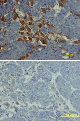
ROBO1 in Rat Brain.
ROBO1 was detected in perfusion fixed frozen sections of rat brain using Goat Anti-Rat ROBO1 Antigen Affinity-purified Polyclonal Antibody (Catalog # AF1749) at 15 µg/mL overnight at 4 °C. Tissue was stained using the Anti-Goat HRP-DAB Cell & Tissue Staining Kit (brown; Catalog # CTS008) and counterstained with hematoxylin (blue). Lower panel shows a lack of labeling if primary antibodies are omitted and tissue is stained only with secondary antibody followed by incubation with detection reagents. View our protocol for Chromogenic IHC Staining of Frozen Tissue Sections.
Rat ROBO1 ELISA Standard Curve
Recombinant Rat ROBO1 Fc Chimera (Catalog # 1749-RB) was serially diluted and captured by Mouse Anti-Rat ROBO1 Monoclonal Antibody (Catalog # MAB17491) coated on a Clear Polystyrene Microplate (Catalog # DY990). Goat Anti-Rat ROBO1 Antigen Affinity-purified Polyclonal Antibody (Catalog # AF1749) was biotinylated and incubated with the protein captured on the plate. Detection of the standard curve was achieved by incubating Streptavidin-HRP (Catalog # DY998)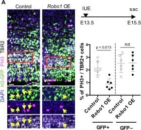
Detection of ROBO1 by Immunohistochemistry
Elevated expression of Robo1 leads to a decrease in IPC proliferation.(A) In utero electroporation (IUE) of either an empty vector (control) or a Robo1 expression vector (Robo1 OE) together with a H3-GFP expression vector was performed into WT cortices at E13.5. The cortical sections are stained with DAPI (blue), anti-GFP (green), anti-PH3 (magenta), and anti-TBR2 (white) antibodies at E15.5. The graph shows the ratio of PH3+ cells in TBR2+ and GFP+ cells (left) or PH3+ cells in TBR2+ and GFP-negative cells (right). Arrows indicate GFP+, PH3+, and TBR2+ cells. (B) IUE of an empty (control) or a Robo1 expression vector (Robo1 OE) and a H3-GFP expression vector into WT cortices at E14.5. The cortical sections are immunostained with DAPI (blue), anti-GFP (green), anti-KI67 (magenta), and anti-TBR2 (white) antibodies at E16.5. The graph shows the percentage of KI67+ and TBR2-negative cells located in the VZ (RGCs), TBR2+ and KI67+ cells (IPCs), and KI67-negative differentiated cells (postmitotic neurons) among total GFP+ cells. The percentage of RGCs and IPCs in electroporated cells was not different between Robo1 OE and control. (C) Auts2fl(ex8)/fl(ex8) or Emx1Cre/+; Auts2fl(ex8)/fl(ex8) (Auts2 cKO) cortices were electroporated with scrambled or Robo1 shRNA vector in utero at E13.5 and immunostained with anti-GFP (green), anti-PH3 (magenta) and anti-TBR2 (white) antibodies at E15.5. The graph shows the percentage of PH3+ cells among TBR2+ and GFP+ cells. Arrows indicate GFP+, PH3+, and TBR2+ cells. Data were presented as the mean ± SD (N = 3 mice, six sections). NS, not significant, unpaired Student’s t-test (A, B) and one-way ANOVA with Turkey’s post hoc test (C). Scale bars, 20 µm (A, C) and 50 µm (B). Source data are available online for this figure. Image collected and cropped by CiteAb from the following open publication (https://pubmed.ncbi.nlm.nih.gov/39815005), licensed under a CC-BY license. Not internally tested by R&D Systems.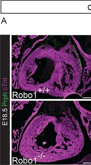
Detection of ROBO1 by Immunohistochemistry
Constitutive knock-out analysis indicates a role for Slit2, Slit3, and Robo1 during cardiac innervation development.(A) Representative images of Robo1+/+ and Robo1-/- sections stained by immunohistochemistry for the myocardium using cardiac Troponin I (cTnI) in purple and total innervation using Peripherin (Prph) in green. Arrowhead points to reduced total innervation staining in green in the Robo1-/-. (B) Quantification of total ventricular innervation corrected for ventricle volume (myocardial marker cTnI) in constitutive knock-outs and wild-type controls. T-test or one-way ANOVA with Tukey’s test per mouse line. (C) Quantification of total ventricular volume based on myocardial marker cTnI in constitutive knock-outs and wild-type controls. T-test or one-way ANOVA with Tukey’s test per mouse line. Scale bars, 100 μm.Figure 1—source data 1.Source data Figure 1B - total innervation measurements.Figure 1—source data 2.Source data Figure 1C - ventricle size measurements.Source data Figure 1B - total innervation measurements.Source data Figure 1C - ventricle size measurements. Image collected and cropped by CiteAb from the following open publication (https://elifesciences.org/articles/105932), licensed under a CC-BY license. Not internally tested by R&D Systems.Applications for Rat ROBO1 Antibody
Immunohistochemistry
Sample: Immersion fixed frozen sections of rat embryo (neural tube) and perfusion fixed frozen sections of rat brain
Western Blot
Sample: Recombinant Rat ROBO1 Fc Chimera (Catalog # 1749-RB)
Formulation, Preparation, and Storage
Purification
Reconstitution
Reconstitute at 0.2 mg/mL in sterile PBS. For liquid material, refer to CoA for concentration.
Formulation
Shipping
Stability & Storage
- 12 months from date of receipt, -20 to -70 °C as supplied.
- 1 month, 2 to 8 °C under sterile conditions after reconstitution.
- 6 months, -20 to -70 °C under sterile conditions after reconstitution.
Calculators
Background: ROBO1
References
- Guthrie, S. (2001) Curr. Biol. 11:R300.
- Guthrie, S. (2004) Curr. Biol. 14:R632.
- Park, K.W. et al. (2003) Dev. Biol. 261:251.
- Seeger, M. et al. (1993) Neuron 10:409.
- Kidd, T. et al. (1998) Cell 92:205.
- Marillat, V. et al. (2002) J. Comp. Neurol. 442:130.
- Bashaw, G.J. et al. (2000) Cell 101:703.
- Jen, J.C. et al. (2004) Science 304:1509.
- Clark, K. et al. (2002) FEBS Lett. 523:12.
- Camurri, L. et al. (2005) Mol. Cell. Neurosci. 30:485.
- Sundaresan, V. et al. (1998) Mol. Cell. Neurosci. 11:29.
- GenBank Accession # O55005.
- Sabatier, C. et al. (2004) Cell 117:157.
- Liu, Z. et al. (2004) Mol. Cell. Neurosci. 26:232.
- Brose, K. et al. (1999) Cell 96:795.
Long Name
Alternate Names
Gene Symbol
UniProt
Additional ROBO1 Products
Product Documents for Rat ROBO1 Antibody
Certificate of Analysis
To download a Certificate of Analysis, please enter a lot or batch number in the search box below.
Note: Certificate of Analysis not available for kit components.
Product Specific Notices for Rat ROBO1 Antibody
For research use only
Citations for Rat ROBO1 Antibody
 Powered by Bioz
Customer Reviews for Rat ROBO1 Antibody
There are currently no reviews for this product. Be the first to review Rat ROBO1 Antibody and earn rewards!
Have you used Rat ROBO1 Antibody?
Submit a review and receive an Amazon gift card!
$25/€18/£15/$25CAN/¥2500 for a review with an image
$10/€7/£6/$10CAN/¥1110 for a review without an image
Submit a review
Protocols
Find general support by application which include: protocols, troubleshooting, illustrated assays, videos and webinars.
- Antigen Retrieval Protocol (PIER)
- Antigen Retrieval for Frozen Sections Protocol
- Appropriate Fixation of IHC/ICC Samples
- Cellular Response to Hypoxia Protocols
- Chromogenic IHC Staining of Formalin-Fixed Paraffin-Embedded (FFPE) Tissue Protocol
- Chromogenic Immunohistochemistry Staining of Frozen Tissue
- ClariTSA™ Fluorophore Kits
- Detection & Visualization of Antibody Binding
- Fluorescent IHC Staining of Frozen Tissue Protocol
- Graphic Protocol for Heat-induced Epitope Retrieval
- Graphic Protocol for the Preparation and Fluorescent IHC Staining of Frozen Tissue Sections
- Graphic Protocol for the Preparation and Fluorescent IHC Staining of Paraffin-embedded Tissue Sections
- Graphic Protocol for the Preparation of Gelatin-coated Slides for Histological Tissue Sections
- IHC Sample Preparation (Frozen sections vs Paraffin)
- Immunofluorescent IHC Staining of Formalin-Fixed Paraffin-Embedded (FFPE) Tissue Protocol
- Immunohistochemistry (IHC) and Immunocytochemistry (ICC) Protocols
- Immunohistochemistry Frozen Troubleshooting
- Immunohistochemistry Paraffin Troubleshooting
- Preparing Samples for IHC/ICC Experiments
- Preventing Non-Specific Staining (Non-Specific Binding)
- Primary Antibody Selection & Optimization
- Protocol for Heat-Induced Epitope Retrieval (HIER)
- Protocol for Making a 4% Formaldehyde Solution in PBS
- Protocol for VisUCyte™ HRP Polymer Detection Reagent
- Protocol for the Preparation & Fixation of Cells on Coverslips
- Protocol for the Preparation and Chromogenic IHC Staining of Frozen Tissue Sections
- Protocol for the Preparation and Chromogenic IHC Staining of Frozen Tissue Sections - Graphic
- Protocol for the Preparation and Chromogenic IHC Staining of Paraffin-embedded Tissue Sections
- Protocol for the Preparation and Chromogenic IHC Staining of Paraffin-embedded Tissue Sections - Graphic
- Protocol for the Preparation and Fluorescent IHC Staining of Frozen Tissue Sections
- Protocol for the Preparation and Fluorescent IHC Staining of Paraffin-embedded Tissue Sections
- Protocol for the Preparation of Gelatin-coated Slides for Histological Tissue Sections
- R&D Systems Quality Control Western Blot Protocol
- TUNEL and Active Caspase-3 Detection by IHC/ICC Protocol
- The Importance of IHC/ICC Controls
- Troubleshooting Guide: Immunohistochemistry
- Troubleshooting Guide: Western Blot Figures
- Western Blot Conditions
- Western Blot Protocol
- Western Blot Protocol for Cell Lysates
- Western Blot Troubleshooting
- Western Blot Troubleshooting Guide
- View all Protocols, Troubleshooting, Illustrated assays and Webinars