
TPX2 Antibody - BSA Free
Novus Biologicals | Catalog # NB500-184


Scientific images may be resized, cropped, or adjusted for brightness or contrast for presentation purposes. These updates are limited to presentation and do not affect the underlying scientific interpretation of the data. Where available, additional source data may be provided upon request.
Key Product Details
Validated by
Species Reactivity
Validated:
Cited:
Applications
Validated:
Cited:
Label
Antibody Source
Format
Product Specifications
Immunogen
Clonality
Host
Isotype
Scientific Data Images for TPX2 Antibody - BSA Free
Western Blot: TPX2 Antibody [NB500-184]
TPX2-Antibody-Western-Blot-NB500-184-img0005.jpg
Western Blot: TPX2 Antibody [NB500-184] -
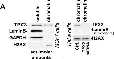
Western Blot: TPX2 Antibody [NB500-184] - TPX2 is constitutively associated with chromatin & impacts the DAPI staining pattern & H4K16ac levels.(A) Although the majority of TPX2 is found in the soluble fraction (see Material & Methods), a small but clearly detectable sub-population of TPX2 constitutively associates with stringent chromatin fractions obtained from MCF7 cells (left panel) or HeLa cells (right panel). These chromatin fractions contain histones but not nuclear LaminB. Upon expression of an inducible TPX2 targeting miRNA (or upon transfection with siRNA; see D) the protein was depleted from chromatin fractions. Ctrl: control cells with no induction of TPX2 miRNA. (B) TPX2 gets enriched in chromatin fractions isolated from HeLa cells after treatment with 10 Gy of ionizing radiation. Note the constitutive association of TPX2 with the chromatin in non-irradiated cells. Levels of H2AX were used as a loading control. (C) Overexpression of GFP-TPX2 or His-TPX2 causes abnormal DAPI staining in MCF7 cells compared to surrounding non-transfected cells or cells transfected with GFP. This is indicative of changes in chromatin structure. Enlargements of white frames are shown. In agreement with previous reports, overexpressed TPX2 is mostly found in the nucleus but also associates with the cytoskeleton [2]. (D-F) Depletion of TPX2 by siRNA (D) or miRNA (F) causes a decrease in H4K16ac levels whereas the levels of H3K9ac & H3K56ac remain unchanged. (E) Quantification of H3K9ac & H3K56ac levels from MCF7 cells transfected with control or TPX2 siRNA (n = 4 independent experiments each; p(t test)>0.05; NS: non significant; Error bars represent SE). Stripping of western blots & re-development with antibodies specific for H3 & H4 ensured equal loading. See text for details. Image collected & cropped by CiteAb from the following publication (https://dx.plos.org/10.1371/journal.pone.0110994), licensed under a CC-BY license. Not internally tested by Novus Biologicals.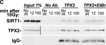
Western Blot: TPX2 Antibody [NB500-184] -
Western Blot: TPX2 Antibody [NB500-184] - SIRT1 modulates the levels of H4K16ac & gamma -H2AX & is in complex with TPX2.(A) siRNA-mediated loss of SIRT1 in HeLa cells increases H4K16ac levels & decreases ionizing radiation-dependent gamma -H2AX levels when compared to controls. (B) Overexpression of SIRT1 in MCF7 cells decreases H4K16ac levels & increases ionizing radiation-dependent gamma -H2AX levels when compared to controls. (C) Co-immunoprecipitations with TPX2 antibodies from HeLa cells with & without ionizing radiation treatment as indicated & in the absence or presence of ethidium bromide (EtBr). (The Input for TPX2 is from a longer exposure of the same blot.) (D) Co-immunoprecipitations with SIRT1 antibodies from HeLa cells with & without ionizing radiation treatment as indicated. Beads without antibodies (No Ab) were used as controls. Cells were treated with 10 Gy (or left untreated) & harvested after 1h recovery (A-B) or as indicated (C-D). See text for details. Levels of H2AX & H4 were used as loading controls. IR: ionizing radiation. Image collected & cropped by CiteAb from the following publication (https://dx.plos.org/10.1371/journal.pone.0110994), licensed under a CC-BY license. Not internally tested by Novus Biologicals.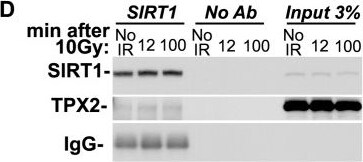
Western Blot: TPX2 Antibody [NB500-184] -
Western Blot: TPX2 Antibody [NB500-184] - SIRT1 modulates the levels of H4K16ac & gamma -H2AX & is in complex with TPX2.(A) siRNA-mediated loss of SIRT1 in HeLa cells increases H4K16ac levels & decreases ionizing radiation-dependent gamma -H2AX levels when compared to controls. (B) Overexpression of SIRT1 in MCF7 cells decreases H4K16ac levels & increases ionizing radiation-dependent gamma -H2AX levels when compared to controls. (C) Co-immunoprecipitations with TPX2 antibodies from HeLa cells with & without ionizing radiation treatment as indicated & in the absence or presence of ethidium bromide (EtBr). (The Input for TPX2 is from a longer exposure of the same blot.) (D) Co-immunoprecipitations with SIRT1 antibodies from HeLa cells with & without ionizing radiation treatment as indicated. Beads without antibodies (No Ab) were used as controls. Cells were treated with 10 Gy (or left untreated) & harvested after 1h recovery (A-B) or as indicated (C-D). See text for details. Levels of H2AX & H4 were used as loading controls. IR: ionizing radiation. Image collected & cropped by CiteAb from the following publication (https://dx.plos.org/10.1371/journal.pone.0110994), licensed under a CC-BY license. Not internally tested by Novus Biologicals.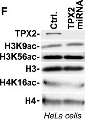
Western Blot: TPX2 Antibody [NB500-184] -
Western Blot: TPX2 Antibody [NB500-184] - TPX2 is constitutively associated with chromatin & impacts the DAPI staining pattern & H4K16ac levels.(A) Although the majority of TPX2 is found in the soluble fraction (see Material & Methods), a small but clearly detectable sub-population of TPX2 constitutively associates with stringent chromatin fractions obtained from MCF7 cells (left panel) or HeLa cells (right panel). These chromatin fractions contain histones but not nuclear LaminB. Upon expression of an inducible TPX2 targeting miRNA (or upon transfection with siRNA; see D) the protein was depleted from chromatin fractions. Ctrl: control cells with no induction of TPX2 miRNA. (B) TPX2 gets enriched in chromatin fractions isolated from HeLa cells after treatment with 10 Gy of ionizing radiation. Note the constitutive association of TPX2 with the chromatin in non-irradiated cells. Levels of H2AX were used as a loading control. (C) Overexpression of GFP-TPX2 or His-TPX2 causes abnormal DAPI staining in MCF7 cells compared to surrounding non-transfected cells or cells transfected with GFP. This is indicative of changes in chromatin structure. Enlargements of white frames are shown. In agreement with previous reports, overexpressed TPX2 is mostly found in the nucleus but also associates with the cytoskeleton [2]. (D-F) Depletion of TPX2 by siRNA (D) or miRNA (F) causes a decrease in H4K16ac levels whereas the levels of H3K9ac & H3K56ac remain unchanged. (E) Quantification of H3K9ac & H3K56ac levels from MCF7 cells transfected with control or TPX2 siRNA (n = 4 independent experiments each; p(t test)>0.05; NS: non significant; Error bars represent SE). Stripping of western blots & re-development with antibodies specific for H3 & H4 ensured equal loading. See text for details. Image collected & cropped by CiteAb from the following publication (https://dx.plos.org/10.1371/journal.pone.0110994), licensed under a CC-BY license. Not internally tested by Novus Biologicals.Applications for TPX2 Antibody - BSA Free
Immunoprecipitation
Western Blot
Formulation, Preparation, and Storage
Purification
Formulation
Format
Preservative
Concentration
Shipping
Stability & Storage
Background: TPX2
Alternate Names
Entrez Gene IDs
Gene Symbol
UniProt
Additional TPX2 Products
Product Documents for TPX2 Antibody - BSA Free
Certificate of Analysis
To download a Certificate of Analysis, please enter a lot or batch number in the search box below.
Product Specific Notices for TPX2 Antibody - BSA Free
This product is for research use only and is not approved for use in humans or in clinical diagnosis. Primary Antibodies are guaranteed for 1 year from date of receipt.
Citations for TPX2 Antibody - BSA Free
 Powered by Bioz
Customer Reviews for TPX2 Antibody - BSA Free
There are currently no reviews for this product. Be the first to review TPX2 Antibody - BSA Free and earn rewards!
Have you used TPX2 Antibody - BSA Free?
Submit a review and receive an Amazon gift card!
$25/€18/£15/$25CAN/¥2500 for a review with an image
$10/€7/£6/$10CAN/¥1110 for a review without an image
Submit a review
Protocols
Find general support by application which include: protocols, troubleshooting, illustrated assays, videos and webinars.
- Appropriate Fixation of IHC/ICC Samples
- Cellular Response to Hypoxia Protocols
- ClariTSA™ Fluorophore Kits
- Detection & Visualization of Antibody Binding
- ICC Cell Smear Protocol for Suspension Cells
- ICC Immunocytochemistry Protocol Videos
- ICC for Adherent Cells
- Immunocytochemistry (ICC) Protocol
- Immunocytochemistry Troubleshooting
- Immunofluorescence of Organoids Embedded in Cultrex Basement Membrane Extract
- Immunohistochemistry (IHC) and Immunocytochemistry (ICC) Protocols
- Immunoprecipitation Protocol
- Preparing Samples for IHC/ICC Experiments
- Preventing Non-Specific Staining (Non-Specific Binding)
- Primary Antibody Selection & Optimization
- Protocol for VisUCyte™ HRP Polymer Detection Reagent
- Protocol for the Fluorescent ICC Staining of Cell Smears - Graphic
- Protocol for the Fluorescent ICC Staining of Cultured Cells on Coverslips - Graphic
- Protocol for the Preparation and Fluorescent ICC Staining of Cells on Coverslips
- Protocol for the Preparation and Fluorescent ICC Staining of Non-adherent Cells
- Protocol for the Preparation and Fluorescent ICC Staining of Stem Cells on Coverslips
- Protocol for the Preparation of a Cell Smear for Non-adherent Cell ICC - Graphic
- R&D Systems Quality Control Western Blot Protocol
- TUNEL and Active Caspase-3 Detection by IHC/ICC Protocol
- The Importance of IHC/ICC Controls
- Troubleshooting Guide: Western Blot Figures
- Western Blot Conditions
- Western Blot Protocol
- Western Blot Protocol for Cell Lysates
- Western Blot Troubleshooting
- Western Blot Troubleshooting Guide
- View all Protocols, Troubleshooting, Illustrated assays and Webinars