
Ubiquilin 2 Antibody (6H9) - BSA Free
Novus Biologicals | Catalog # NBP2-25164

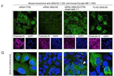
Scientific images may be resized, cropped, or adjusted for brightness or contrast for presentation purposes. These updates are limited to presentation and do not affect the underlying scientific interpretation of the data. Where available, additional source data may be provided upon request.
Key Product Details
Validated by
Species Reactivity
Validated:
Cited:
Applications
Validated:
Cited:
Label
Antibody Source
Format
Product Specifications
Immunogen
Localization
Clonality
Host
Isotype
Theoretical MW
Disclaimer note: The observed molecular weight of the protein may vary from the listed predicted molecular weight due to post translational modifications, post translation cleavages, relative charges, and other experimental factors.
Scientific Data Images for Ubiquilin 2 Antibody (6H9) - BSA Free
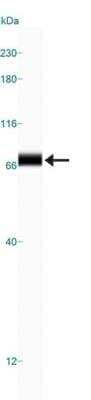
Simple Western: Ubiquilin 2 Antibody (6H9) [NBP2-25164]
Simple Western: Ubiquilin 2 Antibody (6H9) [NBP2-25164] - Simple Western lane view shows a specific band for Ubiquilin 2 in 0.5 mg/ml of HeLa lysate. This experiment was performed under reducing conditions using the 12-230 kDa separation system.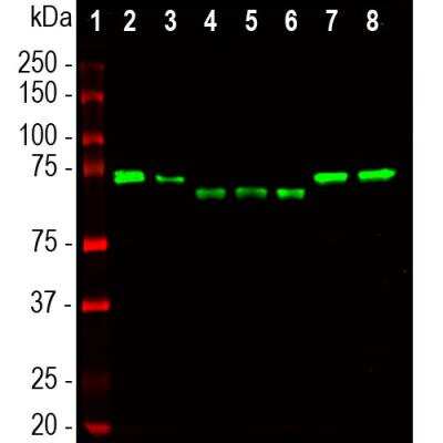
Western Blot: Ubiquilin 2 Antibody (6H9) [NBP2-25164]
Western Blot: Ubiquilin 2 Antibody (6H9) [NBP2-25164] - Analysis of different tissue and cell lysates using mouse mAb to ubiquilin 2, NBP2-25164, dilution 1:1,000 in green: [1] protein standard (red), [2] NIH-3T3, [3] C6, [4] HEK293, [5] HeLa, [6] SH-SY5Y, [7] rat whole brain, and [8] mouse whole brain. The band at 65-70kDa corresponds to ubiquilin 2 protein, which is known to differ between the human and rodent proteins.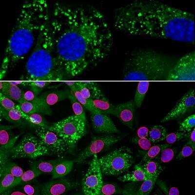
Immunocytochemistry/ Immunofluorescence: Ubiquilin 2 Antibody (6H9) [NBP2-25164]
Immunocytochemistry/Immunofluorescence: Ubiquilin 2 Antibody (6H9) [NBP2-25164] - Analysis of an NIH-3T3 cell culture stained with mouse mAb to ubiquilin 2, NBP2-25164, dilution 1:1,000 in green, and costained with chicken pAb to lamin A/C, dilution 1:5,000 in red. The blue is DAPI staining of nuclear DNA. The cells were treated with 50uM of chloroquine, an inhibitor of autophagy, for 16 hours prior to staining. NBP2-25164 antibody reveals vesicular staining of ubiquilin 2 protein accumulated in lysosomes in the cytoplasm, while the lamin A/C antibody stains the nuclear lamina.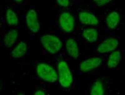
Immunocytochemistry/ Immunofluorescence: Ubiquilin 2 Antibody (6H9) [NBP2-25164]
Immunocytochemistry/Immunofluorescence: Ubiquilin 2 Antibody (6H9) [NBP2-25164] - Analysis of Human U2OS cells stained with Ubiquilin 2 antibody. Primary antibody dilution: 1:1000. Image from verified customer review.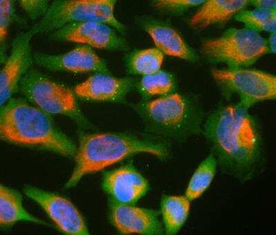
Immunocytochemistry/ Immunofluorescence: Ubiquilin 2 Antibody (6H9) [NBP2-25164]
Immunocytochemistry/Immunofluorescence: Ubiquilin 2 Antibody (6H9) [NBP2-25164] - HeLa cell cultures stained with NBP2-25164 (green) and chicken polyclonal antibody to Vimentin: NB300-223 (red). In most individual cells Ubiquilin 2 is present diffusely in the cytoplasm of cells, though some cells show enrichment of the protein in spherical autophagosome-like structure.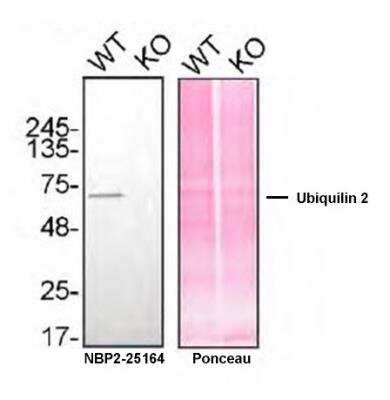
Western Blot: Ubiquilin 2 Antibody (6H9) [NBP2-25164]
Western Blot: Ubiquilin 2 Antibody (6H9) [NBP2-25164] - Lysates of HAP1 WT and Ubiquilin 2 KO were prepared, and 30 ug of protein were processed for immunoblot with NBP2-25164. The Ponceau stained transfer of the blot is shown. Antibody dilution used: 1/2000. Predicted band size: 65 kDa. Image, protocol and testing courtesy of YCharOS Inc. (ycharos.com).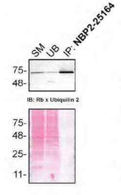
Immunoprecipitation: Ubiquilin 2 Antibody (6H9) [NBP2-25164]
Immunoprecipitation: Ubiquilin 2 Antibody (6H9) [NBP2-25164] - HAP1 lysates were prepared, and immunoprecipitation was performed using 2.0 ug of Ubiquilin 2 antibody (NBP2-25164) pre-coupled to Dynabeads protein G. Samples were washed and processed for immunoblot with NBP2-25164. For immunoblot, Recombinant Ubiquilin 2 (Rb x Ubiquilin 2) was used at 1/1000. The Ponceau stained transfer of the blot is shown. SM=4% starting material; UB=4% unbound fraction. Image, protocol and testing courtesy of YCharOS Inc. (ycharos.com).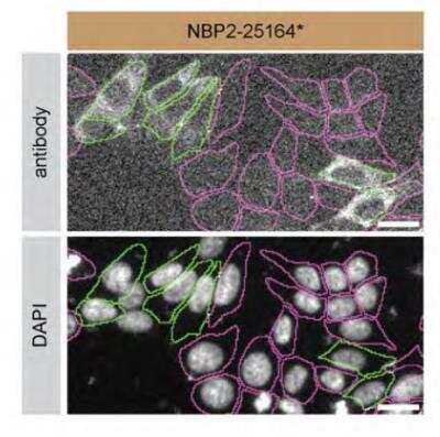
Immunocytochemistry/ Immunofluorescence: Ubiquilin 2 Antibody (6H9) [NBP2-25164]
Immunocytochemistry/ Immunofluorescence: Ubiquilin 2 Antibody (6H9) [NBP2-25164] - HAP1 WT and Ubiquilin 2 KO cells were labeled with a green or a far-red fluorescent dye, respectively. WT and KO cells were mixed and plated to a 1:1 ratio in a 96-well plate with glass bottom. Cells were stained with Ubiquilin-2 antibody (NBP2-25164) and with the corresponding Alexa-fluor 555 coupled secondary antibody including DAPI. Acquisition of the blue (nucleus-DAPI), green (identification of WT cells), red (antibody staining) and far-red (identification of KO cells) channels was performed. Representative image of the blue and red (grayscale) channels is shown. WT and KO cells are outlined with green and magenta dashed line, respectively. Antibody dilution used: 1/1000. Bars = 10 um Image, protocol and testing courtesy of YCharOS Inc. (ycharos.com).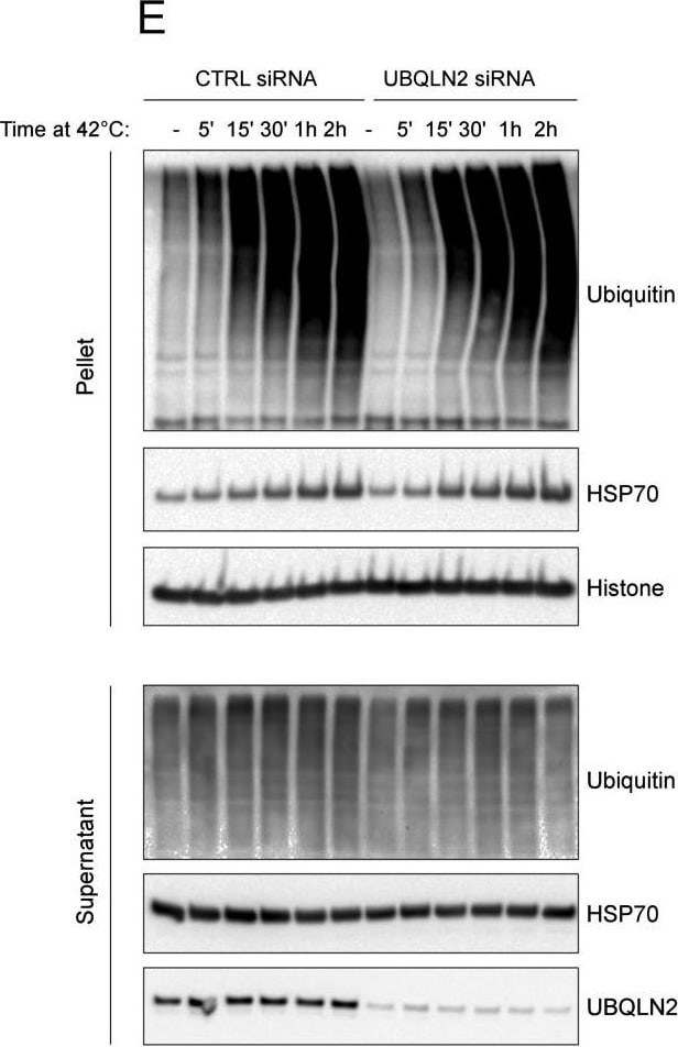
Knockdown Validated: Ubiquilin 2 Antibody (6H9) - BSA Free [NBP2-25164] -
Heat Shock Generates Insoluble Ubiquitin-Positive Aggregates and Does Not Inactive Proteasomes, Related to Figure 1(A) UBQLN2 is not pelleted when cells are heat shocked post lysis. Cell lysates were incubated at 37 or 42C and then fractionated into soluble (S) and pellet (P) fraction. This indicates that UBQLN2 itself does not aggregate as a result of high temperature.(B) UBQLN2 levels are not upregulated in response to heat shock. HSP70 and GAPDH were used as a positive and negative controls, respectively.(C) Heat shock aggregates are insoluble in up to and including 1% SDS but are solubilized in 2% SDS. Blotting of soluble and pellet fractions with anti-ubiquitin and UQBLN2 antibodies confirmed dissolution of the aggregates in 2% SDS.(D) Proteasomes are active after heat shock. To confirm that proteasome activity was not affected by heat shock, we incubated U2OS and MEFs at the indicted temperatures for 2h. Cells were then harvested and cell lysates were incubated with the proteasome inhibitor MG132 or DMSO, followed by incubation with a fluorescent proteasome-activity probe, as indicated. The presence of fluorescently labeled beta-subunits at the same intensity under both heat stress and normal temperature, indicate that proteasome activity is not significantly affected by heat shock.(E) U2OS cells were treated with control or UBQLN2 siRNA and subjected to heat shock for the indicated times. Analysis of the pellet fraction revealed that insoluble ubiquitylated aggregates are generated within 5 min of heat shock, but that depletion of UBQLN2 does not noticeably alter the accumulation of these aggregates at any of the indicated time points. Image collected and cropped by CiteAb from the following open publication (https://pubmed.ncbi.nlm.nih.gov/27477512), licensed under a CC-BY license. Not internally tested by Novus Biologicals.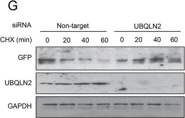
Knockdown Validated: Ubiquilin 2 Antibody (6H9) - BSA Free [NBP2-25164] -
Puromycin Does Not Upregulate UBQLN2 Levels and UBQLN2 Clears Nuclear Aggregated GFP-u, Related to Figure 2(A) Schematic representation on how the three fractions, total insoluble, nuclear soluble and total insoluble were generated.(B) Cells treated with puromycin were fractionated as indicated and treatment did not induce the nuclear localization of UBQLN2.(C) Puromycin treatment did not induce the upregulation of UBQLN2 protein.(D) Schematic showing the GFPu-NLS construct (Bennett et al., 2005).(E) HEK293 cells stably expressing GFPu-NLS were subject to heat shock for 2h at 42C and fractionated into soluble and insoluble fractions. GFPu-NLS recruited to the insoluble fraction after heat shock indicating its heat-induced aggregation.(F) GFPu-NLS cells were subject to heat shock for 2h at 42C and proteasome inhibition with 25 μM MG132 as indicated. UBQLN2 was immunoprecipitated and GFPu-NLS was found to co-immunoprecipitate only upon heat shock, consistent with UBQLN2 nuclear localization. Combined heat shock and proteasome inhibition increased the binding further.(G) GFPu-NLS cells were depleted of UBQLN2 or treated with a control non-targeting siRNA then treated with 50 μg/ml cycloheximide (CHX) for the indicated time to measure turnover. Turnover was quantified using data from three independent experiments.Error bars represent SE. Image collected and cropped by CiteAb from the following open publication (https://pubmed.ncbi.nlm.nih.gov/27477512), licensed under a CC-BY license. Not internally tested by Novus Biologicals.Applications for Ubiquilin 2 Antibody (6H9) - BSA Free
Immunocytochemistry/ Immunofluorescence
Immunohistochemistry
Simple Western
Western Blot
In Simple Western only 10 - 15 uL of the recommended dilution is used per data point.
See Simple Western Antibody Database for Simple Western validation: Tested in HeLa lysate 0.5 mg/mL, separated by Size, antibody dilution of 1:100, apparent MW was 74 kDa. Use in Immunohistochemistry-Frozen reported in scientific literature (PMID:33028421).
Reviewed Applications
Read 2 reviews rated 5 using NBP2-25164 in the following applications:
Formulation, Preparation, and Storage
Purification
Formulation
Format
Preservative
Concentration
Shipping
Stability & Storage
Background: Ubiquilin 2
Alternate Names
Entrez Gene IDs
Gene Symbol
OMIM
UniProt
Additional Ubiquilin 2 Products
Product Documents for Ubiquilin 2 Antibody (6H9) - BSA Free
Certificate of Analysis
To download a Certificate of Analysis, please enter a lot or batch number in the search box below.
Product Specific Notices for Ubiquilin 2 Antibody (6H9) - BSA Free
This product is for research use only and is not approved for use in humans or in clinical diagnosis. Primary Antibodies are guaranteed for 1 year from date of receipt.
Citations for Ubiquilin 2 Antibody (6H9) - BSA Free
 Powered by Bioz
Customer Reviews for Ubiquilin 2 Antibody (6H9) - BSA Free (2)
Have you used Ubiquilin 2 Antibody (6H9) - BSA Free?
Submit a review and receive an Amazon gift card!
$25/€18/£15/$25CAN/¥2500 for a review with an image
$10/€7/£6/$10CAN/¥1110 for a review without an image
Submit a review
Customer Images
-
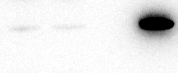
Application: ImmunoprecipitationSample Tested: 293 whole cell lysateSpecies: HumanVerified Customer | Posted 08/19/2022Lane 1,2: HEK293 whole cell lysate input (10 micrograms) Lane 3: HEK293 whole cell lysate immunoprecipitated with normal mouse IgG Lane 4: HEK293 whole cell lysate immunoprecipitated with Mouse anti-Ubiquilin-2 (6H9).Antibody was used at 1:30 dilution and was crosslinked to Protein A/G beads. HEK293 cells were lysed in 1x Tris buffered saline containing 1% NP40 and 5% glycerol. 600 micrograms of whole cell lysate was used for IP. IP was done for 2 hours.

-
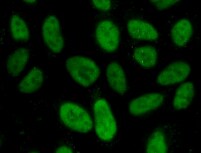
Application: ImmunocytochemistrySample Tested: U2OS cellsSpecies: HumanVerified Customer | Posted 02/09/2022Human U2OS cellsDilution: 1:1000
There are no reviews that match your criteria.
Protocols
Find general support by application which include: protocols, troubleshooting, illustrated assays, videos and webinars.
- Antigen Retrieval Protocol (PIER)
- Antigen Retrieval for Frozen Sections Protocol
- Appropriate Fixation of IHC/ICC Samples
- Cellular Response to Hypoxia Protocols
- Chromogenic IHC Staining of Formalin-Fixed Paraffin-Embedded (FFPE) Tissue Protocol
- Chromogenic Immunohistochemistry Staining of Frozen Tissue
- ClariTSA™ Fluorophore Kits
- Detection & Visualization of Antibody Binding
- Fluorescent IHC Staining of Frozen Tissue Protocol
- Graphic Protocol for Heat-induced Epitope Retrieval
- Graphic Protocol for the Preparation and Fluorescent IHC Staining of Frozen Tissue Sections
- Graphic Protocol for the Preparation and Fluorescent IHC Staining of Paraffin-embedded Tissue Sections
- Graphic Protocol for the Preparation of Gelatin-coated Slides for Histological Tissue Sections
- ICC Cell Smear Protocol for Suspension Cells
- ICC Immunocytochemistry Protocol Videos
- ICC for Adherent Cells
- IHC Sample Preparation (Frozen sections vs Paraffin)
- Immunocytochemistry (ICC) Protocol
- Immunocytochemistry Troubleshooting
- Immunofluorescence of Organoids Embedded in Cultrex Basement Membrane Extract
- Immunofluorescent IHC Staining of Formalin-Fixed Paraffin-Embedded (FFPE) Tissue Protocol
- Immunohistochemistry (IHC) and Immunocytochemistry (ICC) Protocols
- Immunohistochemistry Frozen Troubleshooting
- Immunohistochemistry Paraffin Troubleshooting
- Immunoprecipitation Protocol
- Preparing Samples for IHC/ICC Experiments
- Preventing Non-Specific Staining (Non-Specific Binding)
- Primary Antibody Selection & Optimization
- Protocol for Heat-Induced Epitope Retrieval (HIER)
- Protocol for Making a 4% Formaldehyde Solution in PBS
- Protocol for VisUCyte™ HRP Polymer Detection Reagent
- Protocol for the Fluorescent ICC Staining of Cell Smears - Graphic
- Protocol for the Fluorescent ICC Staining of Cultured Cells on Coverslips - Graphic
- Protocol for the Preparation & Fixation of Cells on Coverslips
- Protocol for the Preparation and Chromogenic IHC Staining of Frozen Tissue Sections
- Protocol for the Preparation and Chromogenic IHC Staining of Frozen Tissue Sections - Graphic
- Protocol for the Preparation and Chromogenic IHC Staining of Paraffin-embedded Tissue Sections
- Protocol for the Preparation and Chromogenic IHC Staining of Paraffin-embedded Tissue Sections - Graphic
- Protocol for the Preparation and Fluorescent ICC Staining of Cells on Coverslips
- Protocol for the Preparation and Fluorescent ICC Staining of Non-adherent Cells
- Protocol for the Preparation and Fluorescent ICC Staining of Stem Cells on Coverslips
- Protocol for the Preparation and Fluorescent IHC Staining of Frozen Tissue Sections
- Protocol for the Preparation and Fluorescent IHC Staining of Paraffin-embedded Tissue Sections
- Protocol for the Preparation of Gelatin-coated Slides for Histological Tissue Sections
- Protocol for the Preparation of a Cell Smear for Non-adherent Cell ICC - Graphic
- R&D Systems Quality Control Western Blot Protocol
- TUNEL and Active Caspase-3 Detection by IHC/ICC Protocol
- The Importance of IHC/ICC Controls
- Troubleshooting Guide: Immunohistochemistry
- Troubleshooting Guide: Western Blot Figures
- Western Blot Conditions
- Western Blot Protocol
- Western Blot Protocol for Cell Lysates
- Western Blot Troubleshooting
- Western Blot Troubleshooting Guide
- View all Protocols, Troubleshooting, Illustrated assays and Webinars