
VAMP-7 Antibody - BSA Free
Novus Biologicals | Catalog # NBP2-32232

![Knockdown Validated: VAMP-7 Antibody [NBP2-32232]](https://resources.rndsystems.com/images/products/VAMP-7-Antibody-Knockdown-Validated-NBP2-32232-img0003.jpg "Western Blot: VAMP-7 Antibody [NBP2-32232]")
Scientific images may be resized, cropped, or adjusted for brightness or contrast for presentation purposes. These updates are limited to presentation and do not affect the underlying scientific interpretation of the data. Where available, additional source data may be provided upon request.
Key Product Details
Validated by
Species Reactivity
Validated:
Cited:
Predicted:
Applications
Validated:
Cited:
Label
Antibody Source
Format
Product Specifications
Immunogen
Clonality
Host
Isotype
Scientific Data Images for VAMP-7 Antibody - BSA Free
![Western Blot: VAMP-7 Antibody [NBP2-32232]](https://resources.rndsystems.com/images/products/VAMP-7-Antibody-Western-Blot-NBP2-32232-img0001.jpg "Western Blot: VAMP-7 Antibody [NBP2-32232]")
Western Blot: VAMP-7 Antibody [NBP2-32232]
Western Blot: VAMP-7 Antibody [NBP2-32232] - Samples: Whole cell lysate (50 ug) from HeLa, 293T, Jurkat, mouse TCMK-1, and mouse NIH3T3 cells. Antibodies: Affinity purified rabbit anti-VAMP7 antibody NBP2-32232 used for WB at 0.1 ug/ml. Detection: Chemiluminescence with an exposure time of 3 minutes.![Immunoprecipitation: VAMP-7 Antibody [NBP2-32232]](https://resources.rndsystems.com/images/products/VAMP-7-Antibody-Immunoprecipitation-NBP2-32232-img0002.jpg "Immunoprecipitation: VAMP-7 Antibody [NBP2-32232]")
Immunoprecipitation: VAMP-7 Antibody [NBP2-32232]
Immunoprecipitation: VAMP-7 Antibody [NBP2-32232] - Samples: Whole cell lysate (0.5 or 1.0 mg per IP reaction; 20% of IP loaded) from 293T cells. Antibodies: Affinity purified rabbit anti-VAMP7 antibody NBP2-32232 used for IP at 6 ug per reaction. VAMP7 was also immunoprecipitated by rabbit anti-VAMP7 antibody BL16273. For blotting immunoprecipitated VAMP7, NBP2-32232 was used at 1 ug/ml. Detection: Chemiluminescence with an exposure time of 10 seconds.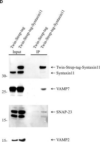
Western Blot: VAMP-7 Antibody [NBP2-32232] -
Western Blot: VAMP-7 Antibody [NBP2-32232] - VAMP7 forms a SNARE complex with SNAP-23 & Syntaxin11. (A) Model illustrating the formation of SNARE complex between VAMP7 (v-SNARE), SNAP-23, & Syntaxin11 (t- SNARE) during cytotoxic granule fusion in human CD8+ T cells. (B) Different Twin-Strep-tag fusion constructs used for pulldown assay. Twin-Strep-tag was fused at the C terminus of VAMP7 & at the N terminus of Syntaxin11 with a (GGS)x3 linker. (C) Western blot of bead stimulated human CD8+ T cells transfected with Twin-Strep-tag-tagged VAMP7, immuno-precipitated with anti-FLAG antibody & detected with antibodies against FLAG, SNAP-23, & Syntaxin11. (D) Western blot of bead stimulated human CD8+ T cells transfected with Twin-Strep-tag-tagged Syntaxin11, immuno-precipitated with anti-Syntaxin antibody & detected with antibodies against Strep-tag, SNAP-23, VAMP7, & VAMP2. As control, cells were transfected with Twin-Strep-tag construct. Ten percentage of the lysates were loaded as input. (E,F) Densitometric quantification of the Western blots shown in (C,D), respectively. Image collected & cropped by CiteAb from the following publication (https://pubmed.ncbi.nlm.nih.gov/31447853), licensed under a CC-BY license. Not internally tested by Novus Biologicals.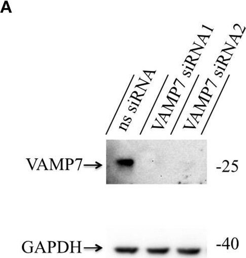
Western Blot: VAMP-7 Antibody [NBP2-32232] -
Western Blot: VAMP-7 Antibody [NBP2-32232] - Knockdown of VAMP7 strongly reduces fusion of cytotoxic granules at the IS. (A) Lysates from bead stimulated human CD8+ T cells transfected with either control or VAMP7 siRNAs (1 or 2, respectively) & blotted for VAMP7 (top) & GAPDH (bottom) as loading control. (B) Quantification of VAMP7 protein expression (in % normalized to control siRNA-treated CTLs) performed by densitometry. Bars indicate SEMs. [VAMP7-siRNA1, N = 3; ***p < 0.001 & VAMP7-siRNA2, N = 3; ***p < 0.001 (t-test)]. (C) Human CD8+ T cells co-transfected with granzyme B-mCherry along with either ns-siRNA or VAMP7-siRNA1 or VAMP7-siRNA2 & imaged 12 h after transfection. Selected live-cell TIRF microscopy images of granzyme B-mCherry in a transfected CTL in contact with an anti-CD3 coated coverslip. Fusion events are indicated with open circles (three frames shown per granule fused). (D) Mean percentage of cytotoxic granule fusion in cells transfected with either ns-siRNA (n = 66 & n = 59, respectively) or VAMP7-siRNA1 [n = 91; **p < 0.01 (t-test)] or VAMP7-siRNA2 [n = 72; ***p < 0.001 (t-test)]. (E) Mean average number of granules fused over time in the TIRF plane per cell p = 0.206 (t-test) for VAMP7-siRNA1 & **p < 0.01 (t-test) for VAMP7-siRNA2. Bars indicate mean ± SEM. Scale bar, 5 μm. (F) Calcein-based killing assay for CTLs transfected with either ns-siRNA, VAMP7-siRNA1, or VAMP7-siRNA2. Experiments were carried out in duplicate [N = 4; ***p < 0.001 (t-test)]. Image collected & cropped by CiteAb from the following publication (https://pubmed.ncbi.nlm.nih.gov/31447853), licensed under a CC-BY license. Not internally tested by Novus Biologicals.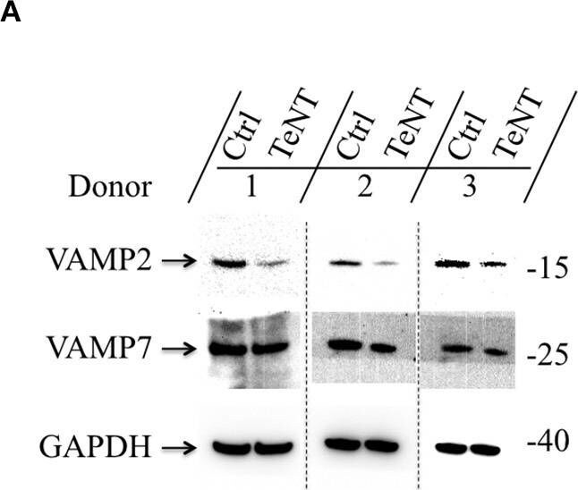
Western Blot: VAMP-7 Antibody [NBP2-32232] -
Western Blot: VAMP-7 Antibody [NBP2-32232] - Fusion of cytotoxic granules with the plasma membrane is insensitive to Tetanus toxin. (A) Bead-stimulated human CD8+ T cells transfected with GFP or TeNT-GFP as indicated. VAMP2 & VAMP7 protein levels were determined by Western blot analysis 12–16 h after transfection. (B) Expression of VAMP2 & VAMP7 protein levels relative to GAPDH in CTLs transfected with GFP or TeNT-GFP as indicated. Graphs represent means [N = 3, ***p < 0.001 (Student's t-test)]. (C) Bead-stimulated human CTLs co-transfected with either GFP or TeNT-GFP along with granzyme B-mCherry & imaged 12 h after transfection. Representative live-cell TIRFM images of CTLs in contact with an anti-CD3 coated coverslip. Fusion events indicated with open arrowheads. (D) Mean percentage of cytotoxic granule fusion in cells transfected with either GFP (n = 50) or TeNT-GFP (n = 40), p = 0.704 (Student's t-test). (E) Mean average number of granules fused per cell over time in the TIRF plane in cells transfected with either GFP (n = 20) or TeNT-GFP (n = 15), p = 0.939 (Student's t-test). Bars show mean ± SEM. Scale bar, 5 μm. Image collected & cropped by CiteAb from the following publication (https://pubmed.ncbi.nlm.nih.gov/31447853), licensed under a CC-BY license. Not internally tested by Novus Biologicals.Applications for VAMP-7 Antibody - BSA Free
Immunoprecipitation
Western Blot
Formulation, Preparation, and Storage
Purification
Formulation
Format
Preservative
Concentration
Shipping
Stability & Storage
Background: VAMP-7
Long Name
Alternate Names
Gene Symbol
UniProt
Additional VAMP-7 Products
Product Documents for VAMP-7 Antibody - BSA Free
Certificate of Analysis
To download a Certificate of Analysis, please enter a lot or batch number in the search box below.
Product Specific Notices for VAMP-7 Antibody - BSA Free
This product is for research use only and is not approved for use in humans or in clinical diagnosis. Primary Antibodies are guaranteed for 1 year from date of receipt.
Related Research Areas
Citations for VAMP-7 Antibody - BSA Free
 Powered by Bioz
Customer Reviews for VAMP-7 Antibody - BSA Free
There are currently no reviews for this product. Be the first to review VAMP-7 Antibody - BSA Free and earn rewards!
Have you used VAMP-7 Antibody - BSA Free?
Submit a review and receive an Amazon gift card!
$25/€18/£15/$25CAN/¥2500 for a review with an image
$10/€7/£6/$10CAN/¥1110 for a review without an image
Submit a review
Protocols
Find general support by application which include: protocols, troubleshooting, illustrated assays, videos and webinars.
- Cellular Response to Hypoxia Protocols
- Immunoprecipitation Protocol
- R&D Systems Quality Control Western Blot Protocol
- Troubleshooting Guide: Western Blot Figures
- Western Blot Conditions
- Western Blot Protocol
- Western Blot Protocol for Cell Lysates
- Western Blot Troubleshooting
- Western Blot Troubleshooting Guide
- View all Protocols, Troubleshooting, Illustrated assays and Webinars