

![Knockdown Validated: Vimentin Antibody [NBP1-31327]](https://resources.rndsystems.com/images/products/Vimentin-Antibody-Western-Blot-NBP1-31327-img0031.jpg "Western Blot: Vimentin Antibody [NBP1-31327]")
Scientific images may be resized, cropped, or adjusted for brightness or contrast for presentation purposes. These updates are limited to presentation and do not affect the underlying scientific interpretation of the data. Where available, additional source data may be provided upon request.
Loading...
Key Product Details
Validated by
Knockout/Knockdown, Orthogonal Validation
Species Reactivity
Validated:
Human, Mouse, Rat, Canine, Equine, Feline
Cited:
Human, Bovine, Guinea Pig
Predicted:
Bovine (98%), Chicken (92%), Chimpanzee (99%), Guinea Pig (97%). Backed by our 100% Guarantee.
Applications
Validated:
Immunohistochemistry, Immunohistochemistry-Paraffin, Immunohistochemistry-Frozen, Western Blot, ELISA, Sandwich ELISA, Immunocytochemistry/ Immunofluorescence, Immunoprecipitation, Knockdown Validated
Cited:
Western Blot, Immunocytochemistry/ Immunofluorescence
Label
Unconjugated
Antibody Source
Polyclonal Rabbit IgG
Loading...
Product Specifications
Immunogen
Recombinant protein encompassing a sequence within the center region of human Vimentin. The exact sequence is proprietary.
Reactivity Notes
Immunogen displays the following percentage of sequence identity for non-tested species: Xenopus laevis (84%). Equine reactivity reported from a verified customer review. Cat (100%).
Marker
Mesenchymal Cells Marker
Clonality
Polyclonal
Host
Rabbit
Isotype
IgG
Theoretical MW
56 kDa.
Disclaimer note: The observed molecular weight of the protein may vary from the listed predicted molecular weight due to post translational modifications, post translation cleavages, relative charges, and other experimental factors.
Disclaimer note: The observed molecular weight of the protein may vary from the listed predicted molecular weight due to post translational modifications, post translation cleavages, relative charges, and other experimental factors.
Scientific Data Images for Vimentin Antibody
![Immunocytochemistry/ Immunofluorescence: Vimentin Antibody [NBP1-31327]](https://resources.rndsystems.com/images/products/Vimentin-Antibody-Immunocytochemistry-NBP1-31327-img0037.jpg "Immunocytochemistry/ Immunofluorescence: Vimentin Antibody [NBP1-31327]")
Immunocytochemistry/ Immunofluorescence: Vimentin Antibody [NBP1-31327]
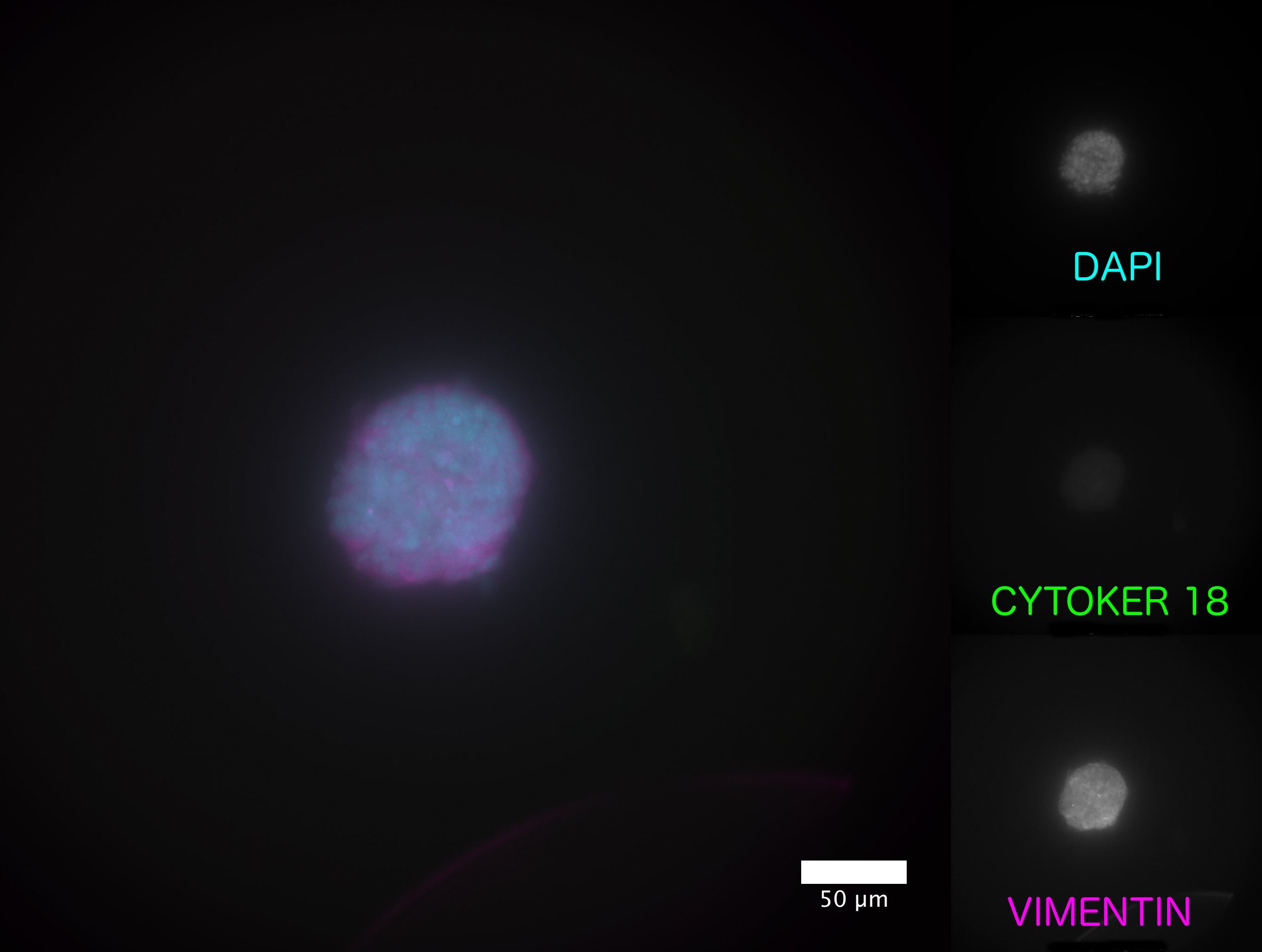
Immunocytochemistry/Immunofluorescence: Vimentin Antibody [NBP1-31327] - Cellular aggregates composed of equine endometrial fibroblast. Co-staining using the vimentin antibody and a cytokeratin antibody (mouse). Image submitted by a verified customer review.![Immunocytochemistry/ Immunofluorescence: Vimentin Antibody [NBP1-31327]](https://resources.rndsystems.com/images/products/Vimentin-Antibody-Immunocytochemistry-Immunofluorescence-NBP1-31327-img0024.jpg "Immunocytochemistry/ Immunofluorescence: Vimentin Antibody [NBP1-31327]")
Immunocytochemistry/ Immunofluorescence: Vimentin Antibody [NBP1-31327]
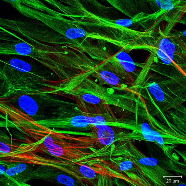
Immunocytochemistry/Immunofluorescence: Vimentin Antibody [NBP1-31327] - Human primary cardiac fibroblasts stained with Vimentin (green) and smooth muscle actin (red) antibodies. Image from verified customer review.![Immunohistochemistry-Paraffin: Vimentin Antibody [NBP1-31327]](https://resources.rndsystems.com/images/products/Vimentin-Antibody-Immunohistochemistry-Paraffin-NBP1-31327-img0036.jpg "Immunohistochemistry-Paraffin: Vimentin Antibody [NBP1-31327]")
Immunohistochemistry-Paraffin: Vimentin Antibody [NBP1-31327]
Immunohistochemistry-Paraffin: Vimentin Antibody [NBP1-31327] - Paraffin-embedded rat ovary. Vimentin antibody at 1:500.![Western Blot: Vimentin Antibody [NBP1-31327]](https://resources.rndsystems.com/images/products/Vimentin-Antibody-Western-Blot-NBP1-31327-img0014.jpg "Western Blot: Vimentin Antibody [NBP1-31327]")
Western Blot: Vimentin Antibody [NBP1-31327]
Western Blot: Vimentin Antibody [NBP1-31327] - 293T cell lysate: A. 20 ug. B. 10 ug. C. 5 ug. D. 1 ug.![Western Blot: Vimentin Antibody [NBP1-31327]](https://resources.rndsystems.com/images/products/Vimentin-Antibody-Western-Blot-NBP1-31327-img0023.jpg "Western Blot: Vimentin Antibody [NBP1-31327]")
Western Blot: Vimentin Antibody [NBP1-31327]
Western Blot: Vimentin Antibody [NBP1-31327] - Various whole cell extracts (30 ug) were separated by 10% SDS-PAGE, membrane was blotted with Vimentin antibody at 1:50000.![Immunohistochemistry-Paraffin: Vimentin Antibody [NBP1-31327]](https://resources.rndsystems.com/images/products/Vimentin-Antibody-Immunohistochemistry-Paraffin-NBP1-31327-img0022.jpg "Immunohistochemistry-Paraffin: Vimentin Antibody [NBP1-31327]")
Immunohistochemistry-Paraffin: Vimentin Antibody [NBP1-31327]
Immunohistochemistry-Paraffin: Vimentin Antibody [NBP1-31327] - Paraffin-embedded human lung adenocarcinoma. Vimentin antibody diluted at 1:500.![Immunohistochemistry-Paraffin: Vimentin Antibody [NBP1-31327]](https://resources.rndsystems.com/images/products/Vimentin-Antibody-Immunohistochemistry-Paraffin-NBP1-31327-img0034.jpg "Immunohistochemistry-Paraffin: Vimentin Antibody [NBP1-31327]")
Immunohistochemistry-Paraffin: Vimentin Antibody [NBP1-31327]
Immunohistochemistry-Paraffin: Vimentin Antibody [NBP1-31327] - Paraffin-embedded rat testis. Vimentin antibody at 1:500.![Immunoprecipitation: Vimentin Antibody [NBP1-31327]](https://resources.rndsystems.com/images/products/Vimentin-Antibody-Immunoprecipitation-NBP1-31327-img0019.jpg "Immunoprecipitation: Vimentin Antibody [NBP1-31327]")
Immunoprecipitation: Vimentin Antibody [NBP1-31327]
Immunoprecipitation: Vimentin Antibody [NBP1-31327] - Vimentin protein from HeLa whole cell extracts using 5 ug of Vimentin antibody. Western blot analysis was performed using Vimentin antibody. EasyBlot anti-Rabbit IgG was used as a secondary reagent.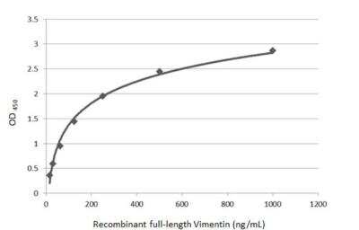
Sandwich ELISA: Vimentin Antibody [NBP1-31327] - Detection of recombinant full-length Vimentin protein using as capture antibody at concentration of 5 ug/mL as detection antibody at concentration of 1 ug/mL. Rabbit IgG antibody (HRP) (NBP2-19301) was diluted at 1:10000 and used to detect the primary antibody.
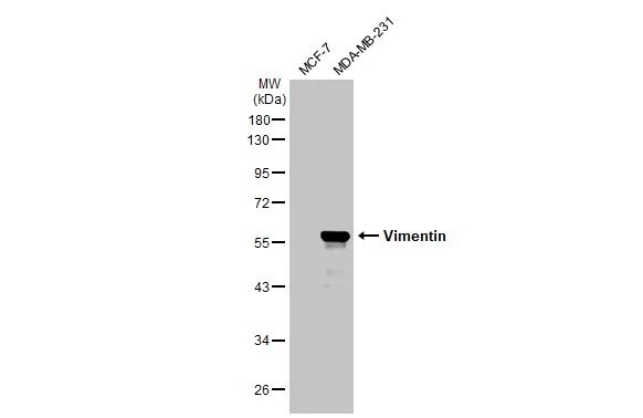
Western Blot: Vimentin Antibody [NBP1-31327] -
Western Blot: Vimentin Antibody [NBP1-31327] - Various whole cell extracts (30 ug) were separated by 10% SDS-PAGE, and the membrane was blotted with Vimentin antibody (NBP1-31327) diluted at 1:5000. The HRP-conjugated anti-rabbit IgG antibody was used to detect the primary antibody.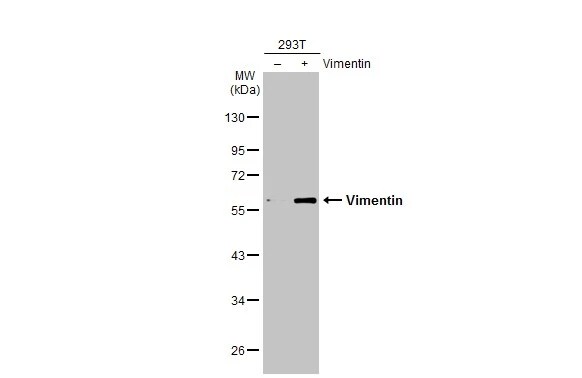
Western Blot: Vimentin Antibody [NBP1-31327] -
Western Blot: Vimentin Antibody [NBP1-31327] - Non-transfected (-) and transfected (+) 293T whole cell extracts (10 ug) were separated by 10% SDS-PAGE, and the membrane was blotted with Vimentin antibody (NBP1-31327) diluted at 1:10000. The HRP-conjugated anti-rabbit IgG antibody was used to detect the primary antibody.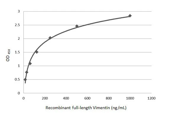
ELISA: Vimentin Antibody [NBP1-31327] -
ELISA: Vimentin Antibody [NBP1-31327] - Sandwich ELISA detection of recombinant full-length Vimentin protein using the capture antibody at concentration of 5 ug/mL and NBP1-31327 as detection antibody at concentration of 1 ug/mL. Rabbit IgG antibody (HRP) was diluted at 1:10000 and used to detect the primary antibody.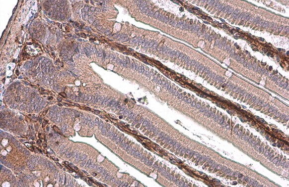
Immunohistochemistry-Paraffin: Vimentin Antibody [NBP1-31327] -
Immunohistochemistry-Paraffin: Vimentin Antibody [NBP1-31327] - Vimentin antibody detects Vimentin protein at cytoplasm by immunohistochemical analysis.Sample: Paraffin-embedded mouse intestine.
Vimentin stained by Vimentin antibody (NBP1-31327) diluted at 1:500.
Antigen Retrieval: Citrate buffer, pH 6.0, 15 min
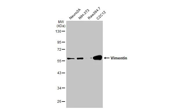
Western Blot: Vimentin Antibody [NBP1-31327] -
Western Blot: Vimentin Antibody [NBP1-31327] - Various whole cell extracts (30 ug) were separated by 10% SDS-PAGE, and the membrane was blotted with Vimentin antibody (NBP1-31327) diluted at 1:5000. The HRP-conjugated anti-rabbit IgG antibody was used to detect the primary antibody.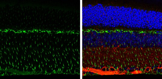
Immunohistochemistry-Paraffin: Vimentin Antibody [NBP1-31327] -
Immunohistochemistry-Paraffin: Vimentin Antibody [NBP1-31327] - Vimentin antibody detects Vimentin protein expression by immunohistochemical analysis.Sample:Paraffin-Embedded adult mouse retina.
Green: Vimentin protein stained by Vimentin antibody (NBP1-31327) diluted at 1:250.
Red: beta Tubulin 3/ TUJ1, stained by beta Tubulin 3/ TUJ1 antibody [GT11710] diluted at 1:500.
Blue: Fluoroshield with DAPI.
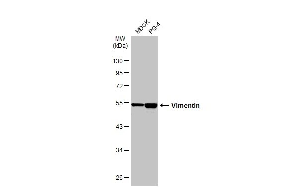
Western Blot: Vimentin Antibody [NBP1-31327] -
Western Blot: Vimentin Antibody [NBP1-31327] - Various whole cell extracts (30 ug) were separated by 10% SDS-PAGE, and the membrane was blotted with Vimentin antibody (NBP1-31327) diluted at 1:20000. The HRP-conjugated anti-rabbit IgG antibody was used to detect the primary antibody.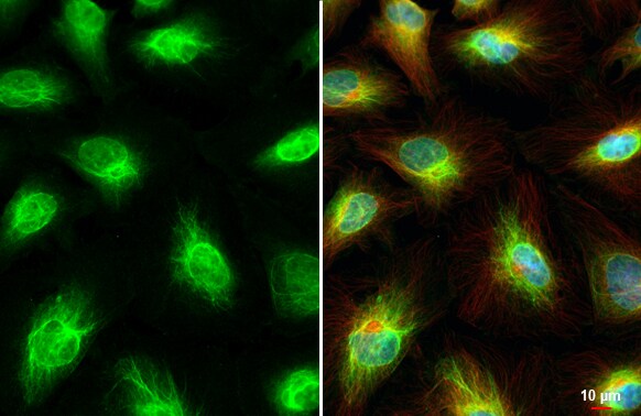
Immunocytochemistry/ Immunofluorescence: Vimentin Antibody [NBP1-31327] -
Immunocytochemistry/ Immunofluorescence: Vimentin Antibody [NBP1-31327] - Vimentin antibody detects Vimentin protein at cytoskeleton and nucleus by immunofluorescent analysis.Sample: HeLa cells were fixed in 4% paraformaldehyde at RT for 15 min.
Green: Vimentin stained by Vimentin antibody (NBP1-31327) diluted at 1:500.
Red: alpha Tubulin, a cytoskeleton marker, stained by alpha Tubulin antibody [GT114] diluted at 1:1000.
Blue: Fluoroshield with DAPI.
Immunohistochemistry-Paraffin: Vimentin Antibody [NBP1-31327] -
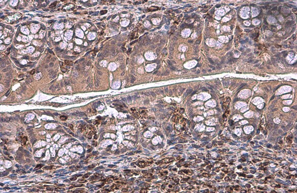
Immunohistochemistry-Paraffin: Vimentin Antibody [NBP1-31327] - Vimentin antibody detects Vimentin protein at cytoplasm by immunohistochemical analysis.Sample: Paraffin-embedded mouse colon.
Vimentin stained by Vimentin antibody (NBP1-31327) diluted at 1:500.
Antigen Retrieval: Citrate buffer, pH 6.0, 15 min
Immunohistochemistry-Paraffin: Vimentin Antibody [NBP1-31327] -
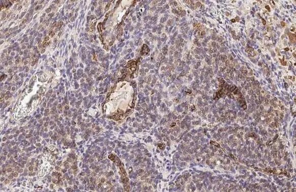
Immunohistochemistry-Paraffin: Vimentin Antibody [NBP1-31327] - Vimentin antibody detects Vimentin protein at cell membrane and cytoplasm by immunohistochemical analysis.Sample: Paraffin-embedded rat ovary.
Vimentin stained by Vimentin antibody (NBP1-31327) diluted at 1:500.
Antigen Retrieval: Citrate buffer, pH 6.0, 15 min
Immunohistochemistry-Paraffin: Vimentin Antibody [NBP1-31327] -
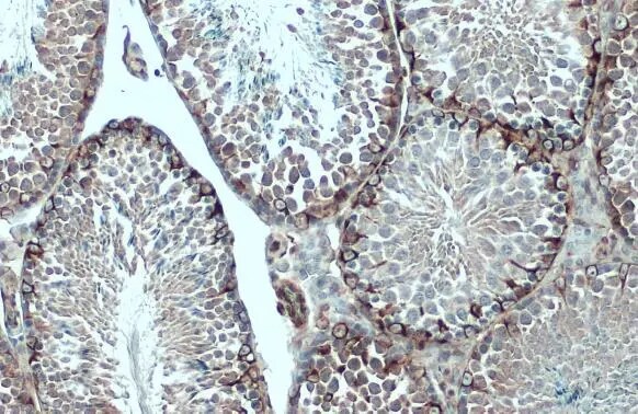
Immunohistochemistry-Paraffin: Vimentin Antibody [NBP1-31327] - Vimentin antibody detects Vimentin protein at cytoplasm by immunohistochemical analysis.Sample: Paraffin-embedded mouse testis.
Vimentin stained by Vimentin antibody (NBP1-31327) diluted at 1:500.
Antigen Retrieval: Citrate buffer, pH 6.0, 15
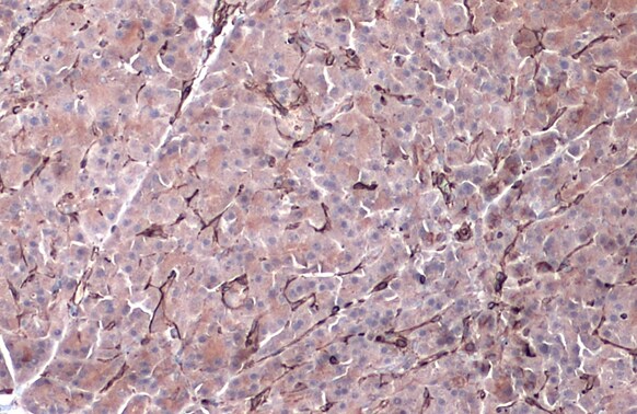
Immunohistochemistry-Paraffin: Vimentin Antibody [NBP1-31327] -
Immunohistochemistry-Paraffin: Vimentin Antibody [NBP1-31327] - Vimentin antibody detects Vimentin protein by immunohistochemical analysis.Sample: Paraffin-embedded dog pancreas.
Vimentin stained by Vimentin antibody (NBP1-31327) diluted at 1:500.
Antigen Retrieval: Citrate buffer, pH 6.0, 15 min
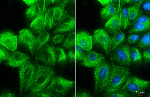
Immunocytochemistry/ Immunofluorescence: Vimentin Antibody [NBP1-31327] -
Immunocytochemistry/ Immunofluorescence: Vimentin Antibody [NBP1-31327] - Vimentin antibody detects Vimentin protein at cytoskeleton by immunofluorescent analysis.Sample: MDCK cells were fixed in 4% paraformaldehyde at RT for 15 min.
Green: Vimentin stained by Vimentin antibody (NBP1-31327) diluted at 1:500.
Blue: Hoechst 33342 staining.
Scale bar= 10 um.
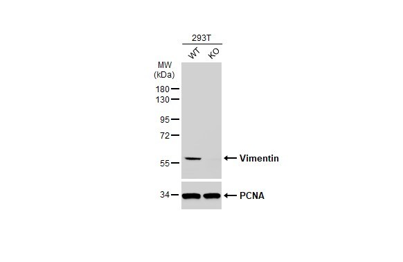
Western Blot: Vimentin Antibody [NBP1-31327] -
Western Blot: Vimentin Antibody [NBP1-31327] - Wild-type (WT) and Vimentin knockout (KO) 293T cell extracts (30 ug) were separated by 10% SDS-PAGE, and the membrane was blotted with Vimentin antibody diluted at 1:50000. The HRP-conjugated anti-rabbit IgG antibody was used to detect the primary antibody.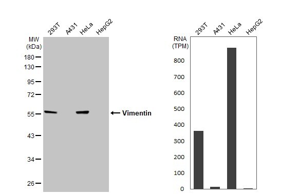
Western Blot: Vimentin Antibody [NBP1-31327] -
Various whole cell extracts (30 ug) were separated by 10% SDS-PAGE, and the membrane was blotted with Vimentin antibody (NBP1-31327) diluted at 1:50000. The HRP-conjugated anti-rabbit IgG antibody was used to detect the primary antibody. Corresponding RNA expression data for the same cell lines are based on Human Protein Atlas program.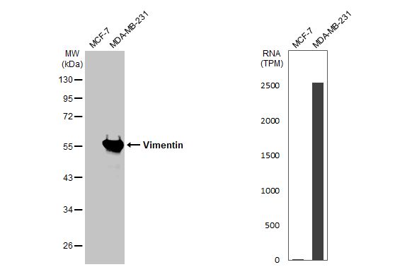
Western Blot: Vimentin Antibody [NBP1-31327] -
Various whole cell extracts (30 ug) were separated by 10% SDS-PAGE, and the membrane was blotted with Vimentin antibody (NBP1-31327) diluted at 1:5000. The HRP-conjugated anti-rabbit IgG antibody was used to detect the primary antibody. Corresponding RNA expression data for the same cell lines are based on Human Protein Atlas program.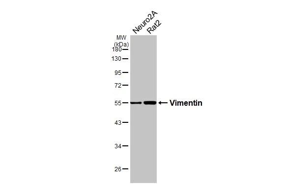
Western Blot: Vimentin Antibody [NBP1-31327] -
Various whole cell extracts (30 ug) were separated by 10% SDS-PAGE, and the membrane was blotted with Vimentin antibody (NBP1-31327) diluted at 1:2000. The HRP-conjugated anti-rabbit IgG antibody was used to detect the primary antibody.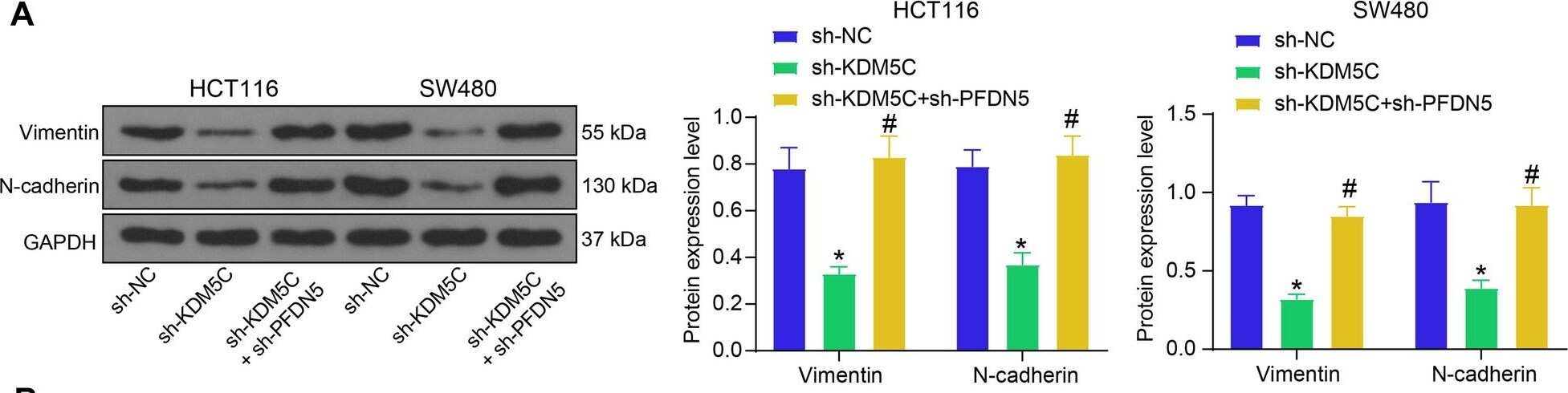
Western Blot: Vimentin Antibody [NBP1-31327] -
KDM5C/PFDN5 affects EMT in CRC. A protein levels of Vimentin and N-cadherin in HCT116 and SW480 cells determined by WB analysis (two-way ANOVA); B protein levels of Vimentin and N-cadherin in xenograft tumors determined by western blot analysis (two-way ANOVA). For cellular experiments, three biological replicates were performed. For animal studies, n = 5 in each group. *p < 0.05 vs. the sh-NC group; #p < 0.05 vs. the sh-KDM5C group Image collected and cropped by CiteAb from the following open publication (https://molmed.biomedcentral.com/articles/10.1186/s10020-023-00775-7), licensed under a CC-BY license. Not internally tested by Novus Biologicals.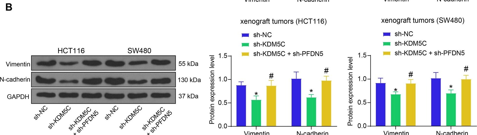
Western Blot: Vimentin Antibody [NBP1-31327] -
KDM5C/PFDN5 affects EMT in CRC. A protein levels of Vimentin and N-cadherin in HCT116 and SW480 cells determined by WB analysis (two-way ANOVA); B protein levels of Vimentin and N-cadherin in xenograft tumors determined by western blot analysis (two-way ANOVA). For cellular experiments, three biological replicates were performed. For animal studies, n = 5 in each group. *p < 0.05 vs. the sh-NC group; #p < 0.05 vs. the sh-KDM5C group Image collected and cropped by CiteAb from the following open publication (https://molmed.biomedcentral.com/articles/10.1186/s10020-023-00775-7), licensed under a CC-BY license. Not internally tested by Novus Biologicals.Applications for Vimentin Antibody
Application
Recommended Usage
ELISA
Assay dependent.
Immunocytochemistry/ Immunofluorescence
1:100-1:1000
Immunohistochemistry
Assay dependent.
Immunohistochemistry-Frozen
Assay dependent.
Immunohistochemistry-Paraffin
Assay dependent.
Immunoprecipitation
1:100-1:500
Sandwich ELISA
Assay dependent.
Western Blot
1:5000-1:50000
Reviewed Applications
Read 3 reviews rated 5 using NBP1-31327 in the following applications:
Formulation, Preparation, and Storage
Purification
Antigen Affinity-purified
Formulation
PBS, 20% Glycerol
Preservative
0.025% Proclin 300
Concentration
Concentrations vary lot to lot. See vial label for concentration. If unlisted please contact technical services.
Shipping
The product is shipped with polar packs. Upon receipt, store it immediately at the temperature recommended below.
Stability & Storage
Aliquot and store at -20C or -80C. Avoid freeze-thaw cycles.
Background: Vimentin
Activated macrophages have been shown to secrete phosphorylated vimentin which can be stimulated by a variety of pathophysiological factors including oxidized low-density lipoproteins and TNF-alpha or inhibited by IL-10 (1). The vimentin protein is often expressed at the cell surface playing a role in cell-cell interactions, tissue damage and repair, immune response, and pathogen recognition (1). Vimentin functions in many cytoskeletal processes including cell migration, which is highlighted by its upregulation during epithelial-to-mesenchymal transition (EMT) (4,5). Vimentin is a commonly used marker for EMT and is expressed by many tumor types (5). For example, high metastasis of oral squamous cell carcinomas also showed high vimentin positive expression in immunohistochemical staining analysis (5). A number of vimentin targeting compounds are in cancer-related clinical trials, however, given the multifunctional role of vimentin, the effect of inhibition on non-malignant cells needs to be thoroughly examined (5).
References
1. Ramos, I., Stamatakis, K., Oeste, C. L., & Perez-Sala, D. (2020). Vimentin as a Multifaceted Player and Potential Therapeutic Target in Viral Infections. International Journal of Molecular Sciences. https://doi.org/10.3390/ijms21134675
2. Uniprot (P08670)
3. Morrow, C. S., & Moore, D. L. (2020). Vimentin's side gig: Regulating cellular proteostasis in mammalian systems. Cytoskeleton (Hoboken, N.J.). https://doi.org/10.1002/cm.21645
4. van Bodegraven, E. J., & Etienne-Manneville, S. (2020). Intermediate filaments against actomyosin: the david and goliath of cell migration. Current Opinion in Cell Biology. https://doi.org/10.1016/j.ceb.2020.05.006
5. Strouhalova, K., Prechova, M., Gandalovicova, A., Brabek, J., Gregor, M., & Rosel, D. (2020). Vimentin Intermediate Filaments as Potential Target for Cancer Treatment. Cancers. https://doi.org/10.3390/cancers12010184
Alternate Names
VIM
Gene Symbol
VIM
Additional Vimentin Products
Product Documents for Vimentin Antibody
Certificate of Analysis
To download a Certificate of Analysis, please enter a lot or batch number in the search box below.
Product Specific Notices for Vimentin Antibody
This product is for research use only and is not approved for use in humans or in clinical diagnosis. Primary Antibodies are guaranteed for 1 year from date of receipt.
Citations for Vimentin Antibody
 Powered by Bioz
Customer Reviews for Vimentin Antibody (3)
5 out of 5
3 Customer Ratings
Have you used Vimentin Antibody?
Submit a review and receive an Amazon gift card!
$25/€18/£15/$25CAN/¥2500 for a review with an image
$10/€7/£6/$10CAN/¥1110 for a review without an image
Submit a review
Customer Images
Showing
1
-
3 of
3 reviews
Showing All
Filter By:
-
Application: ImmunocytochemistrySample Tested: fibroblastsSpecies: EquineVerified Customer | Posted 12/11/2018Co-staining using the vimentin antibody and a cytokeratin antibody (mouse).I will have to look up the lot number and supply tomorrow. Staining material: cellular aggregates composed of equine endometrial fibroblast only. Co-staining using vimentin antibody (purchase order number: 3641031 from NovusBio website directly) and a cytokeratin antibody (mouse monoclonal) we have in the lab. Equine endometrial fibroblast express vimentin only. The image on the left shows the staining for vimentin and DAPI. Cytoplasmic staining is evident colored in red. Nuclei are stained with DAPI colored in blue. No staining for vimentin present. The images on the right hand site depict the negative control (i.e. omission of the primary antibody, DAPI only).

-
Application: ImmunofluorescenceSample Tested: primary human cellsSpecies: HumanVerified Customer | Posted 07/13/2016human primary cardiac fibroblasts immunostained for vimentin (green) and smooth muscle actin (red)
-
Application: Western BlotSample Tested: Human cancer cellSpecies: HumanVerified Customer | Posted 09/02/2015
There are no reviews that match your criteria.
Protocols
Find general support by application which include: protocols, troubleshooting, illustrated assays, videos and webinars.
- Antigen Retrieval Protocol (PIER)
- Antigen Retrieval for Frozen Sections Protocol
- Appropriate Fixation of IHC/ICC Samples
- Cellular Response to Hypoxia Protocols
- Chromogenic IHC Staining of Formalin-Fixed Paraffin-Embedded (FFPE) Tissue Protocol
- Chromogenic Immunohistochemistry Staining of Frozen Tissue
- ClariTSA™ Fluorophore Kits
- Detection & Visualization of Antibody Binding
- ELISA Sample Preparation & Collection Guide
- ELISA Troubleshooting Guide
- Fluorescent IHC Staining of Frozen Tissue Protocol
- Graphic Protocol for Heat-induced Epitope Retrieval
- Graphic Protocol for the Preparation and Fluorescent IHC Staining of Frozen Tissue Sections
- Graphic Protocol for the Preparation and Fluorescent IHC Staining of Paraffin-embedded Tissue Sections
- Graphic Protocol for the Preparation of Gelatin-coated Slides for Histological Tissue Sections
- How to Run an R&D Systems DuoSet ELISA
- How to Run an R&D Systems Quantikine ELISA
- How to Run an R&D Systems Quantikine™ QuicKit™ ELISA
- ICC Cell Smear Protocol for Suspension Cells
- ICC Immunocytochemistry Protocol Videos
- ICC for Adherent Cells
- IHC Sample Preparation (Frozen sections vs Paraffin)
- Immunocytochemistry (ICC) Protocol
- Immunocytochemistry Troubleshooting
- Immunofluorescence of Organoids Embedded in Cultrex Basement Membrane Extract
- Immunofluorescent IHC Staining of Formalin-Fixed Paraffin-Embedded (FFPE) Tissue Protocol
- Immunohistochemistry (IHC) and Immunocytochemistry (ICC) Protocols
- Immunohistochemistry Frozen Troubleshooting
- Immunohistochemistry Paraffin Troubleshooting
- Immunoprecipitation Protocol
- Preparing Samples for IHC/ICC Experiments
- Preventing Non-Specific Staining (Non-Specific Binding)
- Primary Antibody Selection & Optimization
- Protocol for Heat-Induced Epitope Retrieval (HIER)
- Protocol for Making a 4% Formaldehyde Solution in PBS
- Protocol for VisUCyte™ HRP Polymer Detection Reagent
- Protocol for the Fluorescent ICC Staining of Cell Smears - Graphic
- Protocol for the Fluorescent ICC Staining of Cultured Cells on Coverslips - Graphic
- Protocol for the Preparation & Fixation of Cells on Coverslips
- Protocol for the Preparation and Chromogenic IHC Staining of Frozen Tissue Sections
- Protocol for the Preparation and Chromogenic IHC Staining of Frozen Tissue Sections - Graphic
- Protocol for the Preparation and Chromogenic IHC Staining of Paraffin-embedded Tissue Sections
- Protocol for the Preparation and Chromogenic IHC Staining of Paraffin-embedded Tissue Sections - Graphic
- Protocol for the Preparation and Fluorescent ICC Staining of Cells on Coverslips
- Protocol for the Preparation and Fluorescent ICC Staining of Non-adherent Cells
- Protocol for the Preparation and Fluorescent ICC Staining of Stem Cells on Coverslips
- Protocol for the Preparation and Fluorescent IHC Staining of Frozen Tissue Sections
- Protocol for the Preparation and Fluorescent IHC Staining of Paraffin-embedded Tissue Sections
- Protocol for the Preparation of Gelatin-coated Slides for Histological Tissue Sections
- Protocol for the Preparation of a Cell Smear for Non-adherent Cell ICC - Graphic
- Quantikine HS ELISA Kit Assay Principle, Alkaline Phosphatase
- Quantikine HS ELISA Kit Principle, Streptavidin-HRP Polymer
- R&D Systems Quality Control Western Blot Protocol
- Sandwich ELISA (Colorimetric) – Biotin/Streptavidin Detection Protocol
- Sandwich ELISA (Colorimetric) – Direct Detection Protocol
- TUNEL and Active Caspase-3 Detection by IHC/ICC Protocol
- The Importance of IHC/ICC Controls
- Troubleshooting Guide: ELISA
- Troubleshooting Guide: Immunohistochemistry
- Troubleshooting Guide: Western Blot Figures
- Western Blot Conditions
- Western Blot Protocol
- Western Blot Protocol for Cell Lysates
- Western Blot Troubleshooting
- Western Blot Troubleshooting Guide
- View all Protocols, Troubleshooting, Illustrated assays and Webinars

