
Tumor Necrosis Factor Alpha (TNF-alpha ) also known as Cachectin and TNFSF2, is the prototypic ligand of the TNF superfamily. It is a pleiotropic molecule that plays a central role in inflammation, apoptosis, and immune system development. TNF-alpha is produced by a wide variety of immune and epithelial cell types (1, 2). Rat TNF-alpha consisits of a 35 amino acid (aa) cytoplasmic domain, a 21 aa transmembrane segment, and a 179 aa extracellular domain (ECD) (3). Within the ECD, rat TNF-alpha shares 94% aa sequence identity with mouse and 69% - 76% with bovine, canine, cotton rat, equine, feline, human, porcine, and rhesus TNF-alpha. The 26 kDa type 2 transmembrane protein is assembled intracellularly to form a noncovalently linked homotrimer (4). Ligation of this complex induces reverse signaling that promotes lymphocyte co-stimulation but diminishes monocyte responsiveness (5). Cleavage of membrane bound TNF-alpha by TACE/ADAM17 releases a 55 kDa soluble trimeric form of TNF-alpha (6, 7). TNF-alpha trimers bind the ubiquitous TNF RI and the hematopoietic cell-restricted TNF RII, both of which are also expressed as homotrimers (1, 8). TNF-alpha regulates lymphoid tissue development through control of apoptosis (2). It also promotes inflammatory responses by inducing the activation of vascular endothelial cells and macrophages (2). TNF-alpha is a key cytokine in the development of several inflammatory disorders (9). It contributes to the development of type 2 diabetes through its effects on insulin resistance and fatty acid metabolism (10, 11).
Rat TNF-alpha Antibody
R&D Systems | Catalog # AF-510-NA


Scientific images may be resized, cropped, or adjusted for brightness or contrast for presentation purposes. These updates are limited to presentation and do not affect the underlying scientific interpretation of the data. Where available, additional source data may be provided upon request.
Key Product Details
Species Reactivity
Validated:
Cited:
Applications
Validated:
Cited:
Label
Antibody Source
Product Specifications
Immunogen
Leu80-Leu235
Accession # P16599
Specificity
Clonality
Host
Isotype
Endotoxin Level
Scientific Data Images for Rat TNF-alpha Antibody
Cytotoxicity Induced by TNF‑ alpha and Neutralization by Rat TNF‑ alpha Antibody.
Recombinant Rat TNF-a (Catalog # 510-RT) induces cytotoxicity in the the L-929 mouse fibroblast cell line in a dose-dependent manner (orange line), as measured by Resazurin. Cytotoxicity elicited by Recombinant Rat TNF-a (0.25 ng/mL) is neutralized (green line) by increasing concentrations of Goat Anti-Rat TNF-a Antigen Affinity-purified Polyclonal Antibody (Catalog # AF-510-NA). The ND50 is typically 0.15-0.75 µg/mL in the presence of the metabolic inhibitor actinomycin D (1 µg/mL).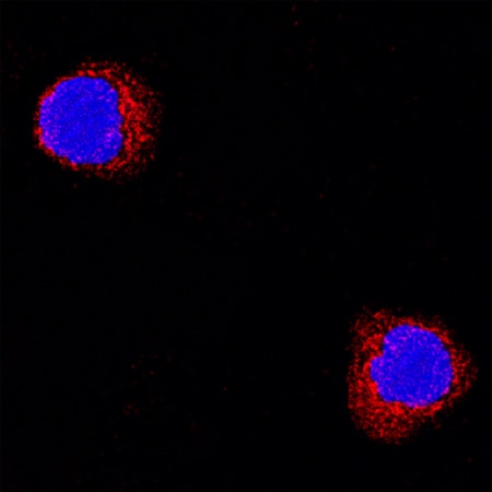
TNF‑ alpha in Rat Splenocytes.
TNF-a was detected in immersion fixed rat splenocytes using Goat Anti-Rat TNF-a Antigen Affinity-purified Polyclonal Antibody (Catalog # AF-510-NA) at 15 µg/mL for 3 hours at room temperature. Cells were stained using the NorthernLights™ 557-conjugated Anti-Goat IgG Secondary Antibody (red; Catalog # NL001) and counterstained with DAPI (blue). Specific staining was localized to cytoplasmic. View our protocol for Fluorescent ICC Staining of Non-adherent Cells.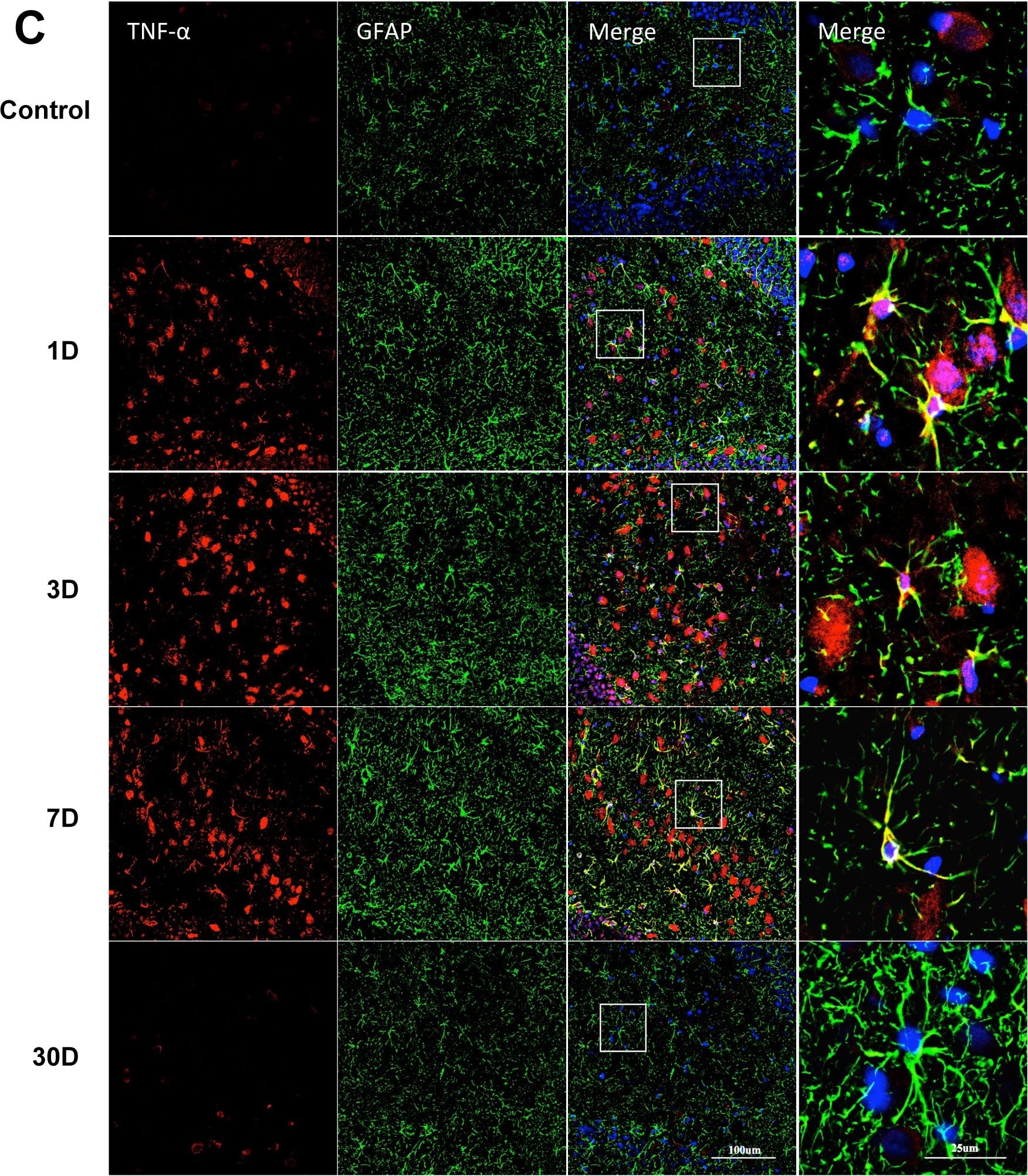
Detection of Porcine TNF-alpha by Immunocytochemistry/ Immunofluorescence
Astrocytes activity and GFAP/TNF-alpha double staining in the DG area.Astrocyte activity was measure with GFAP immunofluorescence. Representative photomicrographs show control and LPS-injected rats at different time points up to day 30. (A) Densitometry of GFAP fluorescence intensity was up-regulated from day 1 to day 7. Following LPS injection, astrocytes showed changes in morphology, which was recognized by decreased ramification and hypertrophy of the soma. (B) Double fluorescence staining of GFAP and TNF-alpha in hippocampal astrocytes. The ratio of TNF-alpha positive astrocytes in total astrocytes (GFAP-positive cells) was significantly increased from day 1 and peaking on day 7. Pictures show DG area, data are expressed as mean ± standard error of the mean (n = 4) and compared by 1-way analysis of variance followed with Boferroni post hoc analysis, **p<0.01, ***p<0.001 vs Control. Image collected and cropped by CiteAb from the following open publication (https://dx.plos.org/10.1371/journal.pone.0106331), licensed under a CC-BY license. Not internally tested by R&D Systems.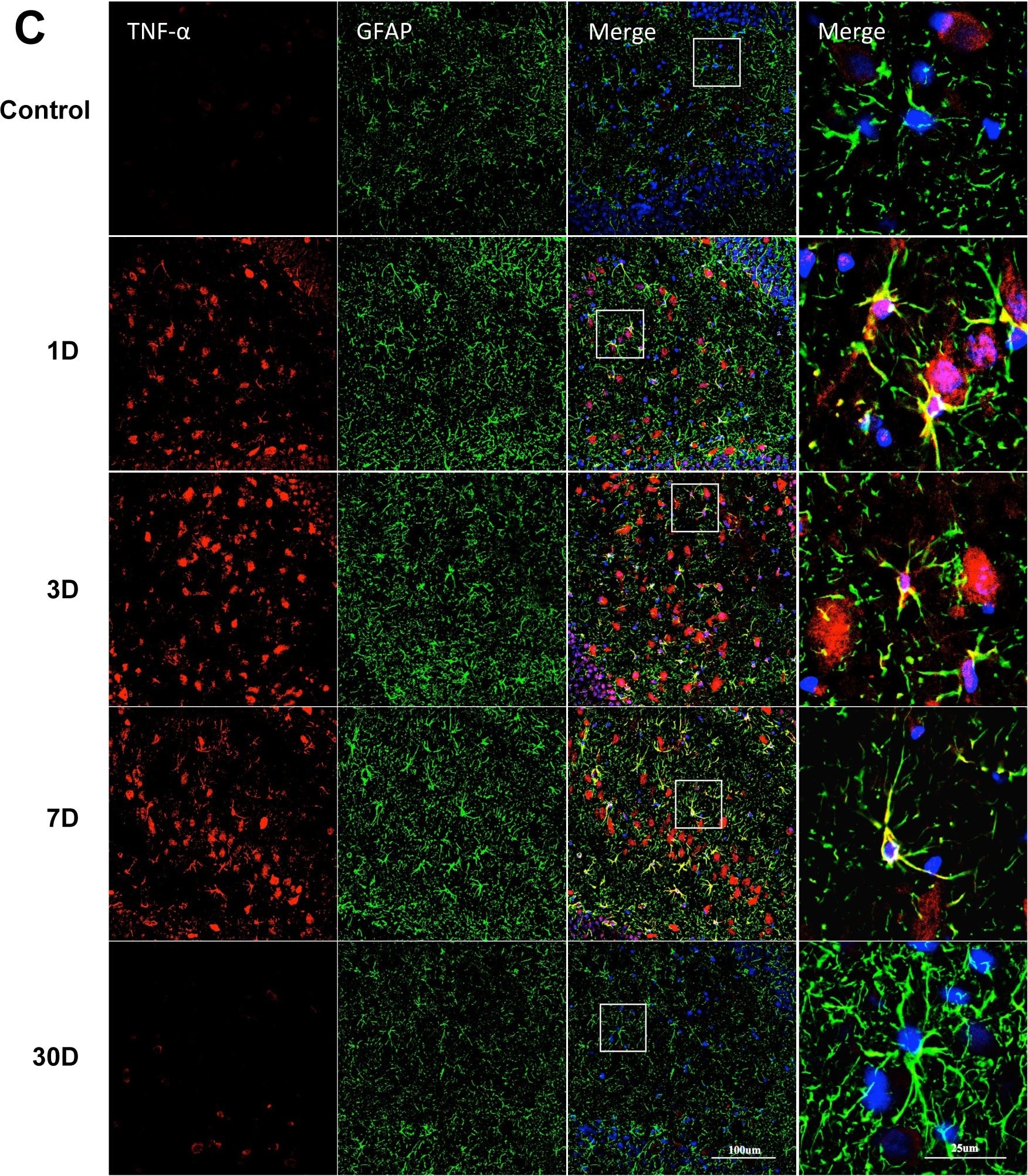
Detection of Rat TNF-alpha by Immunocytochemistry/ Immunofluorescence
Astrocytes activity and GFAP/TNF-alpha double staining in the DG area.Astrocyte activity was measure with GFAP immunofluorescence. Representative photomicrographs show control and LPS-injected rats at different time points up to day 30. (A) Densitometry of GFAP fluorescence intensity was up-regulated from day 1 to day 7. Following LPS injection, astrocytes showed changes in morphology, which was recognized by decreased ramification and hypertrophy of the soma. (B) Double fluorescence staining of GFAP and TNF-alpha in hippocampal astrocytes. The ratio of TNF-alpha positive astrocytes in total astrocytes (GFAP-positive cells) was significantly increased from day 1 and peaking on day 7. Pictures show DG area, data are expressed as mean ± standard error of the mean (n = 4) and compared by 1-way analysis of variance followed with Boferroni post hoc analysis, **p<0.01, ***p<0.001 vs Control. Image collected and cropped by CiteAb from the following open publication (https://dx.plos.org/10.1371/journal.pone.0106331), licensed under a CC-BY license. Not internally tested by R&D Systems.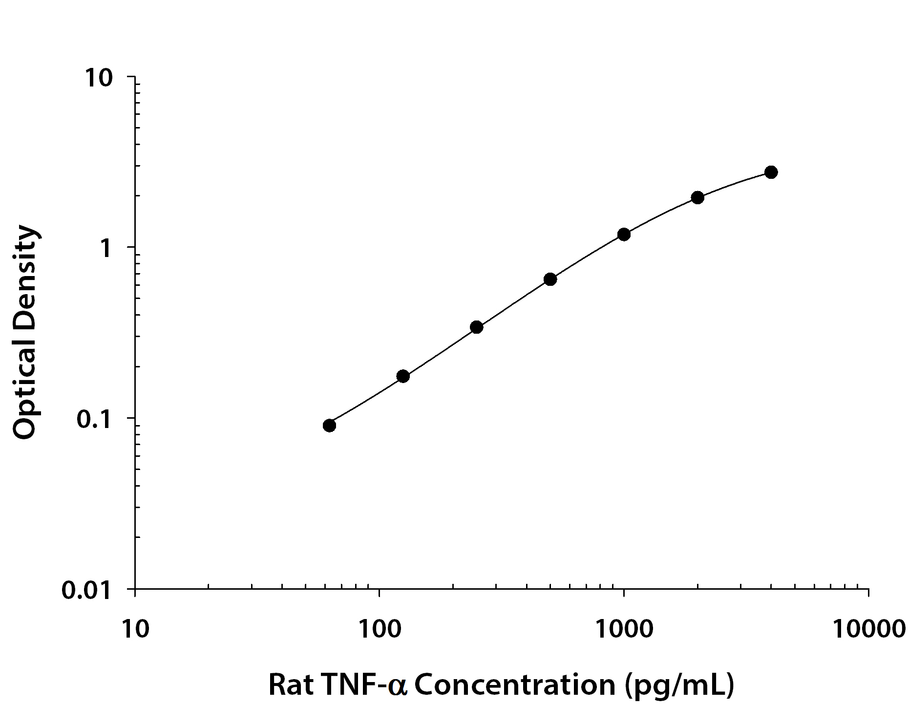
Rat TNF-alpha ELISA Standard Curve
Recombinant Rat TNF‑ alpha (Catalog # 510-RT) was serially diluted and captured by Mouse Anti-Rat TNF‑ alpha Monoclonal Antibody (Catalog # MAB510) coated on a Clear Polystyrene Microplate (Catalog # DY990). Goat Anti-Rat TNF‑ alpha Antigen Affinity-purified Polyclonal Antibody (Catalog # AF-510-NA) was biotinylated and incubated with the protein captured on the plate. Detection of the standard curve was achieved by incubating Streptavidin-HRP (Catalog # DY998)Applications for Rat TNF-alpha Antibody
Immunocytochemistry
Sample: Immersion fixed rat splenocytes
Western Blot
Sample: Recombinant Rat TNF‑ alpha (Catalog # 510-RT)
Neutralization
Formulation, Preparation, and Storage
Purification
Reconstitution
Reconstitute at 0.2 mg/mL in sterile PBS. For liquid material, refer to CoA for concentration.
Formulation
Shipping
Stability & Storage
- 12 months from date of receipt, -20 to -70 °C as supplied.
- 1 month, 2 to 8 °C under sterile conditions after reconstitution.
- 6 months, -20 to -70 °C under sterile conditions after reconstitution.
Calculators
Background: TNF-alpha
References
- Idriss, H.T. and J.H. Naismith (2000) Microsc. Res. Tech. 50:184.
- Hehlgans, T. and K. Pfeffer (2005) Immunology 115:1.
- Estler, H.C. et al. (1992) Biol. Chem. Hoppe-Seyler 373:271.
- Tang, P. et al. (1996) Biochemistry 35:8216.
- Eissner G. et al. (2004) Cytokine Growth Factor Rev. 15:353.
- Black, R.A. et al. (1997) Nature 385:729.
- Moss, M.L. et al. (1997) Nature 385:733.
- Loetscher, H. et al. (1991) J. Biol. Chem. 266:18324.
- Clark, I.A. (2007) Cytokine Growth Factor Rev. 18:335.
- Romanatto, T. et al. (2007) Peptides 28:1050.
- Hector, J. et al. (2007) Horm. Metab. Res. 39:250.
Long Name
Alternate Names
Entrez Gene IDs
Gene Symbol
UniProt
Additional TNF-alpha Products
Product Documents for Rat TNF-alpha Antibody
Certificate of Analysis
To download a Certificate of Analysis, please enter a lot or batch number in the search box below.
Note: Certificate of Analysis not available for kit components.
Product Specific Notices for Rat TNF-alpha Antibody
For research use only
Related Research Areas
Citations for Rat TNF-alpha Antibody
 Powered by Bioz
Customer Reviews for Rat TNF-alpha Antibody
There are currently no reviews for this product. Be the first to review Rat TNF-alpha Antibody and earn rewards!
Have you used Rat TNF-alpha Antibody?
Submit a review and receive an Amazon gift card!
$25/€18/£15/$25CAN/¥2500 for a review with an image
$10/€7/£6/$10CAN/¥1110 for a review without an image
Submit a review
Protocols
Find general support by application which include: protocols, troubleshooting, illustrated assays, videos and webinars.
- Appropriate Fixation of IHC/ICC Samples
- Cellular Response to Hypoxia Protocols
- ClariTSA™ Fluorophore Kits
- Detection & Visualization of Antibody Binding
- ICC Cell Smear Protocol for Suspension Cells
- ICC Immunocytochemistry Protocol Videos
- ICC for Adherent Cells
- Immunocytochemistry (ICC) Protocol
- Immunocytochemistry Troubleshooting
- Immunofluorescence of Organoids Embedded in Cultrex Basement Membrane Extract
- Immunohistochemistry (IHC) and Immunocytochemistry (ICC) Protocols
- Preparing Samples for IHC/ICC Experiments
- Preventing Non-Specific Staining (Non-Specific Binding)
- Primary Antibody Selection & Optimization
- Protocol for VisUCyte™ HRP Polymer Detection Reagent
- Protocol for the Fluorescent ICC Staining of Cell Smears - Graphic
- Protocol for the Fluorescent ICC Staining of Cultured Cells on Coverslips - Graphic
- Protocol for the Preparation and Fluorescent ICC Staining of Cells on Coverslips
- Protocol for the Preparation and Fluorescent ICC Staining of Non-adherent Cells
- Protocol for the Preparation and Fluorescent ICC Staining of Stem Cells on Coverslips
- Protocol for the Preparation of a Cell Smear for Non-adherent Cell ICC - Graphic
- R&D Systems Quality Control Western Blot Protocol
- TUNEL and Active Caspase-3 Detection by IHC/ICC Protocol
- The Importance of IHC/ICC Controls
- Troubleshooting Guide: Western Blot Figures
- Western Blot Conditions
- Western Blot Protocol
- Western Blot Protocol for Cell Lysates
- Western Blot Troubleshooting
- Western Blot Troubleshooting Guide
- View all Protocols, Troubleshooting, Illustrated assays and Webinars
FAQs for Rat TNF-alpha Antibody
-
Q: What is the expected band size when running Western blot under nonreducing conditions with Catalog # AF-510-NA
A: Rat TNF-alpha exists as a homotrimer under nonreducing conditions. Each monomer has a molecular weight of ~17 kDa, so the homotrimer form under nonreducing conditions is expected to band at ~51 kDa.
Associated Pathways





