
TGF-beta 1 (transforming growth factor beta 1) is one of three closely related mammalian members of the large TGF-beta superfamily that share a characteristic cystine knot structure. TGF-beta 1, -2 and -3 are highly pleiotropic cytokines that are proposed to act as cellular switches that regulate processes such as immune function, proliferation and epithelial-mesenchymal transition. Each TGF-beta isoform has some non-redundant functions; for TGF-beta 1, mice with targeted deletion show defects in hematopoiesis and endothelial differentiation, and die of overwhelming inflammation. Human TGF-beta 1 cDNA encodes a 390 amino acid (aa) precursor that contains a 29 aa signal peptide and a 361 aa proprotein. A furin-like convertase processes the proprotein to generate an N-terminal 249 aa latency-associated peptide (LAP) and a C-terminal 112 aa mature TGF- beta 1. Disulfide-linked homodimers of LAP and TGF-beta 1 remain non-covalently associated after secretion, forming the small latent TGF-beta 1 complex. Covalent linkage of LAP to one of three latent TGF-beta binding proteins (LTBPs) creates a large latent complex that may interact with the extracellular matrix. TGF-beta is activated from latency by pathways that include actions of the protease plasmin, matrix metalloproteases, thrombospondin 1 and a subset of integrins. Mature human TGF-beta 1 shares 100% aa identity with pig, dog and cow TGF-beta 1, and 99% aa identity with mouse, rat and horse TGF-beta 1. It demonstrates cross-species activity. TGF-beta 1 signaling begins with high-affinity binding to a type II ser/thr kinase receptor termed TGF-beta RII. This receptor then phosphorylates and activates a second ser/thr kinase receptor, TGF-beta RI (also called activin receptor-like kinase (ALK) -5), or alternatively, ALK‑1. This complex phosphorylates and activates Smad proteins that regulate transcription. Contributions of the accessory receptors betaglycan (also known as TGF-beta RIII) and endoglin, or use of Smad-independent signaling pathways, allow for disparate actions observed in response to TGF-beta in different contexts.

Scientific images may be resized, cropped, or adjusted for brightness or contrast for presentation purposes. These updates are limited to presentation and do not affect the underlying scientific interpretation of the data. Where available, additional source data may be provided upon request.
Key Product Details
Species Reactivity
Validated:
Cited:
Applications
Validated:
Cited:
Label
Antibody Source
Product Specifications
Immunogen
Ala279-Ser390
Accession # P01137
Specificity
Clonality
Host
Isotype
Endotoxin Level
Scientific Data Images for TGF-beta 1/1.2 Antibody
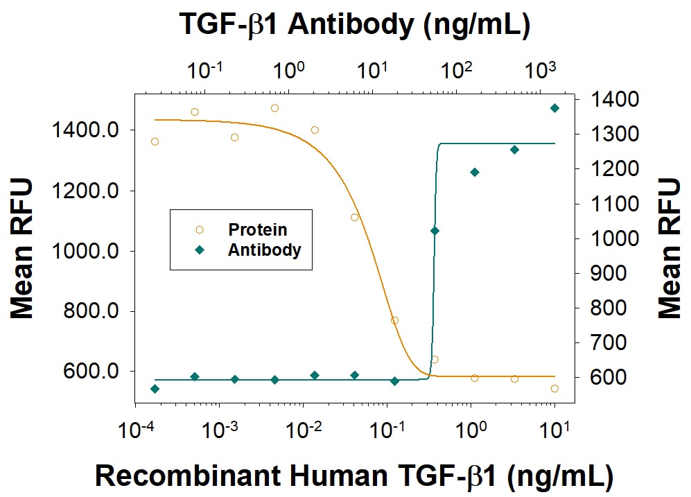
TGF‑ beta 1 Inhibition of IL‑4-dependent Cell Proliferation and Neutralization by TGF‑ beta 1/1.2 Antibody.
Recombinant Human TGF-beta 1 (Catalog # 240-B) inhibits Recombinant Mouse IL-4 (Catalog # 404-ML) induced proliferation in the HT-2 mouse T cell line in a dose-dependent manner (orange line). Inhibition of Recombinant Mouse IL-4 (7.5 ng/mL) activity elicited by Recombinant Human TGF-beta 1 (1 ng/mL) is neutralized (green line) by increasing concentrations of Chicken Anti-TGF-beta 1/1.2 Antigen Affinity-purified Polyclonal Antibody (Catalog # AF-101-NA). The ND50 is typically ≤100 ng/mL.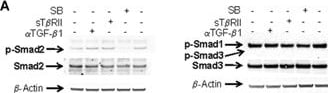
Detection of Rat TGF-beta 1/1.2 by Western Blot
Effect of Alk4-/Alk5-inhibition on Smad-phosphorylation under TGF-beta -free conditions (A) Western blot of p-Smad2, 1, 3 of PC cultured for 24 hrs under complete serum-free conditions and subjected to Alk4/5 Inhibitor (SB-431542, 5 μM), rsT beta RII (1 μg/ml) and neutralizing anti TGF-beta 1 antibody (4 μg/ml), respectively. Lysates were probed for p-Smad2, p-Smad1, and p-Smad3, total Smad2, total Smad3, and beta -actin, respectively. Representative blots of four independent experiments are shown. (B) Western blot of p-Smad3 (cross-reacting with p-Smad1) of PC cultured for 24 hrs under serum-free conditions with or without addition of rhTGF-beta 1 (5ng/ml). The blot is representative for three independent experiments. Image collected and cropped by CiteAb from the following open publication (https://pubmed.ncbi.nlm.nih.gov/18266973), licensed under a CC-BY license. Not internally tested by R&D Systems.Applications for TGF-beta 1/1.2 Antibody
Western Blot
Sample: Recombinant Human TGF-beta 1 (Catalog # 240-B)
Recombinant Human TGF-beta 1.2 (Catalog # 304-B3)
Neutralization
Reviewed Applications
Read 1 review rated 5 using AF-101-NA in the following applications:
Formulation, Preparation, and Storage
Purification
Reconstitution
Reconstitute at 0.2 mg/mL in sterile PBS. For liquid material, refer to CoA for concentration.
Formulation
Shipping
Stability & Storage
- 12 months from date of receipt, -20 to -70 °C as supplied.
- 1 month, 2 to 8 °C under sterile conditions after reconstitution.
- 6 months, -20 to -70 °C under sterile conditions after reconstitution.
Calculators
Background: TGF-beta 1/1.2
Long Name
Alternate Names
Gene Symbol
UniProt
Additional TGF-beta 1/1.2 Products
Product Documents for TGF-beta 1/1.2 Antibody
Certificate of Analysis
To download a Certificate of Analysis, please enter a lot or batch number in the search box below.
Note: Certificate of Analysis not available for kit components.
Product Specific Notices for TGF-beta 1/1.2 Antibody
For research use only
Citations for TGF-beta 1/1.2 Antibody
 Powered by Bioz
Customer Reviews for TGF-beta 1/1.2 Antibody (1)
Have you used TGF-beta 1/1.2 Antibody?
Submit a review and receive an Amazon gift card!
$25/€18/£15/$25CAN/¥2500 for a review with an image
$10/€7/£6/$10CAN/¥1110 for a review without an image
Submit a review
Customer Images
-
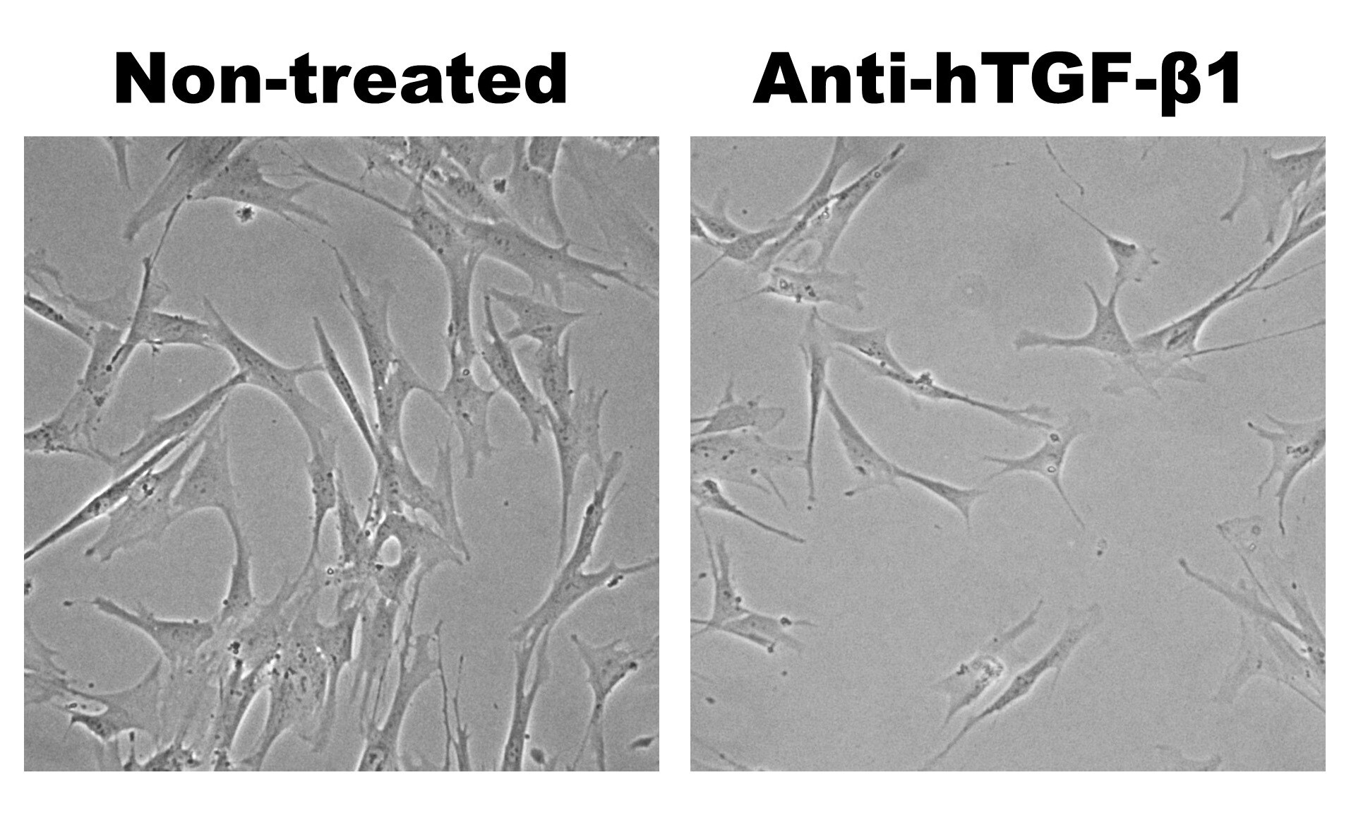
Application: Block/NeutralizeSample Tested: MRC-5 human embryonic lung fibroblast cell lineSpecies: HumanVerified Customer | Posted 06/28/2017

There are no reviews that match your criteria.
Protocols
Find general support by application which include: protocols, troubleshooting, illustrated assays, videos and webinars.
- Cellular Response to Hypoxia Protocols
- R&D Systems Quality Control Western Blot Protocol
- Troubleshooting Guide: Western Blot Figures
- Western Blot Conditions
- Western Blot Protocol
- Western Blot Protocol for Cell Lysates
- Western Blot Troubleshooting
- Western Blot Troubleshooting Guide
- View all Protocols, Troubleshooting, Illustrated assays and Webinars
FAQs for TGF-beta 1/1.2 Antibody
-
Q: Can pro-IL-1 beta be detected by Catalog #s AF-201-NA and MAB601 ?
A: The amino acid sequence present in mature IL-1 beta (Ala117-Ser269) is also present in pro IL-1 beta. So in theory, both Catalog #s AF-201-NA and MAB601 should detect the pro form of IL-1 beta. The results would depend on whether the pro form is abundantly present in the sample type being evaluated.
-
Q:
Does AF-101-NA detect other isoforms of TGF-beta?
A: Cross-reactivity testing results are listed in the Specificity section on the Product Datasheet: less than 15% cross-reactivity with recombinant amphibian TGF-beta 5 and less than 2% with TGF-beta 2 and TGF-beta 3, have been observed, as measured by direct ELISA.
-
Q: Can pro-IL-1 beta be detected by Catalog #s AF-201-NA and MAB601 ?
A: The amino acid sequence present in mature IL-1 beta (Ala117-Ser269) is also present in pro IL-1 beta. So in theory, both Catalog #s AF-201-NA and MAB601 should detect the pro form of IL-1 beta. The results would depend on whether the pro form is abundantly present in the sample type being evaluated.
-
Q:
Does AF-101-NA detect other isoforms of TGF-beta?
A: Cross-reactivity testing results are listed in the Specificity section on the Product Datasheet: less than 15% cross-reactivity with recombinant amphibian TGF-beta 5 and less than 2% with TGF-beta 2 and TGF-beta 3, have been observed, as measured by direct ELISA.