
FLASH-Seq Protocol with SEQURNA for Single-Cell RNA Sequencing
In Brief
SEQURNA (Catalog # 9028) is a synthetic thermostable RNase inhibitor for single-cell RNA-sequencing (scRNAseq), yielding single-cell libraries of equal or superior quality compared to ubiquitously used protein-based recombinant RNase inhibitors (RRIs).
SEQURNA provides additional unique improvements in reproducibility and throughput, enables new experimental workflows including retained RNase inhibition throughout heat cycles, and can reduce the need for dry-ice transport.
FLASH-seq protocol is a fast, highly sensitive, full-length single-cell RNA sequencing (scRNA-seq) protocol designed to profile gene expression and alternative splicing.
The following protocol provides guidelines for using thermostable RNase inhibitor SEQURNA. It was developed by Khven et al.1 and is a modification of the original FLASH-seq protocol by Hahaut et al.2.
Important Information
- No additional RNase inhibitor should be included in the RT mix. SEQURNA in cell lysis buffer remains active throughout cell lysis (by heating) and the reverse transcription (RT) reaction. Adding additional RNase inhibitor in the RT mix may decrease performance of the protocol.
- Dimethylformamide (DMF) is toxic and should be handled under a fume hood, or in accordance to local safety regulations.
- Reagent mixes should be prepared shortly before use.
- Mix thoroughly each mix before dispensing. For higher accuracy use liquid handling robots and/or nano-dispensers whenever possible. In FLASH-seq, the Picelli lab has used the I.DOT (Dispendix) for all the dispensing steps and the Fluent 780 liquid handling robot (Tecan) for sample cleanup, reagent transfers and pooling.
- The protocol described below is meant to be carried out in 384-well plates. There is no need to use mineral oil to prevent evaporation.
- The protocol below uses 1 U/µl of SEQURNA inhibitor. This represents the final concentration in the RT-PCR step. The range 0.75-1 U/µl SEQURNA in the RT-PCR step has been shown by benchmarking to be the most suitable one for the FLASH-seq and results in markedly improved gene detection compared to the use of standard RRIs1. Note that the optimal SEQURNA concentration may differ for other single-cell RNA-seq protocols, even if using the same SMART-seq chemistry, due to volumetric changes, buffer conditions, and enzymes used in the protocols.
- Always use low-binding (LoBind) plates and tubes (especially for long-term storage) to prevent RNA/ cDNA/ amplified DNA from sticking to plastic.
Keep 384-well PCR plates on a thermoconductive cooling module or rack placed on ice (e.g., SEQblock™ PCR 384-well (SQ-PCR-384, Genovis AB)). This maintains optimal low temperatures for samples and reaction mixtures throughout setup, improves handling convenience, and prevents condensation from forming on the exterior of the plates. Water droplets can otherwise introduce lime, salt, or mineral deposits into the thermocycler heat block over time, which gradually impairs heat transfer efficiency and compromises thermal cycling performance of the thermocycler.
Oligonucleotide Sequences (5’ to 3’)
Standard FLASH-Seq RT-PCR
Smart dT30VN: 5’-/5Biosg/AAGCAGTGGTATCAACGCAGAGTACT30VN (desalted or HPLC)
FS TSO: 5’- /5Biosg/AAGCAGTGGTATCAACGCAGAGTACrGrGrG (desalted or HPLC)
1. Preparation of Lysis Mix (15 min.)
1.1 Prepare the following lysis buffer mix
| Reagent | Conc. in lysis buffer | µl per reaction | 384-well plate (422.4 rxns) |
|---|---|---|---|
| Triton-X100 (10% v/v) | 0.2% | 0.020 | 8.448 |
| dNTP mix (25 mM each) | 6 mM | 0.240 | 101.376 |
| SMART dT30VN(100 µM) | 1.8 mM | 0.018 | 7.603 |
| SEQURNA (50 mass units/μl) | 5 U/µl* | 0.100* | 42.240* |
| Betaine (5 M) | 0.2 M | 0.200 | 84.480 |
| Nuclease-free water | - | 0.422 | 178.253 |
| Total | - | 1 µl | 422.4 µl |
* This amount results in 1 mass unit/µl SEQURNA in the downstream RT-PCR reaction.
Table 1: Reagent preparation for lysis buffer: Volumes for 384-well plates
IMPORTANT! NO DTT is used in this version of FLASH-seq. Including DTT will decrease performance of the protocol.
1.2 Add 1 µl Lysis Mix to each well of a 384-well plate.
1.3 Seal the plate with a PCR seal and quickly spin it down to collect the Lysis Mix to the bottom.
1.4 Proceed immediately to the next step or store the plate at -20 °C long-term. Plates that are going to be used on the same day can be stored in the fridge or kept on ice.
SAFE STOPPING POINT – Plates containing lysis buffer can be stored for >6 months at -20 ºC.
2. Sample Collection (10 min.)
2.1 Sort single cells into 384-well plates containing 1 µl of Lysis Mix.
2.2 Seal the plate with an aluminum seal. If processing multiple plates at once, keep each plate on dry ice until ready to transfer them all to -80 °C for long-term storage.
2.3 Even if proceeding with the protocol immediately after sorting, it is advisable to put the plate on dry ice for 5 minutes, followed by heat denaturation (Cell Lysis step), as freeze-thawing facilitates cell lysis.
SAFE STOPPING POINT – Sorted cells in lysis buffer can be stored for >6 months at -80 ºC. Longer storage might lead to lower yield or increased presence of shorter cDNA fragments.
3. Cell Lysis (3 min.)
3.1 Remove the plates from the -80 °C freezer and check that the aluminium seal is still intact. If damaged or not sticking to the plate anymore, wait a few minutes for the plate to partially thaw, remove the damaged foil and replace it with a new one.
3.2 Place the plate in a thermocycler with a heated lid and incubate for 3 minutes at 72 °C, followed by a 4 °C hold step.
3.3 Spin down any condensation droplets that may have formed during the incubation and return the plate to a cooling rack on ice (e.g. SEQblock™ PCR 384-well (SQ-PCR-384,GenovisAB)). Proceed quickly to the next step. If not ready with the RT-PCR mix, keep the plate on the cooling rack on ice at all times.
4. RT-PCR Reaction (3 h 30 min.)
4.1 While the plate is in the thermocycler (Step 3, Cell Lysis), prepare the following RT-PCR Mix:
| Reagent | Conc. in RT | µl per reaction | 384-well plate (422.4 rxns) |
|---|---|---|---|
| MgCl2 (1 M) | 9.2 mM | 0.046 | 19.430 |
| Betaine (5 M) | 1 M | 0.800 | 337.920 |
| Nuclease-free water | - | 0.422 | 178.253 |
| dCTP (100mM) | 1.8 mM | 0.090 | 38.016 |
| Maxima H- RT (200 U/μl) | 2 U/ul | 0.050 | 21.120 |
| KAPA HiFi HotStart ReadyMix (2x) | 1x | 2.500 | 1056.000 |
| FS TSO (100 μM) | 1.84 μM | 0.092 | 38.861 |
| Total volume (µl) | 4.000 | 1689.600 | |
| Total | - | 4 µl | 1689.600 µl |
Table 2. Reagent preparation for reverse transcription reaction: Volumes for 384-well plates
IMPORTANT! NO additional RNase Inhibitor should be included in the RT mix. Adding additional RNase inhibitor in this step will decrease performance of the protocol. NO DTT is used in this version of FLASH-seq. Including DTT will decrease performance of the protocol.
4.2 Add 4 µl of the RT-PCR Mix into each well of the 384-well plate.
4.3 Seal the plate with a PCR seal, gently vortex and spin down to collect the liquid at the bottom.
4.4 Place it in a thermocycler with heated lid and start the following RT-PCR program:
| Condition | Step | Temperature | Time | Cycles |
|---|---|---|---|---|
| RT | 50 ºC | 60 min | 1x | |
| PCR | Initial Denaturation | 98 ºC | 3 min | 1x |
| Denaturation | 98 ºC | 20 sec | 18-21x* | |
| Annealing | 67 ºC | 20 sec | ||
| Elongation | 72 ºC | 5 min | ||
| 15 ºC | Hold |
* Adjust the number of cycles according to the cell type used. We recommend 18-19 cycles for HEK 293T cells and 21 cycles for hPBMC.
Table 3. Thermocycling conditions for reverse transcription/PCR
SAFE STOPPING POINT – Amplified cDNA before purification can be stored for several months at -20 ºC.
5. cDNA Purification (30 min.)
5.1 You can either use AMPure XP beads, SPRI beads, or prepare your own in-house solution of SeraMag beads containing 18% w/v PEG to reduce costs. A detailed protocol for making your own magnetic bead solution is described in Picelli S, Methods ;Mol Biol. 2019:1979:25-445.
5.2 Bring out the magnetic bead working solution from the +4 °C (fridge) storage and equilibrate at room temperature for 15 min. Vortex the bead solution.
5.3 We recommend adding extra nuclease-free water to each sample, to increase the volume, simplify the handling and improve recovery rate from the 384-well plates. We generally add 10 µl of nuclease-free water to 5 µl of amplified cDNA.
5.4 Add a 0.8× volume ratio of magnetic bead working solution to each well (i.e., 12 µl beads for each 15 µl cDNA). Mix thoroughly by pipetting or vortexing.
5.5 Incubate the plate off the magnetic stand for 5 min. at room temperature.
5.6 Place the plate on the magnetic stand and leave it for 5 min. or until the solution appears clear.
5.7 Remove the supernatant without disturbing the beads.
5.8 Performing an ethanol wash is not necessary. We do not recommend it when working in 384-well plates and with liquid handling robots, to avoid cDNA losses.
5.9 Remove the plate from the magnetic stand, add 15 µl of nuclease-free water and mix well by pipetting or vortexing to resuspend the beads. Do not let the bead pellet dry, as it will decrease the final cDNA yield.
5.10 Incubate for 2 min off the magnetic stand.
5.11 Place the plate back on the magnetic stand and incubate for 2 min or until the solution appears clear.
5.12 Remove 14 µl of the supernatant, containing the purified cDNA, and transfer it to a new plate.
SAFE STOPPING POINT – Amplified and purified cDNA can be stored for several months at -20 °C. We recommend using LoBind plates to avoid material losses upon long-term storage.
6. cDNA Quality Control Check (45 min.)
6.1 Check the cDNA quality on Agilent Bioanalyzer High Sensitivity DNA chip, or similar equipment. Follow the instructions described in the user manual. A high-quality cDNA library is characterized by a low proportion of fragments <500 bp, absence of residual primers (ca. 100 bp) and an average cDNA size of 2.0–2.5 Kb. A representative Bioanalyzer trace of successfully amplified FLASH-seq cDNA from a HEK cell (19 cycles) using SEQURNA is shown in Figure 1.
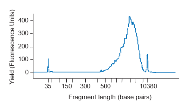
Figure 1. Example of cDNA trace from a HEK cell (19 PCR cycles), using an Agilent Bioanalyzer High Sensitivity DNA Analysis chip (Agilent). x-axis: cDNA yield (fluorescence units), y-axis: fragment length (base pairs). Data obtained from Picelli et al. 2025.3
7. cDNA Quantification (15 min.)
7.1 Allow the Quant-iT PicoGreen reagent to warm to room temperature before opening the vial. PicoGreen is light sensitive; while thawing, wrap in aluminum foil.
7.2 Prepare a 1× working solution of TE using 20× TE (supplied) and nuclease-free water.
7.3 Prepare a 1:800 dilution of PicoGreen solution and always use a plastic vessel (tubes, Falcon, etc.). Do not use glass as PicoGreen may adsorb to glass.
7.4 Prepare the standard curve using Lambda DNA standard (supplied at a concentration of 100ng/µl, with the PicoGreen kit) and 1x TE in 8 tubes, as below. The stock tubes can be used multiple times, keep any leftover in the fridge at +4 °C between experiments.
7.5 Vortex well and spin down the DNA standards before every use. Not vortexing thoroughly the standards is going to negatively affect the standard curve and your readings. Serial dilutions should be prepared as shown in the table below.
| Tube no. | Contents | Concentration | Final volume |
|---|---|---|---|
| 1 | 90 μl TE + 10 μl Lambda DNA stock | 10 ng/μl | 100 μl |
| 2 | 50 μl from Tube 1 + 50 μl TE | 5 ng/μl | 100 μl |
| 3 | 50 μl from Tube 2 + 50 μl TE | 2.5 ng/μl | 100 μl |
| 4 | 50 μl from Tube 3 + 50 μl TE | 1.25 ng/μl | 100 μl |
| 5 | 50 μl from Tube 4 + 50 μl TE | 0.625 ng/μl | 100 μl |
| 6 | 50 μl from Tube 5 + 50 μl TE | 0.3125 ng/μl | 100 μl |
| 7 | 50 μl from Tube 6 + 50 μl TE | 0.15625 ng/μl | 100 μl |
| 8 | TE only | blank | - |
7.6 Prepare the PicoGreen solution by pipetting 0.25 µl of PicoGreen dye + 99.5 µl of 1X TE for each sample. Vortex to mix.
7.7 Pipette 1 µl of each of the 7 standards + 1 Blank into a black, flat-bottom Nunc™ F96 MicroWell™ Plate. Place the standards on one column.
7.8 Pipet 1 µl of your samples into the center of each well of the Nunc™ F96 MicroWell™ Polystyrene Plate.
7.9 Add 99 µl of PicoGreen + TE mix into every well. There is no need to mix.
7.10 Cover the plate with the provided plastic (transparent) lid to prevent possible contaminations.
8. Plate Normalization (10 min.)
8.1 Prepare a normalization plate by adding 1 µl of purified cDNA and nuclease-free water to a final concentration of 200 pg/µl.
SAFE STOPPING POINT – Normalized cDNA can be stored for several months at -20 °C. LoBind plates must be used to avoid material losses upon long-term storage.
9. Tagmentation and Indexing PCR (1 h.)
9.1 Please note that the Tn5 transposase amount indicated below is a suggested starting point for tagmenting 200 pg/µl cDNA. Optimization might be necessary, depending on the specific activity of each batch of Tn5.
9.2 Prepare the Tagmentation Mix as described below:
| Reagent | Final Concentration | Volume (µl) |
|---|---|---|
| TAPS-Mg Buffer, pH=7.3 (5x) | 10 mM TAPS, 5 mM MgCl2 | 0.800 |
| Dimethylformamide (DMF)(100%) | 20% | 0.800 |
| Tn5 transposase (2 µM working dil.) | 62.5 nmol | 0.125 |
| Nuclease-free water | - | 2.275 |
| Total volume (µl) | 3.000 |
IMPORTANT! Dimethylformamide (DMF) is toxic and should be handled under the hood, or according to local safety regulations.
9.3 Dispense 3 µl of Tagmentation Mix in a new 384-well plate.
9.4 Add 1 µl of normalized cDNA (200 pg/µl) to each well containing the Tagmentation Mix.
9.5 Seal the plate, vortex, spin down, and carry out the tagmentation reaction: 55 °C for 8 min., 4 °C hold. Upon completion proceed immediately to the next step.
9.6 Add 1 µl of 0.2% SDS to each well. Seal the plate, vortex, spin down and incubate for 5 min. at room temperature. Do not put the plate back on ice.
9.7 Add 2 µl of prediluted N7xx + S5xx Index Adaptors (5 µM each).
9.8 Add 3 µl of Enrichment PCR Mix to each well:
| Reagent | Final Concentration | Volume (µl) |
|---|---|---|
| KAPA HiFi enzyme (1 U/μl) | 0.02 U/µl | 0.200 |
| KAPA HiFi Buffer (5x) | 1x | 2.000 |
| dNTPs (10 mM) | 300 mM | 0.300 |
| Nuclease-free water | - | 0.500 |
| Total volume (µl) | 3.000 |
9.9 Seal the plate vortex, spin down, and place it in a thermocycler and carry out the Enrichment PCR Reaction. Adjust the number of PCR cycles according to the number of processed cells.
| Step | Temperature | Time | Cycles | |
|---|---|---|---|---|
| Gap filling | 72 ºC | 3 min | 1x | |
| Enrichment PCR | Initial denaturation | 98 ºC | 30 sec | 1x |
| Denaturation | 98 ºC | 10 sec | 12x | |
| Annealing | 55 ºC | 30 sec | ||
| Elongation | 72 ºC | 30 sec | ||
| 15 ºC | hold | |||
SAFE STOPPING POINT - The final unpurified sequencing library can be stored for several months at -20 °C.
10. Library Cleanup and Quantification (45 min.)
10.1 Take an aliquot from each sample for the final library cleanup (i.e., 5 μl) and transfer it to a 1.5 ml Eppendorf tube. The rest of the library can be stored long-term at - 20 °C.
10.2 Remove the Sera-Mag SpeedBeads™ working solution (alternatively: AMPure XP beads or SPRI beads) from the +4 °C storage and equilibrate it at room temperature for 15 min.
10.3 Add Sera -Mag SpeedBeads™ working solution to a final ratio of 0.8× and mix well to homogenization. Use a different ratio if the goal is to recover longer (i.e., lower ratio) or shorter (i.e., higher ratio) fragments.
10.4 Incubate the tube off the magnetic stand for 5 min. at room temperature.
10.5 Place the tube on the magnetic stand and leave it for 5 min. or until the solution appears clear.
10.6 Remove the supernatant without disturbing the beads.
10.7 Recommended: wash the pellet with 1 ml of 80% v/v ethanol. Incubate for 30 sec. without removing the tube from the magnetic stand.
10.8 Remove any trace of ethanol and let the bead pellet dry for 2 min. Do not cap the tube or remove it from the magnetic stand during this time. Do not completely dry the beads.
10.9 Remove the tube from the magnetic stand, add 50 μl of nuclease-free water and mix well by pipetting or vortexing to resuspend the beads.
10.10 Incubate for 2 min. off the magnetic stand.
10.11 Place the tube back on the magnetic stand and incubate for 2 min. or until the solution appears clear.
10.12 Remove 49 µl of the supernatant and transfer it to a new 1.5-ml LoBind tube. Store the cDNA in a -20 °C freezer long-term or until ready for sequencing.
10.13 Use a Qubit fluorometer or a similar fluorimetric assay to quantify the library .Library yield can vary from 1 to 100 ng/μl depending on the number of cells being pooled as well as PCR cycles used.
10.14 Check the final library size on the Agilent Bioanalyzer, or similar equipment. Follow the instructions as described in the High Sensitivity DNA chip user manual.
10.15 Use the average size indicated on the Bioanalyzer and the concentration reported after Qubit measurement to determine the exact molarity required for sequencing. Example of trace shown in Figure 2.
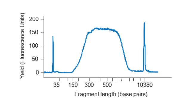
Figure 2. Example of trace of amplified library from a single HEK293 cell (19 cycles). Data obtained from Picelli et al. 2025.3
SAFE STOPPING POINT – The final purified sequencing library can be stored for several months at -20 °C.
11. Pooling and Sequencing
11.1 The purified library can be sequenced on any Illumina, Element Bio, or equivalent NGS sequencer. Follow the specifications reported for each instrument.
12. References
1. Khven et al . (2025) SEQURNA enhances FLASH-seq gene detection while eliminating DTT dependence. bioRxiv.12.12.693841; Benchmarking study demonstrating superior performance of SEQURNA Thermostable RNase inhibitor in FLASH-seq.
2. Hahaut et al. (2022) Fast and highly sensitive full-length single-cell RNA sequencing using FLASH-seq. Nat. Biotechnol. 40, 1447. Original FLASH-seq publication.
3. Picelli S. (2025) FLASH-seq with SEQURNA RNAse Inhibitor. Protocols.io. for FLASH-seq with SEQURNA, maintained by the Picelli lab.
4. Noble et al. (2024). Introducing synthetic thermostable RNase inhibitors to single-cell RNA-seq. Nat. Commun. 15, 8373. Original publication of the SEQURNA Thermostable RNase inhibitor.
5. Picelli S. (2019) Full-Length Single-Cell RNA Sequencing with Smart-seq2. Methods Mol. Biol. 1979, 25. Protocol including description of in-house preparation of SeraMag beads.
13. Frequently Asked Questions about SEQURNA
- What is SEQURNA?
SEQURNA is a synthetic thermostable RNase inhibitor made from a mixture of non-toxic organic molecules.
- What is SEQURNA used for?
It helps protect RNA from degradation during the preparation of single-cell libraries, leading to higher quality RNA-seq data. By improving the stability and integrity of RNA throughout the protocol, SEQURNA enhances reproducibility and throughput in single-cell transcriptomic studies.
- What are the advantages of using SEQURNA?
- It produces single-cell libraries of equal or superior quality compared to protein-based recombinant RNase inhibitors (RRIs).
- It shows robustness to various harsh treatments, such as pH changes, heating, freeze-thaw and vortexing.
- It does not require toxic reducing agents (like DTT or beta-mercaptoethanol)
- Why should I use SEQURNA instead of a recombinant RNase inhibitor (RRI)?
SEQURNA eliminates the need for any toxic reducing agents that are needed for conventional RRI functionality (e.g., DTT or beta-mercaptoethanol). SEQURNA is robust to various harsh treatments, such as pH changes, heating, freeze-thaw and vortexing.
- Can SEQURNA be used with protocols other than FLASH-seq?
SEQURNA can be used also with Smart-seq2 protocol, Smart-seq3 protocol and Smart-seq3xpress protocol.
- How does SEQURNA improve gene detection?
SEQURNA improves gene detection by acting as a synthetic thermostable RNase inhibitor that increases sensitivity and eliminates the need for destabilizing reducing agents.