
Mouse CXCL1/GRO alpha/KC/CINC-1 Antibody
R&D Systems | Catalog # AF-453-NA


Scientific images may be resized, cropped, or adjusted for brightness or contrast for presentation purposes. These updates are limited to presentation and do not affect the underlying scientific interpretation of the data. Where available, additional source data may be provided upon request.
Key Product Details
Species Reactivity
Validated:
Cited:
Applications
Validated:
Cited:
Label
Antibody Source
Product Specifications
Immunogen
Arg20-Lys96
Accession # P12850
Specificity
Clonality
Host
Isotype
Endotoxin Level
Scientific Data Images for Mouse CXCL1/GRO alpha/KC/CINC-1 Antibody
Chemotaxis Induced by CXCL1/KC and Neutralization by Mouse CXCL1/KC Antibody.
Recombinant Mouse CXCL1/KC (453-KC) chemoattracts the BaF3 mouse pro-B cell line transfected with human CXCR2 in a dose-dependent manner (orange line). The amount of cells that migrated through to the lower chemotaxis chamber was measured by Resazurin (AR002). Chemotaxis elicited by Recombinant Mouse CXCL1/KC (30 ng/mL) is neutralized (green line) by increasing concentrations of Goat Anti-Mouse CXCL1/KC Antigen Affinity-purified Polyclonal Antibody (Catalog # AF-453-NA). The ND50 is typically 0.3-1.5 µg/mL.
Mouse CXCL1 / GRO alpha / KC / CINC-1 ELISA Standard Curve
Recombinant Mouse CXCL1/GRO alpha /KC/CINC‑1 aa 20-96 (Catalog # 453-KC) was serially diluted and captured by Rat Anti-Mouse CXCL1/GRO alpha /KC/CINC‑1 Monoclonal Antibody (Catalog # MAB453) coated on a Clear Polystyrene Microplate (Catalog # DY990). Goat Anti-Mouse CXCL1/GRO alpha /KC/CINC‑1 Antigen Affinity-purified Polyclonal Antibody (Catalog # AF-453-NA) was biotinylated and incubated with the protein captured on the plate. Detection of the standard curve was achieved by incubating Streptavidin-HRP (Catalog # DY998)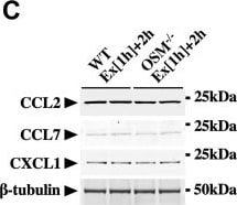
Detection of jn090xqgax6wba8rkwla.jpg by Western Blot
Effects of OSM on the protein expression of exercise-induced chemokines in the skeletal muscle.A, Western blot analysis of CCL2, CCL7, and CXCL1 in the skeletal muscle of sedentary mice (Sed [1 h] + 2 h) and mice at 2 h after the exercise (Ex [1 h] + 2 h). The apparent molecular weights are indicated on the right. B, quantitative analysis of the protein expression of CCL2, CCL7, and CXCL1. The band intensities of CCL2, CCL7, and CXCL1 were normalized to beta -tubulin and are represented as the fold induction relative to the intensities of sedentary mice in the bar graph. C, Western blot analysis of CCL2, CCL7, and CXCL1 in the skeletal muscle of WT and OSM−/− mice at 2 h after the exercise. The apparent molecular weights are indicated on the right. D, quantitative analysis of the protein expression of CCL2, CCL7, and CXCL1. The band intensities of CCL2, CCL7, and CXCL1 were normalized to beta -tubulin and are represented as the fold induction relative to the intensities of WT mice in the bar graph. E, Western blot analysis of CCL2, CCL7, and CXCL1 in the skeletal muscle of mice at 2 h after the injection of vehicle (Veh) or OSM. F, quantitative analysis of the protein expression of CCL2, CCL7, and CXCL1. The band intensities of CCL2, CCL7, and CXCL1 were normalized to beta -tubulin and are represented as the fold induction relative to the intensities of Veh group in the bar graph. Data are expressed as mean ± SD; n = 5 per group. ∗p < 0.05, ∗∗p < 0.01, and ∗∗∗p < 0.001. Student’s t test. CCL, CC chemokine ligand; CXCL1, CXC chemokine ligand 1; OSM, oncostatin M. Image collected and cropped by CiteAb from the following open publication (https://linkinghub.elsevier.com/retrieve/pii/S0021925822011292), licensed under a CC-BY license. Not internally tested by R&D Systems.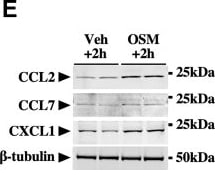
Detection of CXCL1/KC by Western Blot
Effects of OSM on the protein expression of exercise-induced chemokines in the skeletal muscle.A, Western blot analysis of CCL2, CCL7, and CXCL1 in the skeletal muscle of sedentary mice (Sed [1 h] + 2 h) and mice at 2 h after the exercise (Ex [1 h] + 2 h). The apparent molecular weights are indicated on the right. B, quantitative analysis of the protein expression of CCL2, CCL7, and CXCL1. The band intensities of CCL2, CCL7, and CXCL1 were normalized to beta -tubulin and are represented as the fold induction relative to the intensities of sedentary mice in the bar graph. C, Western blot analysis of CCL2, CCL7, and CXCL1 in the skeletal muscle of WT and OSM−/− mice at 2 h after the exercise. The apparent molecular weights are indicated on the right. D, quantitative analysis of the protein expression of CCL2, CCL7, and CXCL1. The band intensities of CCL2, CCL7, and CXCL1 were normalized to beta -tubulin and are represented as the fold induction relative to the intensities of WT mice in the bar graph. E, Western blot analysis of CCL2, CCL7, and CXCL1 in the skeletal muscle of mice at 2 h after the injection of vehicle (Veh) or OSM. F, quantitative analysis of the protein expression of CCL2, CCL7, and CXCL1. The band intensities of CCL2, CCL7, and CXCL1 were normalized to beta -tubulin and are represented as the fold induction relative to the intensities of Veh group in the bar graph. Data are expressed as mean ± SD; n = 5 per group. ∗p < 0.05, ∗∗p < 0.01, and ∗∗∗p < 0.001. Student’s t test. CCL, CC chemokine ligand; CXCL1, CXC chemokine ligand 1; OSM, oncostatin M. Image collected and cropped by CiteAb from the following open publication (https://linkinghub.elsevier.com/retrieve/pii/S0021925822011292), licensed under a CC-BY license. Not internally tested by R&D Systems.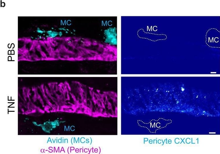
Detection of CXCL1/KC by Immunohistochemistry
Pericyte ICAM-1 and CXCL1 expression is increased in vessels with MCs but total expression is not impacted by IL-17A.a–d Cremaster muscles of WT or IL-17AKO mice were stimulated with TNF (300 ng) or PBS for 4 h. Cremaster muscles were collected and immunostained for MCs (avidin), pericytes ( alpha -SMA) and ICAM-1 or CXCL1. a–b Images of venules showing a ICAM-1 or b CXCL1 expression (pseudocolour intensity gradient) on pericytes (purple) and MCs (cyan), dashed lines the limit of MC, scale bars, 5 µm. c, d Quantification of c ICAM-1 MFI (n = 6 WT, n = 5 IL-17AKO mice) and d CXCL1 MFI (n = 5 mice) on pericytes. e, f Cremaster muscles of WT mice were stimulated with TNF (300 ng) or PBS in combination with a blocking anti-IL-17A or control mAbs (50 µg) for 4 h. e, f Quantification of e ICAM-1 MFI (n = 6 PBS Ctrl mAb, n = 9 TNF Ctrl mAb, n = 6 PBS Anti-IL-17A mAb, n = 9 TNF anti-IL-17A mAb mice) and f CXCL1 MFI (n = 4 PBS Ctrl mAb, n = 6 TNF Ctrl mAb, n = 4 PBS Anti-IL-17A mAb, n = 6 TNF anti-IL-17A mAb mice). Mean ± SEM (each mouse represents one independent experiment). c–f two-way ANOVA followed by Sidak’s post-hoc test. *p < 0.05, **p < 0.01, ***p < 0.001 as compared to WT or ctrl mAb (ns = not significant). Source data are provided as a Source Data file. Image collected and cropped by CiteAb from the following open publication (https://pubmed.ncbi.nlm.nih.gov/36396641), licensed under a CC-BY license. Not internally tested by R&D Systems.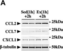
Detection of CXCL1/KC by Western Blot
Effects of OSM on the protein expression of exercise-induced chemokines in the skeletal muscle.A, Western blot analysis of CCL2, CCL7, and CXCL1 in the skeletal muscle of sedentary mice (Sed [1 h] + 2 h) and mice at 2 h after the exercise (Ex [1 h] + 2 h). The apparent molecular weights are indicated on the right. B, quantitative analysis of the protein expression of CCL2, CCL7, and CXCL1. The band intensities of CCL2, CCL7, and CXCL1 were normalized to beta -tubulin and are represented as the fold induction relative to the intensities of sedentary mice in the bar graph. C, Western blot analysis of CCL2, CCL7, and CXCL1 in the skeletal muscle of WT and OSM−/− mice at 2 h after the exercise. The apparent molecular weights are indicated on the right. D, quantitative analysis of the protein expression of CCL2, CCL7, and CXCL1. The band intensities of CCL2, CCL7, and CXCL1 were normalized to beta -tubulin and are represented as the fold induction relative to the intensities of WT mice in the bar graph. E, Western blot analysis of CCL2, CCL7, and CXCL1 in the skeletal muscle of mice at 2 h after the injection of vehicle (Veh) or OSM. F, quantitative analysis of the protein expression of CCL2, CCL7, and CXCL1. The band intensities of CCL2, CCL7, and CXCL1 were normalized to beta -tubulin and are represented as the fold induction relative to the intensities of Veh group in the bar graph. Data are expressed as mean ± SD; n = 5 per group. ∗p < 0.05, ∗∗p < 0.01, and ∗∗∗p < 0.001. Student’s t test. CCL, CC chemokine ligand; CXCL1, CXC chemokine ligand 1; OSM, oncostatin M. Image collected and cropped by CiteAb from the following open publication (https://linkinghub.elsevier.com/retrieve/pii/S0021925822011292), licensed under a CC-BY license. Not internally tested by R&D Systems.Applications for Mouse CXCL1/GRO alpha/KC/CINC-1 Antibody
Western Blot
Sample: Recombinant Mouse CXCL1/GRO alpha /KC/CINC‑1 aa 20-96 (Catalog # 453-KC)
Neutralization
Formulation, Preparation, and Storage
Purification
Reconstitution
Reconstitute at 0.2 mg/mL in sterile PBS. For liquid material, refer to CoA for concentration.
Formulation
Shipping
Stability & Storage
- 12 months from date of receipt, -20 to -70 °C as supplied.
- 1 month, 2 to 8 °C under sterile conditions after reconstitution.
- 6 months, -20 to -70 °C under sterile conditions after reconstitution.
Calculators
Background: CXCL1/GRO alpha/KC/CINC-1
Alternate Names
Gene Symbol
UniProt
Additional CXCL1/GRO alpha/KC/CINC-1 Products
Product Documents for Mouse CXCL1/GRO alpha/KC/CINC-1 Antibody
Certificate of Analysis
To download a Certificate of Analysis, please enter a lot or batch number in the search box below.
Note: Certificate of Analysis not available for kit components.
Product Specific Notices for Mouse CXCL1/GRO alpha/KC/CINC-1 Antibody
For research use only
Related Research Areas
Citations for Mouse CXCL1/GRO alpha/KC/CINC-1 Antibody
 Powered by Bioz
Customer Reviews for Mouse CXCL1/GRO alpha/KC/CINC-1 Antibody
There are currently no reviews for this product. Be the first to review Mouse CXCL1/GRO alpha/KC/CINC-1 Antibody and earn rewards!
Have you used Mouse CXCL1/GRO alpha/KC/CINC-1 Antibody?
Submit a review and receive an Amazon gift card!
$25/€18/£15/$25CAN/¥2500 for a review with an image
$10/€7/£6/$10CAN/¥1110 for a review without an image
Submit a review
Protocols
Find general support by application which include: protocols, troubleshooting, illustrated assays, videos and webinars.
- Cellular Response to Hypoxia Protocols
- R&D Systems Quality Control Western Blot Protocol
- Troubleshooting Guide: Western Blot Figures
- Western Blot Conditions
- Western Blot Protocol
- Western Blot Protocol for Cell Lysates
- Western Blot Troubleshooting
- Western Blot Troubleshooting Guide
- View all Protocols, Troubleshooting, Illustrated assays and Webinars
Associated Pathways
