Human/Mouse/Rat SOX2 Antibody Summary
Gly135-Met317
Accession # P48431
Applications
Please Note: Optimal dilutions should be determined by each laboratory for each application. General Protocols are available in the Technical Information section on our website.
Scientific Data
 View Larger
Detection of Human, Mouse, and Rat SOX2 by Western Blot. Western blot shows lysates of D3 mouse embryonic stem cell line, NTera-2 human testicular embryonic carcinoma cell line, F9 mouse teratocarcinoma stem cells, and rat cortical stem cells. PVDF membrane was probed with 1 µg/mL of Goat Anti-Human/Mouse/Rat SOX2 Antigen Affinity-purified Polyclonal Antibody (Catalog # AF2018) followed by HRP-conjugated Anti-Goat IgG Secondary Antibody (HAF017). A specific band was detected for SOX2 at approximately 36 kDa (as indicated). This experiment was conducted under reducing conditions and using Immunoblot Buffer Group 1.
 View Larger
Detection of SOX2-regulated Genes by Chromatin Immunoprecipitation. BG01V human embryonic stem cells were fixed using formaldehyde, resuspended in lysis buffer, and sonicated to shear chromatin. SOX2/DNA complexes were immunoprecipitated using 5 µg Goat Anti-Human/Mouse/Rat SOX2 Antigen Affinity-purified Polyclonal Antibody (Catalog # AF2018) or control antibody (AB-108-C) for 15 minutes in an ultrasonic bath, followed by Biotinylated Anti-Goat IgG Secondary Antibody (BAF109). Immunocomplexes were captured using 50 µL of MagCellect Streptavidin Ferrofluid (MAG999) and DNA was purified using chelating resin solution. Thenanogpromoter was detected by standard PCR.
.") View Larger
View Larger
SOX2 in Mouse Cortical Stem Cells. SOX2 was detected in immersion fixed undifferentiated mouse cortical stem cells using Goat Anti-Human/Mouse/Rat SOX2 Antigen Affinity-purified Polyclonal Antibody (Catalog # AF2018) at 10 µg/mL for 3 hours at room temperature. Cells were stained using the NorthernLights™ 557-conjugated Anti-Goat IgG Secondary Antibody (red; (NL001) and counterstained with DAPI (blue). Nestin was also detected in stem cells using Mouse Anti-Mouse/Rat Nestin Monoclonal Antibody (MAB2736) and co-stained using the NorthernLights™ 493-conjugated Anti-Mouse IgG Secondary Antibody (green; NL009). Specific staining of SOX2 was localized to nuclei. View our protocol for Fluorescent ICC Staining of Stem Cells on Coverslips.
.") View Larger
View Larger
SOX2 in Rat Cortical Stem Cells. SOX2 was detected in immersion fixed undifferentiated rat cortical stem cells using Goat Anti-Human/Mouse/Rat SOX2 Antigen Affinity-purified Polyclonal Antibody (Catalog # AF2018) at 10 µg/mL for 3 hours at room temperature. Cells were stained using the NorthernLights™ 557-conjugated Anti-Goat IgG Secondary Antibody (red; NL001) and counterstained with DAPI (blue). Nestin was also detected using Mouse Anti-Mouse/Rat Nestin Monoclonal Antibody (MAB2736) and stained using the NorthernLights™ 493-conjugated Anti-Mouse IgG Secondary Antibody (green; NL009). Specific staining of SOX2 was localized to nuclei. View our protocol for Fluorescent ICC Staining of Stem Cells on Coverslips.
 View Larger
Detection of Human SOX2 by Simple WesternTM. Simple Western lane view shows lysates of BG01V human embryonic stem cells and NTera-2 human testicular embryonic carcinoma cell line, loaded at 0.2 mg/mL. A specific band was detected for SOX2 at approximately 53 kDa (as indicated) using 5 µg/mL of Goat Anti-Human/Mouse/Rat SOX2 Antigen Affinity-purified Polyclonal Antibody (Catalog # AF2018) followed by 1:50 dilution of HRP-conjugated Anti-Goat IgG Secondary Antibody (HAF109). This experiment was conducted under reducing conditions and using the 12-230 kDa separation system.
.") View Larger
View Larger
SOX2 in ADLF1 and FAB2 Stem Cell Lines. SOX2 was detected in immersion fixed ADLF1 (top panel) and FAB2 (bottom panel) induced pluripotent stem cell lines using Goat Anti-Human/Mouse/Rat SOX2 Antigen Affinity-purified Polyclonal Antibody (Catalog # AF2018) at 10 µg/mL for 3 hours at room temperature. Cells were stained using the NorthernLights™ 557-conjugated Anti-Goat IgG Secondary Antibody (red; NL001) and counterstained with DAPI (blue). Specific staining was localized to nuclei. View our protocol for Fluorescent ICC Staining of Stem Cells on Coverslips.
 View Larger
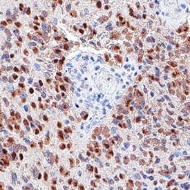
Detection of SOX2 in glioblastoma. SOX2 was detected in immersion fixed paraffin-embedded sections of glioblastoma using Goat Anti-Human/Mouse/Rat SOX2 Antigen Affinity-purified Polyclonal Antibody (Catalog # AF2018) at 3 µg/mL for 1 hour at room temperature followed by incubation with the Anti-Goat IgG VisUCyte™ HRP Polymer Antibody (Catalog # VC004). Before incubation with the primary antibody, tissue was subjected to heat-induced epitope retrieval using VisUCyte Antigen Retrieval Reagent-Basic (Catalog # VCTS021). Tissue was stained using DAB (brown) and counterstained with hematoxylin (blue). Specific staining was localized to nuclei in cancer cells. View our protocol for IHC Staining with VisUCyte HRP Polymer Detection Reagents.
 View Larger
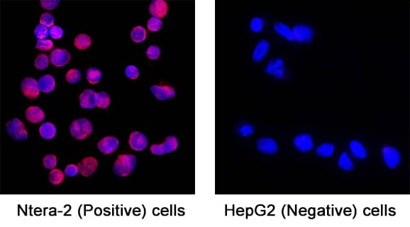
Detection of SOX2 in NTera‑2 cells (Positive) & HepG2 cells (Negative). SOX2 was detected in immersion fixed NTera‑2 human testicular embryonic carcinoma cells (Positive) & absent in HepG2 human hepatocellular carcinoma cells (Negative) using Goat Anti-Human/Mouse/Rat SOX2 Antigen Affinity-purified Polyclonal Antibody (Catalog # AF2018) at 5 µg/mL for 3 hours at room temperature. Cells were stained using the NorthernLights™ 557-conjugated Anti-Goat IgG Secondary Antibody (red; Catalog # NL001) and counterstained with DAPI (blue). Specific staining was localized to Cell surface and nuclear. View our protocol for Fluorescent ICC Staining of Cells on Coverslips.
 View Larger
Detection of Human SOX2 by Western Blot Knockdown OGT inhibits proliferation and tumorsphere formation of hepatoma cell through reducing eIF4E expression. A, Huh7 and PLC/PRF/5 cells were infected with control shRNA, OGT shRNA2 alone, or with wild‐type eIF4E lentivirus. The cell lysates were harvested for western blotting analysis using indicated antibodies. beta ‐actin expression was served as a loading control. B, Cell proliferation of Huh7 and PLC/PRF/5 cells infected with lentiviruses as in panel (A) were measured with CCK8 assay. (C‐H) Huh7 and PLC/PRF/5 cells infected with lentiviruses as in panel (A) were seeded into 96‐well plates. After 12 d, tumorsphere were counted and quantified. Representative images of sphere (scale bars, 100 μm) were shown (C, F). The diameter of sphere (D, G) and number of sphere (E, H) were count. Data represent mean ± SD of at least three independent experiments. The two‐tailed Student's t tests were used. **P < 0.01. I, Huh7 cells expressing either OGT shRNA2 alone or with wild‐type eIF4E lentivirus were incubated with PE‐labelled anti‐AC133 antibody. The percentages of CD133+ cells in graphs were analysed by flow cytometry. Black line, control IgG staining; red line, CD133 staining. J, Cell lysates were examined by western blotting with indicated antibodies. The right panel showcases relative protein amounts of different groups. Error bars represent ±SD of triplicate experiments. **P < 0.01; n.s, no significance. K, Huh7 cells were collected and subjected to immunoprecipitation with antibody against eIF4E or normal mouse IgG. Total RNAs were purified from immunocomplexes and subjected to RT‐PCR to measure Sox2, OCT4, and KLF4 mRNAs associated with eIF4E Image collected and cropped by CiteAb from the following publication (https://pubmed.ncbi.nlm.nih.gov/30677218), licensed under a CC-BY license. Not internally tested by R&D Systems.
 View Larger
Detection of Human SOX2 by Immunohistochemistry TP63 and SOX2 staining in cervical biopsies.Immunohistochemical staining of cervical biopsies. Bars = 50µm.(A) normal cervix no primary control; (B) normal cervix stained with anti-SOX2; (C) Squamous cell cervical carcinoma no primary control; (D) representative TP63, and (E) representative SOX2, staining of tumour cells. For both TP63 and SOX2 staining was seen in the nucleus of positive cells (examples indicated by solid arrows); negative cells were a minority of tumour cells (examples indicated by unfilled arrows). (F) Image analysis results of % nuclear +ve tumour cells in biopsies. Parallel sections from 11 cases were stained with SOX2 and TP63. Tumour cells were evaluated for their nuclear expression of the transcription factors. There was no significant difference between the data for SOX2 and TP63 (Wilcoxon signed rank test). Image collected and cropped by CiteAb from the following publication (https://pubmed.ncbi.nlm.nih.gov/25531390), licensed under a CC-BY license. Not internally tested by R&D Systems.
 View Larger
Detection of Zebrafish SOX2 by Immunohistochemistry Direct effects of glucocorticoids in ependymal glia. (A–I) Zebrafish spinal cord transverse sections. (A–C) Immunostaining of wild-types for Nr3c1 (green) and Gfap (red), plus DAPI staining (blue) with Dex treatment at 24 h post sham injury (A), 24 h (B) and 48 h post SCI (C). Nuclear Nr3c1 (active) localization in Gfap-positive cells around the central canal is stimulated with Dex treatment. (D–F) Immunostaining for EGFP (pan-cytoplasmic) in the SR4G transgenic reporter line (green) and filamentous Gfap (red) at 24 h post sham injury (D) and SCI with no treatment (E) or 24 h post SCI +Dex (F). Nr3c1 signaling is constitutively active in ependymal glia in uninjured controls (D), diminished after SCI (E), and was maintained after SCI + Dex. (G–I) Immunostaining for Sox2 (green), PCNA (red) and Gfap (blue) from 48 h post sham injury (G), 48 h post SCI followed by no treatment (H), or 48 h post SCI +Dex (I). Sox2 immunoreactivity is reduced in ependymal glia and neural precursor cells after Dex treatment. Representative images from 10 spinal cords per time point in each condition. Scale bars, 20 μm (A is the same as B,C; D is the same as E,F; G is the same as H,I). ∗denotes central canal. Image collected and cropped by CiteAb from the following publication (https://pubmed.ncbi.nlm.nih.gov/31069223), licensed under a CC-BY license. Not internally tested by R&D Systems.
 View Larger
Detection of Mouse SOX2 by Chromatin Immunoprecipitation Epigenetic perturbation of enhancer clustering and genome-widebinding.(A) The fluctuation range (x) and amplitude (y) were obtainedby fitting the pair-correlation function of the indicated dataset with thefluctuation model. Figure 2 and Figure 2—figure supplement 1,Equations10–13. Supplementary file 1. Data from the same conditionwere grouped in separate ellipses. (B) Sox2 ChIP-exo peakdensity distribution in the wild-type and TSA treated (red dotted) cellsacross chromosome 1, 2, 3. In the upper panels, each chromosome was dividedto 500 bins. The color map correlates with the number of peaks in each bin.Top 7000 binding sites were considered in each condition. (C)Cumulative density histogram of the distances to transcription start sites(TSS's) of Sox2 ChIP-exo peaks in WT, Sox2 ChIP-exo peaks in the TSA treatedcells (red dotted), and random genomic positions (gray).DOI:https://dx.doi.org/10.7554/eLife.04236.013 Image collected and cropped by CiteAb from the following publication (https://pubmed.ncbi.nlm.nih.gov/25537195), licensed under a CC-BY license. Not internally tested by R&D Systems.
 View Larger
Detection of Human SOX2 by Immunohistochemistry TP63 and SOX2 staining in cervical biopsies.Immunohistochemical staining of cervical biopsies. Bars = 50µm.(A) normal cervix no primary control; (B) normal cervix stained with anti-SOX2; (C) Squamous cell cervical carcinoma no primary control; (D) representative TP63, and (E) representative SOX2, staining of tumour cells. For both TP63 and SOX2 staining was seen in the nucleus of positive cells (examples indicated by solid arrows); negative cells were a minority of tumour cells (examples indicated by unfilled arrows). (F) Image analysis results of % nuclear +ve tumour cells in biopsies. Parallel sections from 11 cases were stained with SOX2 and TP63. Tumour cells were evaluated for their nuclear expression of the transcription factors. There was no significant difference between the data for SOX2 and TP63 (Wilcoxon signed rank test). Image collected and cropped by CiteAb from the following publication (https://pubmed.ncbi.nlm.nih.gov/25531390), licensed under a CC-BY license. Not internally tested by R&D Systems.
 View Larger
Detection of Human SOX2 by Immunocytochemistry/Immunofluorescence Generation of KCNA5 knockout model. (A) Illustration of polycistronic self-replicating RNA used for reprogramming (Yoshioka et al., 2013). Reading frames for OCT4, KLF4, SOX2, GLIS1, and puromycin resistance are separated by 2A peptide-encoding or IRES sequences. (B) Phase contrast morphology (5 x) and alkaline phosphatase staining of the F1 wild-type hiPSC line generated using the RNA vector shown in (A). (C) Representative F1 karyogram indicating a normal male genotype. (D) The RNA vector is undetectable in established F1 hiPSCs following puromycin withdrawal, whereas freshly transfected parental fibroblasts display robust transgene expression (RT-qPCR data, n = 2). (E) Bisulfite sequencing of OCT4 and NANOG promoter regions of the indicated samples showing an hESC-like hypomethylated state in F1 cells. (F) RT-qPCR analysis of F1 hiPSCs in comparison to the original fibroblasts and a hESC control (n = 2). (G) Immunostains showing robust pluripotency factor expression in F1 hiPSCs at the protein level. (H) Illustration of the Kv1.5 channel. The voltage sensor in transmembrane helix S4 is indicated by symbols. The ion pore region is in-between segments S5 and S6. (I)KCNA5 knockout strategy based on simultaneously transfecting 4 CRISPR/Cas9 nickase vectors targeting the 5' end of the 1-exon gene body. A mutant clone generated this way displayed homozygous deletions causing a frame shift and premature stop as illustrated at the bottom. (J) PCR spanning the mutation site validates the sequencing-verified frame shift-causing deletion in KCNA5fs/fs cells at the DNA and RNA levels. Image collected and cropped by CiteAb from the following publication (https://journal.frontiersin.org/article/10.3389/fphys.2017.00469/full), licensed under a CC-BY license. Not internally tested by R&D Systems.
 View Larger
Detection of Human SOX2 by Western Blot Construction and expression of srRNA.A: Scheme of Self-Replicating srRNA constructs. B: Expression of reprogramming factors in OKS-iM 4F-srRNA, OKS-iG 4F-srRNA, OKS-iGM 5F-srRNA, and OKS-iGML 6F-srRNA on day 2 (left) and day 10 (right). BJ cells were co-transfected with srRNAs plus B18R mRNA (1:1 srRNA:B18R mRNA), selected with puromycin and cultured in Advanced DMEM/10% FBS for 10 days. C: qRT-PCR analysis of LIN28A and NANOG from 4F-srRNA and 5F-srRNA transfected BJ cells normalized to GAPDH. Cells were selected with puromycin after the transfection, and cultured in Advanced DMEM/10% FBS (Ad) or ES medium. Cells were collected on day 2, 4, 6, 8 and 10 for 5F-srRNA, and day 10 for 4F-srRNA (OKS-iM and OKS-iG). D: Immunoblot analysis of LIN28A and NANOG in 5F-srRNA transfected BJ cells on day 6 and 10. iPSC: iPSC clone generated with 5F-srRNA from BJ cells. Image collected and cropped by CiteAb from the following publication (https://pubmed.ncbi.nlm.nih.gov/28750082), licensed under a CC-BY license. Not internally tested by R&D Systems.
 View Larger
Detection of Human Human/Mouse/Rat SOX2 Antibody by Immunocytochemistry/ Immunofluorescence The Majority of Ferret and Human, but Not Mouse, Sox2-Positive Tbr2-Negative BPs Exhibit Nuclear YAP. (G and H) that are shown at higher magnification (F′, F″, G′, G″, H′, and H″), as indicated; selected Tbr2-positive nuclei that are YAP negative in mouse, ferret, and human are outlined by white lines.(I–K) Quantification of the percentage of Tbr2-negative nuclei in the VZ (I), Tbr2-positive nuclei in the VZ (J), and Tbr2-positive nuclei in the SVZ (K) that are YAP positive in mouse E14.5, ferret E36, and human 11 wpc neocortex. Two or three images per embryo-fetus were taken, 30 randomly picked Tbr2-negative nuclei in the VZ (I) and Tbr2-positive nuclei in the VZ (J) and SVZ (K) were scored per image, and the values obtained were averaged for each embryo-fetus. Data are the mean of three or four embryos-fetuses.(A–C and F–H) Images are 1-μm optical sections. Scale bars represent 50 μm (A–C and F–H), 10 μm (A′, B′, and C′), and 20 μm (F′, F″, G′, G″, H′, and H″).(D, E, and I–K) Error bars indicate SEM; ∗∗p < 0.01, ∗∗∗p < 0.001 (one-way ANOVA, post hoc Tukey HSD).See also Figures S1 and S2. Image collected and cropped by CiteAb from the following publication (https://pubmed.ncbi.nlm.nih.gov/31018127), licensed under a CC-BY license. Not internally tested by R&D Systems.
 View Larger
Detection of Human Human/Mouse/Rat SOX2 Antibody by Immunocytochemistry/ Immunofluorescence The Majority of Ferret and Human, but Not Mouse, Sox2-Positive Tbr2-Negative BPs Exhibit Nuclear YAP(A–C) Double immunofluorescence for YAP (green) and Sox2 (magenta), combined with DAPI staining (white), of mouse E14.5 (A), ferret E36 (B), and human 14 wpc (C) neocortex. Boxes indicate areas in the SVZ (A) and oSVZ (B and C) that are shown at higher magnification (A′, B′, and C′); selected Sox2-positive nuclei that are YAP negative in mouse and YAP positive in ferret and human are outlined by white lines; arrowheads indicate a YAP-positive basal process of a bRG. Image collected and cropped by CiteAb from the following publication (https://pubmed.ncbi.nlm.nih.gov/31018127), licensed under a CC-BY license. Not internally tested by R&D Systems.
 View Larger
Detection of Mouse Human/Mouse/Rat SOX2 Antibody by Immunocytochemistry/ Immunofluorescence The Majority of Ferret and Human, but Not Mouse, Sox2-Positive Tbr2-Negative BPs Exhibit Nuclear YAP.(F–H) Double immunofluorescence for YAP (green) and Tbr2 (magenta), combined with DAPI staining (white), of mouse E14.5 (F), ferret E36 (G), and human 11 wpc (H) neocortex. Boxes indicate areas in the VZ and SVZ (F) or iSVZ (G and H) that are shown at higher magnification (F′, F″, G′, G″, H′, and H″), as indicated; selected Tbr2-positive nuclei that are YAP negative in mouse, ferret, and human are outlined by white lines.(I–K) Quantification of the percentage of Tbr2-negative nuclei in the VZ (I), Tbr2-positive nuclei in the VZ (J), and Tbr2-positive nuclei in the SVZ (K) that are YAP positive in mouse E14.5, ferret E36, and human 11 wpc neocortex. Two or three images per embryo-fetus were taken, 30 randomly picked Tbr2-negative nuclei in the VZ (I) and Tbr2-positive nuclei in the VZ (J) and SVZ (K) were scored per image, and the values obtained were averaged for each embryo-fetus. Data are the mean of three or four embryos-fetuses.(A–C and F–H) Images are 1-μm optical sections. Scale bars represent 50 μm (A–C and F–H), 10 μm (A′, B′, and C′), and 20 μm (F′, F″, G′, G″, H′, and H″).(D, E, and I–K) Error bars indicate SEM; ∗∗p < 0.01, ∗∗∗p < 0.001 (one-way ANOVA, post hoc Tukey HSD).See also Figures S1 and S2. Image collected and cropped by CiteAb from the following publication (https://pubmed.ncbi.nlm.nih.gov/31018127), licensed under a CC-BY license. Not internally tested by R&D Systems.
 View Larger
Detection of Human Human/Mouse/Rat SOX2 Antibody by Immunocytochemistry/ Immunofluorescence The Majority of Ferret and Human, but Not Mouse, Sox2-Positive Tbr2-Negative BPs Exhibit Nuclear YAP(A–C) Double immunofluorescence for YAP (green) and Sox2 (magenta), combined with DAPI staining (white), of mouse E14.5 (A), ferret E36 (B), and human 14 wpc (C) neocortex. Boxes indicate areas in the SVZ (A) and oSVZ (B and C) that are shown at higher magnification (A′, B′, and C′); selected Sox2-positive nuclei that are YAP negative in mouse and YAP positive in ferret and human are outlined by white lines; arrowheads indicate a YAP-positive basal process of a bRG.Image collected and cropped by CiteAb from the following publication (https://pubmed.ncbi.nlm.nih.gov/31018127), licensed under a CC-BY license. Not internally tested by R&D Systems.
 View Larger
Detection of Mouse SOX2 by Immunohistochemistry Immunohistochemical analysis of Trp53-/-Notch1-/-Notch2-/- and Trp53-/-Rbpj-/- tumors. (A) A Trp53-/-Notch1-/-Notch2-/- tumor in the SVZ. Streams of tumor cells invading the brain parenchyma are indicated by arrowheads. The GFP expression from the Rosa-CAG::GFP Cre-reporter indicates derivation from Hes5+ cells. (B) Expression of GFP, progenitor (SOX2) and glial markers (OLIG2, GFAP), as well as staining for mitotically active cells (PCNA) and immature neurons (DCX) in Trp53-/-Notch1-/-Notch2-/- and Trp53-/-Rbpj-/- tumors. Image collected and cropped by CiteAb from the following open publication (https://pubmed.ncbi.nlm.nih.gov/36358826), licensed under a CC-BY license. Not internally tested by R&D Systems.
 View Larger
Detection of Mouse SOX2 by Immunocytochemistry/ Immunofluorescence Deregulation of SOX2, ASCL1&OLIG2 TFs in p27KO cultures at the onset of differentiation. e Immunocytochemistry for p27 (red) in combination with ASCL1, OLIG2 or SOX2 (green) in 2 + 1DIV NPCs. Image collected & cropped by CiteAb from the following open publication (https://pubmed.ncbi.nlm.nih.gov/36627412), licensed under a CC-BY license. Not internally tested by R&D Systems.
 View Larger
Detection of Mouse SOX2 by Immunocytochemistry/ Immunofluorescence Padi2/3 DKO TSCs exhibit diminished stem cell potential&are prone to differentiate into large trophoblast giant cells (TGCs). (D) Immunofluorescence staining on WT&Padi2/3 DKO TSCs for trophoblast stem cell markers CDX2&SOX2. Image collected & cropped by CiteAb from the following open publication (https://pubmed.ncbi.nlm.nih.gov/36010543), licensed under a CC-BY license. Not internally tested by R&D Systems.
 View Larger
Detection of Xenopus SOX2 by Immunohistochemistry Whole-Mount Unedited images from samples featured in Figure 6.Stage 39–40 larvae were amputated and processed for whole-mount immunostaining at the indicated hour post amputation (hpa), with the exception of the 0 h group, which was first fixed and then amputated to represent the pre-amputation group. Images show representative total z-projections of whole-mount immunostained samples for each group corresponding to the edited images shown in Figure 6. Transverse red dashed line indicates amputation plane. Scale bar, 100 µm. Image collected and cropped by CiteAb from the following open publication (https://pubmed.ncbi.nlm.nih.gov/33955353), licensed under a CC-BY license. Not internally tested by R&D Systems.
 View Larger
Detection of Mouse SOX2 by Immunohistochemistry Notch1 and Trp53 deletion in Hes5+ cells leads to formation of forebrain tumors in adult mice. (A) Kaplan–Meier curves showing survival of Hes5::CreERT2 Trp53-/-Notch1-/- and Trp53-/-Notch2-/- mutant mice. (B) A Trp53-/-Notch1-/- tumor in the forebrain. The GFP expression from the Rosa-CAG::GFP Cre-reporter indicates derivation from Hes5+ cells. (C) Percentages of Trp53-/-Notch1-/- and Trp53-/-Notch2-/- mice that developed hyperplasia or tumors in the brain. (D) A Trp53-/-Notch1-/- tumor (dashed line) in the anterior forebrain. In contrast, the forebrain of Trp53-/-Notch2-/- mice appears grossly normal. (E) Expression of GFP, progenitor (SOX2) and glial markers (OLIG2, GFAP), as well as staining for mitotically active cells (PCNA) and immature neurons (DCX) in Trp53-/-Notch1-/- tumors. Image collected and cropped by CiteAb from the following open publication (https://pubmed.ncbi.nlm.nih.gov/36358826), licensed under a CC-BY license. Not internally tested by R&D Systems.
 View Larger
Detection of Mouse SOX2 by Western Blot Wnt signaling downregulates Tcf3 expression in mouse ESCs.A. qRT-PCR analysis of Tcf3 in wild type, ApcNN and ApcMin/Min ESCs. Actb was used as an internal control; bars represent n = 2 ± SD. B. Western blot analysis of the core pluripotency markers Oct4, Nanog, Sox2 and Tcf3 on protein lysates isolated from two independent ApcNN clones and wild type control ESCs. Actb and Tubulin were used as an internal control. C–D. qRT-PCR analysis of Tcf3 in wild type ESCs treated for different time intervals with Wnt3a conditioned medium (C), or with the GSK-inhibitor SB-216763 (D). L-medium and DMSO were employed as controls, respectively. Actb was used as an internal control; bars represent n = 2 ± SD. E. Time course western blot analysis of Tcf3 expression in wild type ESCs treated with Wnt3a conditioned medium (Wnt3a CM) or control L-medium (LM). Actb was used as an internal control. F. qRT-PCR analysis of Tcf3 and Wnt target genes Axin2 and Cdx1 in wild type ESC treated for 48 h with different concentrations of GSK-inhibitor SB-216763 or DMSO as control. Actb was used as an internal control; bars represent n = 2 ± SD. Image collected and cropped by CiteAb from the following open publication (https://pubmed.ncbi.nlm.nih.gov/23658527), licensed under a CC-BY license. Not internally tested by R&D Systems.
 View Larger
Detection of Human SOX2 by Immunohistochemistry Fetal rat lung stained for Sox2 (red), co-stained with DAPI (blue). Image from a verified customer review.
 View Larger
Detection of SOX2 by Flow Cytometry SOX2, OCT2, and PRRX1 are downstream effectors of the SETD2‐regulated EMT program. (A) Expression of SOX2, OCT2, and PRRX1 in TGF‐ beta ‐treated WT (72 h), SETD2 KO, and SETD2 rescue tested by RT‐qPCR (run in triplicate). (B) Migration capacity by wound healing assay, (C) invasiveness by transwell assay, and (D) stemness by 3D spheroid formation assay in RPTEC WT GFP (control vector), SETD2 KO1 and KO2, and SOX2/OCT2/PRRX1‐transduced WT RPTEC lines. Images are taken at 4× magnification, scale bar: 1000 μm for (B) and (D) and at 2.5× magnification, scale bar: 1200 μm for (C). Data are represented as mean ± SEM for triplicate reactions for B–D. P‐value is calculated by one‐way ANOVA in (A), (B), and (D). Two‐way ANOVA is used for statistical test for (C). ****P < 0.0001; ***P < 0.001; **P < 0.01; *P < 0.05; ns, P ≥ 0.05. (E) Model of the SETD2 loss‐driven EMT program through cell intrinsic (transcriptional) and cell extrinsic (paracrine) mechanisms. Image collected and cropped by CiteAb from the following open publication (https://pubmed.ncbi.nlm.nih.gov/37418588), licensed under a CC-BY license. Not internally tested by R&D Systems.
 View Larger
Detection of SOX2 by Flow Cytometry Temporally coordinated expression of Sox2, Tbr2, NueroD, and Prox1 in gfap-GFP+ GNPs and differentiating granule neurons Gfap-GFP+/Sox2+ progenitor cells were found at the dentate neuroepithelium at E14 (arrow in A1). Sox2 expression was found at the VZ. Tbr2+ GNPs are then observed (red, arrow in A2). Tbr2 expression is followed by NeuroD expression (A3). Tbr2+/NeuroD+ GNPs are found (arrow, A3). The Tbr2+ GNPs (red) migrate toward the DG primordium and co-express Prox1 (green), a marker for early differentiating dentate granule neurons (arrow in A4). Gfap-GFP+ progenitors sequentially express Sox2, Tbr2, NeuroD1, and then, Prox1 in the developing DG at E16 (arrows in B1–B4, respectively). Some Sox2+/Tbr2+, Tbr2+/NeuroD+, and Tbr2+/Prox1+ GNPs are found. Similarly, gfap-GFP+ progenitor cells contribute to Tbr2+/NeuroD+ (arrows in D1,D2), Prox1+ GNPs (arrows in D3,D4) at P3. Schematic drawing of the expression patterns of gfap-GFP and transcription factors during the differentiation of GNPs (E). Scale bars; 200 μm in (A1–A4, B1–B4, C1–C3, D1,D3); 50 μm in (D2,D4). Image collected and cropped by CiteAb from the following open publication (https://www.frontiersin.org/articles/10.3389/fnins.2024.1425849/full), licensed under a CC-BY license. Not internally tested by R&D Systems.
Reconstitution Calculator
Preparation and Storage
- 12 months from date of receipt, -20 to -70 °C as supplied.
- 1 month, 2 to 8 °C under sterile conditions after reconstitution.
- 6 months, -20 to -70 °C under sterile conditions after reconstitution.
Background: SOX2
SOX2 belongs to the SOX (SRY-like HMG box) family of transcription factors with diverse roles in development. SOX2 functions in specifying the first three lineages present at implantation and in regulating proliferation and differentiation in the developing peripheral nervous system (1-5).
- Graham, V. et al. (2003) Neuron 39:749.
- Avilion, A.A. et al. (2003) Genes Dev. 17:126.
- Kishi, M. et al. (2000) Development 127:791.
- Yuan, H. et al. (1995) Genes Dev. 9:2635.
- Uwanogho, D. et al. (1995) Mech. Dev. 49:23.
- Stevanovic, M. (2003) Mol. Biol. Rep. 30:127.
Product Datasheets
Citations for Human/Mouse/Rat SOX2 Antibody
R&D Systems personnel manually curate a database that contains references using R&D Systems products. The data collected includes not only links to publications in PubMed, but also provides information about sample types, species, and experimental conditions.
430
Citations: Showing 1 - 10
Filter your results:
Filter by:
-
Asynchronous excitatory neuron development in an isogenic cortical spheroid model of Down syndrome
Authors: Z Li, JA Klein, S Rampam, R Kurzion, NB Campbell, Y Patel, TF Haydar, E Zeldich
Frontiers in Neuroscience, 2022-09-07;16(0):932384.
-
The Proliferation of Dentate Gyrus Progenitors in the Ferret Hippocampus by Neonatal Exposure to Valproic Acid
Authors: Kazuhiko Sawada, Shiori Kamiya, Ichio Aoki
Frontiers in Neuroscience
-
Isogenic Pairs of Wild Type and Mutant Induced Pluripotent Stem Cell (iPSC) Lines from Rett Syndrome Patients as In Vitro Disease Model
Authors: Gene Ananiev, Emily Cunningham Williams, Hongda Li, Qiang Chang
PLoS ONE
-
Intermediate Progenitors Facilitate Intracortical Progression of Thalamocortical Axons and Interneurons through CXCL12 Chemokine Signaling
Authors: Philipp Abe, Zoltán Molnár, Yi-Shiuan Tzeng, Dar-Ming Lai, Sebastian J. Arnold, Ralf Stumm
The Journal of Neuroscience
-
Vascularized Tissue-Engineered Model for Studying Drug Resistance in Neuroblastoma
Authors: A. Villasante, K. Sakaguchi, J. Kim, N.K. Cheung, M. Nakayama, H. Parsa et al.
Theranostics
-
Distinct functions of Sox2 control self-renewal and differentiation in the osteoblast lineage.
Authors: Seo E, Basu-Roy U, Zavadil J et al.
Mol Cell Biol.
-
Sociosexual behavior requires both activating and repressive roles of Tfap2e/AP-2 epsilon in vomeronasal sensory neurons
Authors: Jennifer M Lin, Tyler A Mitchell, Megan Rothstein, Alison Pehl, Ed Zandro M Taroc, Raghu R Katreddi et al.
eLife
-
In-depth comparison of Anc80L65 and AAV9 retinal targeting and characterization of cross-reactivity to multiple AAV serotypes in humans
Authors: Maura K. Schwartz, Shibi Likhite, Tatyana A. Vetter, Megan C. Baird, Vicki McGovern, Andrea Sierra Delgado et al.
Molecular Therapy - Methods & Clinical Development
-
Axud1 Integrates Wnt Signaling and Transcriptional Inputs to Drive Neural Crest Formation
Authors: Marcos Simões-Costa, Michael Stone, Marianne E. Bronner
Developmental Cell
-
Generation of mouse hippocampal brain organoids from primary embryonic neural stem cells
Authors: Francesca Ciarpella, Raluca Georgiana Zamfir, Alessandra Campanelli, Giulia Pedrotti, Marzia Di Chio, Emanuela Bottani et al.
STAR Protocols
-
The subcommissural organ maintains features of neuroepithelial cells in the adult mouse
Authors: Laarni Grace Corales, Hitoshi Inada, Kotaro Hiraoka, Shun Araki, Shinya Yamanaka, Takako Kikkawa et al.
Journal of Anatomy
-
Rewiring of human neurodevelopmental gene regulatory programs by human accelerated regions
Authors: Girskis K, Stergachis A, DeGennaro E Et al.
Neuron
-
Injectable polypeptide hydrogels via methionine modification for neural stem cell delivery
Authors: Wollenberg AL, O'Shea TM, Kim JH et al.
Biomaterials
-
Irx1 regulates dental outer enamel epithelial and lung alveolar type II epithelial differentiation
Authors: Wenjie Yu, Xiao Li, Steven Eliason, Miguel Romero-Bustillos, Ryan J. Ries, Huojun Cao et al.
Developmental Biology
-
Loss of SRY-box2 (SOX2) expression and its impact on survival of patients with oesophageal adenocarcinoma
Authors: F. J. C. ten Kate, S. H. van Olphen, M. J. Bruno, B. P. L. Wijnhoven, J. J. B. van Lanschot, L. H. J. Looijenga et al.
British Journal of Surgery
-
DOT1L activity affects neural stem cell division mode and reduces differentiation and ASNS expression
Authors: Bismark Appiah, Camila L Fullio, Chiara Ossola, Ilaria Bertani, Elena Restelli, Arquimedes Cheffer et al.
EMBO reports
-
Human-specific ARHGAP11B is necessary and sufficient for human-type basal progenitor levels in primate brain organoids
Authors: Jan Fischer, Eduardo Fernández Ortuño, Fabio Marsoner, Annasara Artioli, Jula Peters, Takashi Namba et al.
EMBO Rep
-
Subventricular zone/white matter microglia reconstitute the empty adult microglial niche in a dynamic wave
Authors: Lindsay A Hohsfield, Allison R Najafi, Yasamine Ghorbanian, Neelakshi Soni, Joshua Crapser, Dario X Figueroa Velez et al.
eLife
-
Piezo1 forms a slowly-inactivating mechanosensory channel in mouse embryonic stem cells
Authors: Josefina Inés del Mármol, Kouki K Touhara, Gist Croft, Roderick MacKinnon
eLife
-
Transplantable human motor networks as a neuron-directed strategy for spinal cord injury
Authors: Zachary T. Olmsted, Cinzia Stigliano, Annalisa Scimemi, Tatiana Wolfe, Jose Cibelli, Philip J. Horner et al.
iScience
-
Directed differentiation of human pluripotent stem cells into diverse organ-specific mesenchyme of the digestive and respiratory systems
Authors: K Kishimoto, K Iwasawa, A Sorel, C Ferran-Her, L Han, M Morimoto, JM Wells, T Takebe, AM Zorn
Nature Protocols, 2022-08-17;0(0):.
-
Stat1 is an inducible transcriptional repressor of neural stem cells self-renewal program during neuroinflammation
Authors: Jaime Imitola, Ethan W. Hollingsworth, Fumihiro Watanabe, Marta Olah, Wassim Elyaman, Sarah Starossom et al.
Frontiers in Cellular Neuroscience
-
Chromatin dynamics through mouse preimplantation development revealed by single molecule localisation microscopy
Authors: Marta Portela, Daniel Jimenez-Carretero, Veronica Labrador, Maria Jose Andreu, Elvira Arza, Valeria R. Caiolfa et al.
Biology Open
-
A hypomorphic mutation in Pold1 disrupts the coordination of embryo size expansion and morphogenesis during gastrulation
Authors: Tingxu Chen, Heather Alcorn, Sujan Devbhandari, Dirk Remus, Elizabeth Lacy, Danwei Huangfu et al.
Biology Open
-
Horizontal Basal Cell-Specific Deletion of Pax6 Impedes Recovery of the Olfactory Neuroepithelium Following Severe Injury
Authors: Jun Suzuki, Katsuyasu Sakurai, Maya Yamazaki, Manabu Abe, Hitoshi Inada, Kenji Sakimura et al.
Stem Cells and Development
-
Crosstalk between Glioma-Initiating Cells and Endothelial Cells Drives Tumor Progression
Authors: Hye-Min Jeon, Sung-Hak Kim, Xun Jin, Jong Bae Park, Se Hoon Kim, Kaushal Joshi et al.
Cancer Research
-
Treatment and Prevention of Lung Cancer Using a Virus-Infected Reprogrammed Somatic Cell-Derived Tumor Cell Vaccination (VIReST) Regime
Authors: Zhe Zhang, Shuangshuang Lu, Louisa S. Chard Dunmall, Zhizhong Wang, Zhenguo Cheng, Zhongxian Zhang et al.
Frontiers in Immunology
-
The aryl hydrocarbon receptor directs the differentiation of murine progenitor blastomeres
Authors: Chia-I. Ko, Jacek Biesiada, Hesbon A. Zablon, Xiang Zhang, Mario Medvedovic, Alvaro Puga
Cell Biology and Toxicology
-
Breaking Mental Barriers Promotes Recovery After Spinal Cord Injury
Authors: Haven I. Rodocker, Arman Bordbar, Molly J. E. Larson, Rebecca G. Biltz, Lynde Wangler, Paolo Fadda et al.
Frontiers in Molecular Neuroscience
-
Enhanced generation of iPSCs from older adult human cells by a synthetic five-factor self-replicative RNA
Authors: Naohisa Yoshioka, Steven F. Dowdy
PLOS ONE
-
Induction of myelinating oligodendrocytes in human cortical spheroids
Authors: M Madhavan, ZS Nevin, HE Shick, E Garrison, C Clarkson-P, M Karl, BLL Clayton, DC Factor, KC Allan, L Barbar, T Jain, P Douvaras, V Fossati, RH Miller, PJ Tesar
Nat. Methods, 2018-07-25;0(0):.
-
Fgf and Esrrb integrate epigenetic and transcriptional networks that regulate self-renewal of trophoblast stem cells
Authors: Paulina A. Latos, Angela Goncalves, David Oxley, Hisham Mohammed, Ernest Turro, Myriam Hemberger
Nature Communications
-
A brain precursor atlas reveals the acquisition of developmental-like states in adult cerebral tumours
Authors: Hamed AA, Kunz DJ, El-Hamamy I et al.
Nature communications
-
The Purinergic Receptor P2rx3 is Required for Spiral Ganglion Neuron Branch Refinement during Development
Authors: Zhirong Wang, Johnny S. Jung, Talya C. Inbar, Katherine M. Rangoussis, Christian Faaborg-Andersen, Thomas M. Coate
eNeuro
-
R-SPONDIN2+ mesenchymal cells form the bud tip progenitor niche during human lung development
Authors: Renee F.C. Hein, Joshua H. Wu, Emily M. Holloway, Tristan Frum, Ansley S. Conchola, Yu-Hwai Tsai et al.
Developmental Cell
-
SOX2 gene amplification and protein overexpression are associated with better outcome in squamous cell lung cancer
Authors: Theresia Wilbertz, Patrick Wagner, Karen Petersen, Ann-Cathrin Stiedl, Veit J Scheble, Sebastian Maier et al.
Modern Pathology
-
Testicular somatic cell-like cells derived from embryonic stem cells induce differentiation of epiblasts into germ cells
Authors: Rore H, Owen N, Pina-Aguilar Re Et Al.
Communications biology
-
Generation of esophageal organoids and organotypic raft cultures from human pluripotent stem cells
Authors: Vered Shacham-Silverberg, James M. Wells
Methods in Cell Biology
-
Genomic Rewiring of SOX2 Chromatin Interaction Network during Differentiation of ESCs to Postmitotic Neurons
Authors: Daria Bunina, Nade Abazova, Nichole Diaz, Kyung-Min Noh, Jeroen Krijgsveld, Judith B. Zaugg
Cell Systems
-
CRIPTO Is a Marker of Chemotherapy-Induced Stem Cell Expansion in Non-Small Cell Lung Cancer
Authors: Federica Francescangeli, Maria Laura De Angelis, Rachele Rossi, Giovanni Sette, Adriana Eramo, Alessandra Boe et al.
Frontiers in Oncology
-
Sox-2 Positive Neural Progenitors in the Primate Striatum Undergo Dynamic Changes after Dopamine Denervation
Authors: Cristina Ordoñez, Paz Moreno-Murciano, Maria Hernandez, Carla Di Caudo, Iñaki-Carril Mundiñano, Iñaki Carril-Mundiñano et al.
PLoS ONE
-
A combined human gastruloid model of cardiogenesis and neurogenesis
Authors: Olmsted ZT, Paluh JL.
iScience
-
Patterning and gastrulation defects caused by the tw18 lethal are due to loss of Ppp2r1a
Authors: Lisette Lange, Matthias Marks, Jinhua Liu, Lars Wittler, Hermann Bauer, Sandra Piehl et al.
Biology Open
-
The pathogenesis of common Gjb2 mutations associated with human hereditary deafness in mice
Authors: Qing Li, Chong Cui, Rongyu Liao, Xidi Yin, Daqi Wang, Yanbo Cheng et al.
Cellular and Molecular Life Sciences
-
Optimized protocol for analysis of neural stem proliferation in human-pluripotent-stem-cell-derived cerebral organoids
Authors: Xiao-Yan Tang, Da Wang, Xin-Yue Zhang, Min Xu, Yan Liu
STAR Protocols
-
The miR-200 family is required for ectodermal organ development through the regulation of the epithelial stem cell niche
Authors: Mason Sweat, Yan Sweat, Wenjie Yu, Dan Su, Riley J. Leonard, Steven L. Eliason et al.
Stem Cells
-
A robust protocol for the generation of human midbrain organoids
Authors: Alise Zagare, Matthieu Gobin, Anna S. Monzel, Jens C. Schwamborn
STAR Protocols
-
In vitro Differentiation of TERT-Transfected Multi-Lineage Progenitor Cells (MLPC) into Immortalized Hepatocyte-Like Cells
Authors: Collins D, Hapke J, Aravalli R, Steer C
HMER
-
Isolation of Neural Stem Cells from Whole Brain Tissues of Adult Mice
Authors: Krutika Deshpande, Behnaz Saatian, Vahan Martirosian, Michelle Lin, Alex Julian, Josh Neman
Current Protocols in Stem Cell Biology
-
SOX2 Drives Bronchial Dysplasia in a Novel Organotypic Model of Early Human Squamous Lung Cancer
Authors: Lúcia L. Correia, Jo-Anne Johnson, Peter McErlean, Julien Bauer, Hassan Farah, Doris M. Rassl et al.
American Journal of Respiratory and Critical Care Medicine
-
Wnt Signaling Regulates the Lineage Differentiation Potential of Mouse Embryonic Stem Cells through Tcf3 Down-Regulation
Authors: Yaser Atlasi, Rubina Noori, Claudia Gaspar, Patrick Franken, Andrea Sacchetti, Haleh Rafati et al.
PLoS Genetics
-
Recapitulating porcine cardiac development in vitro: from expanded potential stem cell to embryo culture models
Authors: Hilansi Rawat, Jessica Kornherr, Dorota Zawada, Sara Bakhshiyeva, Christian Kupatt, Karl-Ludwig Laugwitz et al.
Frontiers in Cell and Developmental Biology
-
Neural presbycusis at ultra-high frequency in aged common marmosets and rhesus monkeys
Authors: Sun Z, Cheng Z, Gong N et al.
Aging (Albany NY)
-
Characterization of Strip1 Expression in Mouse Cochlear Hair Cells
Authors: Shasha Zhang, Ying Dong, Ruiying Qiang, Yuan Zhang, Xiaoli Zhang, Yin Chen et al.
Frontiers in Genetics
-
Analysis of SOX2 expression in developing human testis and germ cell neoplasia
Authors: Si B. Sonne, Rebecca M. Perrett, John E. Nielsen, Melissa A. Baxter, David M. Kristensen, Henrik Leffers et al.
The International Journal of Developmental Biology
-
TFAP2C regulates transcription in human naive pluripotency by opening enhancers
Authors: WA Pastor, W Liu, D Chen, J Ho, R Kim, TJ Hunt, A Lukianchik, X Liu, JM Polo, SE Jacobsen, AT Clark
Nat. Cell Biol., 2018-04-25;20(5):553-564.
-
Glucocorticoids Target Ependymal Glia and Inhibit Repair of the Injured Spinal Cord
Authors: Craig M. Nelson, Vanda A. Lennon, Han Lee, Randall G. Krug, Aichurok Kamalova, Nicolas N. Madigan et al.
Frontiers in Cell and Developmental Biology
-
NF-?B Activity Initiates Human ESC-Derived Neural Progenitor Cell Differentiation by Inducing a Metabolic Maturation Program
Authors: LM FitzPatric, KE Hawkins, JMKM Delhove, E Fernandez, C Soldati, LF Bullen, A Nohturfft, SN Waddington, DL Medina, JP Bolaños, TR McKay
Stem Cell Reports, 2018-04-19;0(0):.
-
Pannexin 1 Influences Lineage Specification of Human iPSCs
Authors: Rebecca J. Noort, Grace A. Christopher, Jessica L. Esseltine
Frontiers in Cell and Developmental Biology
-
Single-cell atlas of early human brain development highlights heterogeneity of human neuroepithelial cells and early radial glia
Authors: Ugomma C. Eze, Aparna Bhaduri, Maximilian Haeussler, Tomasz J. Nowakowski, Arnold R. Kriegstein
Nature Neuroscience
-
Comparative regenerative biology of spiny ( Acomys cahirinus) and laboratory ( Mus musculus) mouse skin
Authors: Ting‐Xin Jiang, Hans I‐Chen Harn, Kuang‐Ling Ou, Mingxing Lei, Cheng‐Ming Chuong
Experimental Dermatology
-
Generation of Human iPSC-derived Neural Progenitor Cells (NPCs) as Drug Discovery Model for Neurological and Mitochondrial Disorders
Authors: Annika Zink, Pawel Lisowski, Alessandro Prigione
BIO-PROTOCOL
-
Single-cell multiome sequencing clarifies enteric glial diversity and identifies an intraganglionic population poised for neurogenesis
Authors: Richard A. Guyer, Rhian Stavely, Keiramarie Robertson, Sukhada Bhave, Jessica L. Mueller, Nicole M. Picard et al.
Cell Reports
-
The Pluripotency Factor-Bound Intron 1 of Xist Is Dispensable for X Chromosome Inactivation and Reactivation In Vitro and In Vivo
Authors: Alissa Minkovsky, Tahsin Stefan Barakat, Nadia Sellami, Mark Henry Chin, Nilhan Gunhanlar, Joost Gribnau et al.
Cell Reports
-
Inhibiting USP16 rescues stem cell aging and memory in an Alzheimer’s model
Authors: Felicia Reinitz, Elizabeth Y Chen, Benedetta Nicolis di Robilant, Bayarsaikhan Chuluun, Jane Antony, Robert C Jones et al.
eLife
-
Evolution and cell-type specificity of human-specific genes preferentially expressed in progenitors of fetal neocortex
Authors: Marta Florio, Michael Heide, Anneline Pinson, Holger Brandl, Mareike Albert, Sylke Winkler et al.
eLife
-
Glycolysis-Independent Glucose Metabolism Distinguishes TE from ICM Fate during Mammalian Embryogenesis.
Authors: Chi F, Sharpley M, Nagaraj R et al.
Dev Cell.
-
Müller glia-derived PRSS56 is required to sustain ocular axial growth and prevent refractive error
Authors: Seyyedhassan Paylakhi, Cassandre Labelle-Dumais, Nicholas G Tolman, Michael A. Sellarole, Yusef Seymens, Joseph Saunders et al.
PLOS Genetics
-
Haemorrhage of human foetal cortex associated with SARS-CoV-2 infection
Authors: Marco Massimo, Carlotta Barelli, Catalina Moreno, Chiara Collesi, Rebecca K Holloway, Berta Crespo et al.
Brain
-
A Micropatterned Human‐Specific Neuroepithelial Tissue for Modeling Gene and Drug‐Induced Neurodevelopmental Defects
Authors: Geetika Sahni, Shu‐Yung Chang, Jeremy Teo Choon Meng, Jerome Zu Yao Tan, Jean Jacques Clement Fatien, Carine Bonnard et al.
Advanced Science
-
Modeling post-gastrula development via bidirectional pluripotent stem cells
Authors: Liu, K;Yan, Z;Bai, D;Jiang, R;Bi, Y;Ma, X;Xiang, J;Sheng, Y;Dong, B;Ning, Z;Yi, S;Liu, Y;Lei, X;Jia, Y;Zhang, Y;Zhang, Y;Li, Y;Wu, T;Xi, C;Liu, S;Liu, S;Chen, J;Yin, J;Kou, X;Zhao, Y;Wang, H;Wang, Y;Wei, K;Gao, S;Liu, W;
Cell research
Species: Human
Sample Types: Whole Tissue
Applications: Immunohistochemistry -
MAPK/ERK signaling blocks ectopic H3K9me3 heterochromatin formation to confer mesoderm and endoderm developmental competence
Authors: Matsui, S;Granitto, M;Sampson, S;Mirizio, G;Maeda, R;Tachibana, M;Ahn, C;Lim, HW;Iwafuchi, M;
bioRxiv : the preprint server for biology
Species: Human
Sample Types: Whole Cells
Applications: Immunocytochemistry -
An inducible model of human post-implantation development derived from primed and naive stem cells
Authors: Oura, S;Li, L;Wu, J;
Cell stem cell
Species: Human
Sample Types: Whole Tissue
Applications: Immunohistochemistry -
Novel Roles of Sonic Hedgehog Signaling in Retinal Patterning and Neurogenesis During Mammalian Eye Development
Authors: Krueger, MR;Cheema, SK;Simo, S;Levine, EM;Brown, NL;La Torre, A;
bioRxiv : the preprint server for biology
Species: Mouse, Transgenic Mouse
Sample Types: Whole Tissue
Applications: Immunohistochemistry -
An increase in reactive oxygen species underlies neonatal cerebellum repair
Authors: Pakula, A;El Nagar, S;Bayin, NS;Christensen, JB;Stephen, D;Reid, AJ;Koche, RP;Joyner, AL;
eLife
Species: Mouse
Sample Types: Whole Tissue
Applications: Immunohistochemistry -
Aberrant pace of cortical neuron development in brain organoids from patients with 22q11.2 deletion syndrome-associated schizophrenia
Authors: Rao, SB;Sun, Z;Brundu, F;Chen, Y;Sun, Y;Zhu, H;Shprintzen, RJ;Tomer, R;Rabadan, R;Leong, KW;Markx, S;Kushner, SA;Xu, B;Gogos, JA;
Nature communications
Species: Human
Sample Types: Whole Cells, Organoid
Applications: Flow Cytometry, Immunohistochemistry -
Dicer is essential for proper maturation, composition, and function in the postnatal retina
Authors: Kang, S;Larbi, D;Bruns, E;Hahne, K;Khodadadi-Jamayran, A;Sreenivasaiah, C;Carneiro, ML;Andrade, M;Batsuuri, K;Chen, S;Jager, J;Viswanathan, S;Clark, BS;Wohl, SG;
bioRxiv : the preprint server for biology
Species: Mouse
Sample Types: Whole Tissue
Applications: Immunohistochemistry -
Prdm16 regulates the postnatal fate of embryonic radial glia via Vcam1-dependent mechanisms
Authors: Li, J;Godoy, MI;Lu, Y;Zhang, AJ;Diamante, G;Rathbun, E;Tian, M;Ahn, IS;Cebrian-Silla, A;Alvarez-Buylla, A;Yang, X;Novitch, BG;Carmichael, ST;Zhang, Y;
Nature communications
Species: Mouse
Sample Types: Whole Tissue
Applications: Immunohistochemistry -
Fibroblasts fluctuating between mesenchyme and epithelium are involved in hair follicle mesenchyme development
Authors: Hirose, Y;Miura, A;Ouchi, Y;Kitayama, T;Omura, S;Shimbo, T;Tanaka, A;Fujimoto, M;Saga, K;Tamai, K;
Biochemistry and biophysics reports
Species: Transgenic Mouse
Sample Types: Whole Tissue
Applications: Immunohistochemistry -
Single-nucleus profiling of mouse inner ear aging uncovers cell type heterogeneity and hair cell subtype-specific age-related signatures
Authors: Xia, M;Zhang, F;Ma, J;Li, Y;Jia, G;Wu, M;Lou, Y;Liu, Y;Li, L;Li, H;Li, W;
Cell reports
Species: Mouse
Sample Types: Whole Cells
Applications: Immunocytochemistry -
Viral-mediated Pou5f1 (Oct4) overexpression and inhibition of Notch signaling synergistically induce neurogenic competence in mammalian Müller glia
Authors: Le, N;Awad, S;Palazzo, I;Hoang, T;Blackshaw, S;
eLife
Species: Human
Sample Types: Cell Culture Supernates
-
Reprogramming of two induced pluripotent stem cell clones from a patient with a novel MT-ATP6/8 mutation (m.8570 T > C)
Authors: Haschke, AM;Diecke, S;Stachelscheid, H;Schuelke, M;
Stem cell research
Species: Human
Sample Types: Whole Cells
Applications: Immunocytochemistry -
Proteomic profiling of primary cilia in the developing brain uncovers new regulators of cortical development
Authors: Liu, X;Gutierrez, OT;Kaur, G;Al-Issa, Y;Baboo, S;Diedrich, JK;Cai, E;Yates, JR;Ge, X;
bioRxiv : the preprint server for biology
Species: Human, Mouse
Sample Types: Whole Cells
Applications: Immunocytochemistry -
EIF3D safeguards the homeostasis of key signaling pathways in human primed pluripotency
Authors: Okubo, C;Nakamura, M;Sato, M;Shichino, Y;Mito, M;Takashima, Y;Iwasaki, S;Takahashi, K;
Science advances
Species: Human
Sample Types: Cell Lysates
Applications: Simple Western -
Modification and validation of a GAD-GFP mouse line without accelerated aging-related hearing loss
Authors: Graves, K;Dai, W;Ortgiesen, K;Llano, DA;
bioRxiv : the preprint server for biology
Species: Mouse
Sample Types: Whole Tissue
Applications: Immunohistochemistry -
Shaping early neural development by timed elevated tissue oxygen tension: Insights from multiomic analysis on human cerebral organoids
Authors: Liu, YH;Chung, MT;Lin, HC;Lee, TA;Cheng, YJ;Huang, CC;Wu, HM;Tung, YC;
Science advances
Species: Human
Sample Types: Organoid, Whole Tissue
Applications: Immunohistochemistry -
Costunolide normalizes neuroinflammation and improves neurogenesis deficits in a mouse model of depression through inhibiting microglial Akt/mTOR/NF-?B pathway
Authors: Zhang, SQ;Deng, Q;Tian, C;Zhao, HH;Yang, LY;Cheng, XW;Wang, GP;Liu, D;
Acta pharmacologica Sinica
Species: Mouse
Sample Types: Whole Tissue
Applications: Immunohistochemistry -
OLIG2 mediates a rare targetable stem cell fate transition in sonic hedgehog medulloblastoma
Authors: Desai, K;Wanggou, S;Luis, E;Whetstone, H;Yu, C;Vanner, RJ;Selvadurai, HJ;Lee, L;Vijay, J;Jaramillo, JE;Fan, J;Guilhamon, P;Kushida, M;Li, X;Stein, G;Kesari, S;Simons, BD;Huang, X;Dirks, PB;
Nature communications
Species: Mouse
Sample Types: Whole Cells, Whole Tissue
Applications: Immunohistochemistry, Immunocytochemistry -
Sox2 interacts with Atoh1 and Huwe1 Loci to regulate Atoh1 transcription and stability during hair cell differentiation
Authors: Cheng, YF;Kempfle, JS;Chiang, H;Tani, K;Wang, Q;Chen, SH;Lenz, D;Chen, WY;Wu, W;Petrillo, M;Edge, ASB;
PLoS genetics
Species: Human, Transgenic Mouse
Sample Types: Cell Lysates, Organoid
Applications: Western Blot, Chromatin Immunoprecipitation (ChIP) -
Single-cell RNA sequencing identifies CXADR as a fate determinant of the placental exchange surface
Authors: Angelova, D;Tsolova, A;Prater, M;Ballasy, N;Bacon, W;Hamilton, R;Blackwell, D;Yu, Z;Li, X;Liu, X;Hemberger, M;Charnock?Jones, D;
Nature communications
Species: Mouse
Sample Types: Whole Cells
Applications: Immunocytochemistry -
Stem cell expressed CXCR4 regulates tissue composition in the vomeronasal organ
Authors: Dietz, A;Senf, K;Neuhaus, EM;
Journal of cell science
Species: Mouse
Sample Types: Whole Cells
Applications: Immunocytochemistry -
Expanding GABAergic Neuronal Diversity in iPSC-Derived Disease Models
Authors: Hu, R;Boshans, LL;Zhu, B;Cai, P;Tao, Y;Youssef, M;Girrbach, GI;Song, Y;Wang, X;Tsankov, A;Buxbaum, JD;Ma, S;Yang, N;
bioRxiv : the preprint server for biology
Species: Human
Sample Types: Whole Cells
Applications: Immunocytochemistry -
Reciprocal inhibition of NOTCH and SOX2 shapes tumor cell plasticity and therapeutic escape in triple-negative breast cancer
Authors: Fournier, M;Javary, J;Roh, V;Fournier, N;Radtke, F;
EMBO molecular medicine
Species: Mouse
Sample Types: Whole Tissue
Applications: Immunohistochemistry -
m6A/YTHDF2-mediated mRNA decay targets TGF-? signaling to suppress the quiescence acquisition of early postnatal mouse hippocampal NSCs
Authors: Zhang, F;Fu, Y;Jimenez-Cyrus, D;Zhao, T;Shen, Y;Sun, Y;Zhang, Z;Wang, Q;Kawaguchi, R;Geschwind, DH;He, C;Ming, GL;Song, H;
Cell stem cell
Species: Mouse
Sample Types: Whole Cells
Applications: Immunocytochemistry -
SRF promotes long-range chromatin loop formation and stem cell pluripotency
Authors: Tsaytler, P;Blaess, G;Scholze-Wittler, M;Meierhofer, D;Wittler, L;Koch, F;Herrmann, BG;
Cell reports
Species: Mouse
Sample Types: Whole Cells
Applications: Immunoprecipitation, Mass Spectrometry -
The chromodomain protein CDYL confers forebrain identity to human cortical organoids by inhibiting neuronatin
Authors: Yang, Y;Chen, BR;Ye, XC;Ni, LY;Zhang, XY;Liu, YZ;Lyu, TJ;Tian, Y;Fu, YJ;Wang, Y;
Cell reports
Species: Human
Sample Types: Organoid, Whole Tissue
Applications: Immunohistochemistry -
Mini-heterochromatin domains constrain the cis-regulatory impact of SVA transposons in human brain development and disease
Authors: Horváth, V;Garza, R;Jönsson, ME;Johansson, PA;Adami, A;Christoforidou, G;Karlsson, O;Castilla Vallmanya, L;Koutounidou, S;Gerdes, P;Pandiloski, N;Douse, CH;Jakobsson, J;
Nature structural & molecular biology
Species: Human
Sample Types: Whole Cells
Applications: Immunocytochemistry -
Temporal BMP4 effects on mouse embryonic and extraembryonic development
Authors: Hadas, R;Rubinstein, H;Mittnenzweig, M;Mayshar, Y;Ben-Yair, R;Cheng, S;Aguilera-Castrejon, A;Reines, N;Orenbuch, AH;Lifshitz, A;Chen, DY;Elowitz, MB;Zernicka-Goetz, M;Hanna, JH;Tanay, A;Stelzer, Y;
Nature
Species: Mouse
Sample Types: Embryo
Applications: Immunohistochemistry -
Contributions of mirror-image hair cell orientation to mouse otolith organ and zebrafish neuromast function
Authors: Ono, K;Jarysta, A;Hughes, NC;Jukic, A;Chang, HHV;Deans, MR;Eatock, RA;Cullen, KE;Kindt, K;Tarchini, B;
bioRxiv : the preprint server for biology
Species: Mouse
Sample Types: Whole Tissue
Applications: Immunohistochemistry -
Characterization of two iPSC lines from patients with maternally inherited leigh (MILS) and neuropathy, ataxia, and retinitis pigmentosa (NARP) syndrome carrying the MT-ATP6 m.8993 T>G mutation at different degrees of heteroplasmy
Authors: Haschke, AM;Diecke, S;Schuelke, M;
Stem cell research
Species: Human
Sample Types: Whole Cells
Applications: Immunocytochemistry -
Morphogenic, molecular, and cellular adaptations for unidirectional airflow in the chicken lung
Authors: Gerner-Mauro, KN;Ellis, LV;Wang, G;Nayak, R;Lwigale, PY;Poché, RA;Chen, J;
bioRxiv : the preprint server for biology
Species: Chicken, Mouse
Sample Types: Whole Tissue
Applications: Immunohistochemistry -
Dopaminergic progenitors generated by small molecule approach survived, integrated, and promoted functional recovery in (6-OHDA) mouse model of Parkinson's disease
Authors: Alexanian, AR;Sorokin, A;Duersteler, M;
Journal of the neurological sciences
Species: Human
Sample Types: Whole Cells
Applications: Immunocytochemistry -
Engineering Toxoplasma gondii secretion systems for intracellular delivery of multiple large therapeutic proteins to neurons
Authors: Bracha, S;Johnson, HJ;Pranckevicius, NA;Catto, F;Economides, AE;Litvinov, S;Hassi, K;Rigoli, MT;Cheroni, C;Bonfanti, M;Valenti, A;Stucchi, S;Attreya, S;Ross, PD;Walsh, D;Malachi, N;Livne, H;Eshel, R;Krupalnik, V;Levin, D;Cobb, S;Koumoutsakos, P;Caporale, N;Testa, G;Aguzzi, A;Koshy, AA;Sheiner, L;Rechavi, O;
Nature microbiology
Species: Human
Sample Types: Organoid
Applications: Immunohistochemistry -
A multiomic atlas of the aging hippocampus reveals molecular changes in response to environmental enrichment
Authors: Pérez, RF;Tezanos, P;Peñarroya, A;González-Ramón, A;Urdinguio, RG;Gancedo-Verdejo, J;Tejedor, JR;Santamarina-Ojeda, P;Alba-Linares, JJ;Sainz-Ledo, L;Roberti, A;López, V;Mangas, C;Moro, M;Cintado Reyes, E;Muela Martínez, P;Rodríguez-Santamaría, M;Ortea, I;Iglesias-Rey, R;Castilla-Silgado, J;Tomás-Zapico, C;Iglesias-Gutiérrez, E;Fernández-García, B;Sanchez-Mut, JV;Trejo, JL;Fernández, AF;Fraga, MF;
Nature communications
Species: Mouse
Sample Types: Whole Tissue
Applications: Immunohistochemistry -
Early human fetal lung atlas reveals the temporal dynamics of epithelial cell plasticity
Authors: Quach, H;Farrell, S;Wu, MJM;Kanagarajah, K;Leung, JW;Xu, X;Kallurkar, P;Turinsky, AL;Bear, CE;Ratjen, F;Kalish, B;Goyal, S;Moraes, TJ;Wong, AP;
Nature communications
Species: Human
Sample Types: Whole Tissue
Applications: Immunohistochemistry -
Impact of subthalamic nucleus deep brain stimulation at different frequencies on neurogenesis in a rat model of Parkinson's disease
Authors: Wu, Z;Ren, Z;Gao, R;Sun, K;Sun, F;Liu, T;Zheng, S;Wang, W;Zhang, G;
Heliyon
Species: Rat
Sample Types: Whole Tissue
Applications: Immunohistochemistry -
Translocation of telomerase reverse transcriptase coincided with ATP release in postnatal cochlear supporting cells
Authors: Zhang, Y;Tian, K;Wei, W;Mi, W;Lu, F;Liu, Z;Zhu, Q;Zhang, X;Geng, P;Qiu, J;Song, Y;Zha, D;
Neural regeneration research
Species: Mouse
Sample Types: Whole Cells
Applications: ICC -
IFI35 regulates non-canonical NF-?B signaling to maintain glioblastoma stem cells and recruit tumor-associated macrophages
Authors: Li, D;Wang, X;Chen, K;Shan, D;Cui, G;Yuan, W;Lin, Q;Gimple, RC;Dixit, D;Lu, C;Gu, D;You, H;Gao, J;Li, Y;Kang, T;Yang, J;Yu, H;Song, K;Shi, Z;Fan, X;Wu, Q;Gao, W;Zhu, Z;Man, J;Wang, Q;Lin, F;Tao, W;Mack, SC;Chen, Y;Zhang, J;Li, C;Zhang, N;You, Y;Qian, X;Yang, K;Rich, JN;Zhang, Q;Wang, X;
Cell death and differentiation
Species: Xenograft
Sample Types: Whole Cells, Whole Tissue
Applications: Immunohistochemistry, Immunocytochemistry -
Rbm8a regulates neurogenesis and reduces Alzheimer's disease-associated pathology in the dentate gyrus of 5×FAD mice
Authors: Zhu, C;Ren, X;Liu, C;Liu, Y;Wang, Y;
Neural regeneration research
Species: Mouse
Sample Types: Whole Tissue
Applications: IHC -
Generation of three isogenic gene-edited Huntington's disease human embryonic stem cell lines with DOX-inducible NGN2 expression cassette in the AAVS1 safe locus
Authors: Villegas, LD;Chandrasekaran, A;Andersen, SAF;Nørremølle, A;Schmid, B;Pouladi, MA;Freude, K;
Stem cell research
Species: Human
Sample Types: Whole Cells
Applications: Flow Cytometry, Immunocytochemistry -
Noncanonical function of folate through folate receptor 1 during neural tube formation
Authors: Balashova, OA;Panoutsopoulos, AA;Visina, O;Selhub, J;Knoepfler, PS;Borodinsky, LN;
Nature communications
Species: Human, Xenopus
Sample Types: Embryo, Organoid
Applications: Immunohistochemistry -
Neonatal brain injury unravels transcriptional and signaling changes underlying the reactivation of cortical progenitors
Authors: Foucault, L;Capeliez, T;Angonin, D;Lentini, C;Bezin, L;Heinrich, C;Parras, C;Donega, V;Marcy, G;Raineteau, O;
Cell reports
Species: Mouse, Transgenic Mouse
Sample Types: Whole Cells
Applications: Immunocytochemistry -
Human cerebellar organoids with functional Purkinje cells
Authors: Atamian, A;Birtele, M;Hosseini, N;Nguyen, T;Seth, A;Del Dosso, A;Paul, S;Tedeschi, N;Taylor, R;Coba, MP;Samarasinghe, R;Lois, C;Quadrato, G;
Cell stem cell
Species: Human
Sample Types: Organoid
Applications: Immunohistochemistry -
Stabilization of Pin1 by USP34 promotes Ubc9 isomerization and protein sumoylation in glioma stem cells
Authors: Zhu, Q;Liang, P;Meng, H;Li, F;Miao, W;Chu, C;Wang, W;Li, D;Chen, C;Shi, Y;Yu, X;Ping, Y;Niu, C;Wu, HB;Zhang, A;Bian, XW;Zhou, W;
Nature communications
Species: Human, Xenograft
Sample Types: Whole Tissue
Applications: Immunohistochemistry -
Fatty acid binding protein type 7 deficiency preserves auditory function in noise-exposed mice
Authors: Suzuki, J;Hemmi, T;Maekawa, M;Watanabe, M;Inada, H;Ikushima, H;Oishi, T;Ikeda, R;Honkura, Y;Kagawa, Y;Kawase, T;Mano, N;Owada, Y;Osumi, N;Katori, Y;
Scientific reports
Species: Mouse
Sample Types: Whole Tissue
Applications: Immunohistochemistry -
Brain-Specific Angiogenesis Inhibitor 3 Is Expressed in the Cochlea and Is Necessary for Hearing Function in Mice
Authors: Saegusa, C;Kakegawa, W;Miura, E;Aimi, T;Mogi, S;Harada, T;Yamashita, T;Yuzaki, M;Fujioka, M;
International journal of molecular sciences
Species: Mouse
Sample Types: Whole Tissue
Applications: IHC -
Generation of locus coeruleus norepinephrine neurons from human pluripotent stem cells
Authors: Tao, Y;Li, X;Dong, Q;Kong, L;Petersen, AJ;Yan, Y;Xu, K;Zima, S;Li, Y;Schmidt, DK;Ayala, M;Mathivanan, S;Sousa, AMM;Chang, Q;Zhang, SC;
Nature biotechnology
Species: Mouse
Sample Types: Whole Cells, Whole Tissue
Applications: IHC, Flow Cytometry -
Generation of the iPSC line FINi002-A from a male Parkinson's disease patient carrying compound heterozygous mutations in the PRKN gene
Authors: Pavan, C;Jin, J;Jong, S;Strbenac, D;Davis, RL;Sue, CM;Johnston, J;Lynch, T;Halliday, G;Kirik, D;Parish, CL;Thompson, LH;Ovchinnikov, DA;
Stem cell research
Species: Human
Sample Types: Whole Cells
Applications: Immunocytochemistry -
Wolf-Hirschhorn Syndrome Candidate 1 (Whsc1) Methyltransferase Signals Via a Pitx2-miR-23/24 Axis to Effect Tooth Development
Authors: Su, D;Eliason, S;Sun, Z;Shao, F;Amendt, BA;
The Journal of biological chemistry
Species: Mouse
Sample Types: Whole Tissue
Applications: IHC -
SOX9-positive pituitary stem cells differ according to their position in the gland and maintenance of their progeny depends on context
Authors: Rizzoti, K;Chakravarty, P;Sheridan, D;Lovell-Badge, R;
Science advances
Species: Mouse
Sample Types: Whole Tissue
Applications: IHC -
Aberrant pace of cortical neuron development in brain organoids from patients with 22q11.2 deletion syndrome and schizophrenia
Authors: Rao, SB;Brundu, F;Chen, Y;Sun, Y;Zhu, H;Shprintzen, RJ;Tomer, R;Rabadan, R;Leong, KW;Markx, S;Xu, B;Gogos, JA;
bioRxiv : the preprint server for biology
Species: Human
Sample Types: Whole Cells
Applications: Flow Cytometry -
The Semaphorin 3A-Neuropilin-1 Pathway Promotes Clonogenic Growth of Glioblastoma via Activation of TGF? Signaling
Authors: Jeon, HM;Shin, YJ;Lee, J;Chang, N;Woo, DH;Lee, WJ;Nguyen, D;Kang, W;Cho, HJ;Yang, H;Lee, JK;Sa, JK;Lee, Y;Kim, D;Purow, BW;Yoon, Y;Nam, DH;Lee, J;
JCI insight
Species: Human
Sample Types: Tissue Microarray
Applications: IHC-P -
Crosstalk between PARN and EGFR-STAT3 Signaling Facilitates Self-Renewal and Proliferation of Glioblastoma Stem Cells
Authors: Yin, J;Seo, Y;Rhim, J;Jin, X;Kim, TH;Kim, SS;Hong, JH;Gwak, HS;Yoo, H;Park, JB;Kim, JH;
Cancer research
Species: Human
Sample Types: Cell Lysates
Applications: Western Blot -
RBL2-E2F-GCN5 guide cell fate decisions during tissue specification by regulating cell-cycle-dependent fluctuations of non-cell-autonomous signaling
Authors: Militi, S;Nibhani, R;Jalali, M;Pauklin, S;
Cell reports
Species: Human
Sample Types: Whole Cells
Applications: ICC -
Generation of an induced pluripotent stem cell line XJHi001-A from a fibronectin glomerulopathy patient carrying a heterozygous NM_212482.2(C.5888-1G > C) mutation in the FN1 gene
Authors: She, L;Li, X;Bai, M;
Stem cell research
Species: Human
Sample Types: Transfected Whole Cells
Applications: ICC -
Generation of mouse hippocampal brain organoids from primary embryonic neural stem cells
Authors: Francesca Ciarpella, Raluca Georgiana Zamfir, Alessandra Campanelli, Giulia Pedrotti, Marzia Di Chio, Emanuela Bottani et al.
STAR Protocols
Species: Mouse
Sample Types: Organoid
Applications: Immunohistochemistry -
Engineered 3D Immuno-Glial-Neurovascular Human Brain Model
Authors: Stanton, AE;Bubnys, A;Agbas, E;James, B;Park, DS;Jiang, A;Pinals, RL;Truong, N;Loon, A;Staab, C;Liu, L;Cerit, O;Wen, HL;Kellis, M;Blanchard, JW;Langer, RS;Tsai, LH;
bioRxiv : the preprint server for biology
Species: Human
Sample Types: Whole Tissue
Applications: IHC -
DOT1L activity affects neural stem cell division mode and reduces differentiation and ASNS expression
Authors: Bismark Appiah; Camila L Fullio; Chiara Ossola; Ilaria Bertani; Elena Restelli; Arquimedes Cheffer et al.
EMBO reports
Species: Transgenic Mouse
Sample Types: Whole Tissue
Applications: Immunohistochemistry -
CD200 in dentate gyrus improves depressive-like behaviors of mice through enhancing hippocampal neurogenesis via alleviation of microglia hyperactivation
Authors: Chen, X;Cui, QQ;Hu, XH;Ye, J;Liu, ZC;Mei, YX;Wang, F;Hu, ZL;Chen, JG;
Journal of neuroinflammation
Species: Mouse
Sample Types: Whole Tissue
Applications: IHC -
Scalable projected Light Sheet Microscopy for high-resolution imaging of large samples
Authors: Chen, Y;Gong, C;Chauhan, S;De La Cruz, ED;Datta, MS;Tomer, R;
bioRxiv : the preprint server for biology
Species: Human
Sample Types: Organoid
Applications: Immunohistochemistry -
Zinc finger and SCAN domain-containing 18 suppresses the proliferation, self-renewal, and drug resistance of glioblastoma cells
Authors: Wang, Y;Peng, J;Song, C;Yang, Y;Qin, T;
Heliyon
Species: Human
Sample Types: Cell Lysates
Applications: Western Blot -
Mitotic bookmarking by SWI/SNF subunits
Authors: Zhu, Z;Chen, X;Guo, A;Manzano, T;Walsh, PJ;Wills, KM;Halliburton, R;Radko-Juettner, S;Carter, RD;Partridge, JF;Green, DR;Zhang, J;Roberts, CWM;
Nature
Species: Mouse
Sample Types: Cell Lysates, Whole Cells
Applications: Bioassay, Western Blot, ICC -
Early radial positional information in the cochlea is optimized by a precise linear BMP gradient and enhanced by SOX2
Authors: Thompson, MJ;Young, CA;Munnamalai, V;Umulis, DM;
Scientific reports
Species: Mouse
Sample Types: Whole Tissue
Applications: IHC -
Cell Specificity of Manganese-enhanced MRI Signal in the Cerebellum
Authors: Rallapalli, H;Bayin, NS;Goldman, H;Maric, D;Nieman, BJ;Koretsky, AP;Joyner, AL;Turnbull, DH;
NeuroImage
Species: Mouse
Sample Types: Whole Tissue
Applications: IHC -
Recapitulating porcine cardiac development in vitro: from expanded potential stem cell to embryo culture models
Authors: Hilansi Rawat, Jessica Kornherr, Dorota Zawada, Sara Bakhshiyeva, Christian Kupatt, Karl-Ludwig Laugwitz et al.
Frontiers in Cell and Developmental Biology
Species: Human, Porcine
Sample Types: Whole Cells
Applications: Immunocytochemistry -
Integrated transcriptome and proteome analysis reveals posttranscriptional regulation of ribosomal genes in human brain organoids
Authors: J Sidhaye, P Trepte, N Sepke, M Novatchkov, M Schutzbier, G Dürnberger, K Mechtler, JA Knoblich
Elife, 2023-03-29;12(0):.
Species: Human
Sample Types: Transfected Whole Cells, Whole Tissue
Applications: ICC, IHC -
Single-cell multiome sequencing clarifies enteric glial diversity and identifies an intraganglionic population poised for neurogenesis
Authors: Richard A. Guyer, Rhian Stavely, Keiramarie Robertson, Sukhada Bhave, Jessica L. Mueller, Nicole M. Picard et al.
Cell Reports
Species: Mouse
Sample Types: Whole Tissue
Applications: Immunohistochemistry -
H3.3 contributes to chromatin accessibility and transcription factor binding at promoter-proximal regulatory elements in embryonic stem cells
Authors: A Tafessu, R O'Hara, S Martire, AL Dube, P Saha, VU Gant, LA Banaszynsk
Genome Biology, 2023-02-13;24(1):25.
Species: Mouse
Sample Types: Whole Cells
Applications: Binding Assay -
Epitranscriptomic regulation of cortical neurogenesis via Mettl8-dependent mitochondrial tRNA m3C modification
Authors: F Zhang, K Yoon, DY Zhang, NS Kim, GL Ming, H Song
Cell Stem Cell, 2023-02-09;0(0):.
Species: Human
Sample Types: Organoid
Applications: IHC -
Extracellular Vesicle Treatment Alleviates Neurodevelopmental and Neurodegenerative Pathology in Cortical Spheroid Model of Down Syndrome
Authors: NB Campbell, Y Patel, TL Moore, M Medalla, E Zeldich
International Journal of Molecular Sciences, 2023-02-09;24(4):.
Species: Human
Sample Types: Spheroid
Applications: IHC -
A whole-genome scan for Artemisinin cytotoxicity reveals a novel therapy for human brain tumors
Authors: J Taubenschm, M Orthofer, A Laemmerer, C Krauditsch, M Rózsová, C Studer, D Lötsch, J Gojo, L Gabler, M Dyczynski, T Efferth, A Hagelkruys, G Widhalm, A Peyrl, S Spiegl-Kre, D Hoepfner, S Bian, W Berger, JA Knoblich, U Elling, M Horn, JM Penninger
Embo Molecular Medicine, 2023-02-06;0(0):e16959.
Species: Human
Sample Types: Organoids
Applications: IHC -
Epiblast-like stem cells established by Wnt/ beta -catenin signaling manifest distinct features of formative pluripotency and germline competence
Authors: Qing Luo, Han-pin Pui, Jiayu Chen, Leqian Yu, Paulo R. Jannig, Yu Pei et al.
Cell Reports
Species: Mouse
Sample Types: Whole Tissue
Applications: Immunohistochemistry -
ZFP462 safeguards neural lineage specification by targeting G9A/GLP-mediated heterochromatin to silence enhancers
Authors: R Yelagandul, K Stecher, M Novatchkov, L Michetti, G Michlits, J Wang, P Hofbauer, G Vainorius, C Pribitzer, L Isbel, S Mendjan, D Schübeler, U Elling, J Brennecke, O Bell
Nature Cell Biology, 2023-01-05;0(0):.
Species: Mouse
Sample Types: Chromatin
Applications: ChIP -
Advanced Maternal Age Differentially Affects Embryonic Tissues with the Most Severe Impact on the Developing Brain
Authors: C Kokorudz, BN Radford, W Dean, M Hemberger
Cells, 2022-12-24;12(1):.
Species: Mouse
Sample Types: Whole Tissue
Applications: IHC -
Organoid modeling of human fetal lung alveolar development reveals mechanisms of cell fate patterning and neonatal respiratory disease
Authors: K Lim, APA Donovan, W Tang, D Sun, P He, JP Pett, SA Teichmann, JC Marioni, KB Meyer, AH Brand, EL Rawlins
Cell Stem Cell, 2022-12-08;30(1):20-37.e9.
Species: Human
Sample Types: Organoid
Applications: IHC -
Simultaneous depletion of RB, RBL1 and RBL2 affects endoderm differentiation of human embryonic stem cells
Authors: S Nakanoh, J Kadiwala, L Pinte, CM Morell, AS Lenaerts, L Vallier
PLoS ONE, 2022-11-22;17(11):e0269122.
Species: Human
Sample Types: Whole Cells
Applications: ICC -
The oncogenic JAG1 intracellular domain is a transcriptional cofactor that acts in concert with DDX17/SMAD3/TGIF2
Authors: EJ Kim, JY Kim, SO Kim, N Hong, SH Choi, MG Park, J Jang, SW Ham, S Seo, SY Lee, K Lee, HJ Jeong, SJ Kim, S Jeong, K Min, SC Kim, X Jin, SH Kim, SH Kim, H Kim
Cell Reports, 2022-11-22;41(8):111626.
Species: Transgenic Mouse
Sample Types: Cell Lysates
Applications: Western Blot -
BNIP3-dependent mitophagy safeguards ESC genomic integrity via preventing oxidative stress-induced DNA damage and protecting homologous recombination
Authors: Q Zhao, K Liu, L Zhang, Z Li, L Wang, J Cao, Y Xu, A Zheng, Q Chen, T Zhao
Cell Death & Disease, 2022-11-19;13(11):976.
Species: Transgenic Mouse
Sample Types: Whole Cells
Applications: IHC -
Hierarchical deployment of Tbx3 dictates the identity of hypothalamic KNDy neurons to control puberty onset
Authors: X Shi, Y Zhuang, Z Chen, M Xu, J Kuang, XL Sun, L Gao, X Kuang, H Zhang, W Li, SZH Wong, C Liu, L Liu, D Jiang, D Pei, Y Lin, QF Wu
Science Advances, 2022-11-16;8(46):eabq2987.
Species: Mouse
Sample Types: Whole Cells, Whole Tissue
Applications: ICC, IHC -
Vestibular Nuclei: A New Neural Stem Cell Niche?
Authors: G Rastoldo, I Watabe, A Lapotre, A Tonetto, A López-Juár, B Tighilet
Cells, 2022-11-14;11(22):.
Species: Rat
Sample Types: Whole Tissue
Applications: IHC -
A single-cell transcriptome atlas of glial diversity in the human hippocampus across the postnatal lifespan
Authors: Y Su, Y Zhou, ML Bennett, S Li, M Carceles-C, L Lu, S Huh, D Jimenez-Cy, BC Kennedy, SK Kessler, AN Viaene, I Helbig, X Gu, JE Kleinman, TM Hyde, DR Weinberger, DW Nauen, H Song, GL Ming
Cell Stem Cell, 2022-11-03;29(11):1594-1610.e8.
Species: Human
Sample Types: Whole Tissue
Applications: IHC -
Hypoxia induces an early primitive streak signature, enhancing spontaneous elongation and lineage representation in gastruloids
Authors: Natalia López-Anguita, Seher Ipek Gassaloglu, Maximilian Stötzel, Adriano Bolondi, Deniz Conkar, Marina Typou et al.
Development
Species: Mouse
Sample Types: Organoid
Applications: Immunohistochemistry -
Immediate protein expression from exogenous mRNAs in embryonic brain
Authors: M Naruse, T Saito
Scientific Reports, 2022-10-13;12(1):17145.
Species: Mouse
Sample Types: Whole Tissue
Applications: IHC -
Human PSCs determine the competency of cerebral organoid differentiation via FGF signaling and epigenetic mechanisms.
Authors: Ideno H, Imaizumi K, Shimada H, Sanosaka T, Nemoto A, Kohyama J, Okano H
iScience, 2022-09-16;25(10):105140.
Species: Human
Sample Types: Organoid
Applications: IHC -
beta2-microglobulin maintains glioblastoma stem cells and induces M2-like polarization of tumor-associated macrophages
Authors: D Li, Q Zhang, L Li, K Chen, J Yang, D Dixit, RC Gimple, S Ci, C Lu, L Hu, J Gao, D Shan, Y Li, J Zhang, ZM Shi, D Gu, W Yuan, Q Wu, K Yang, L Zhao, Z Qiu, D Lv, W Gao, H Yang, F Lin, Q Wang, J Man, C Li, W Tao, S Agnihotri, X Qian, Y Shi, Y You, N Zhang, JN Rich, X Wang
Oncogene, 2022-09-16;0(0):.
Species: Human
Sample Types: Whole Cells
Applications: ICC -
Generation of a human induced pluripotent stem cell line (LZUSHi002-A) from a MADD patient with ETFDH mutation
Authors: Y Li, Y Li, H Liu, L Cheng, H Ma, X Xu, N Zhang
Stem Cell Research, 2022-09-14;64(0):102914.
Species: Human
Sample Types: Whole Cells
Applications: IHC -
Progesterone Receptor Modulates Extraembryonic Mesoderm and Cardiac Progenitor Specification during Mouse Gastrulation
Authors: AM Drozd, L Mariani, X Guo, V Goitea, NA Menezes, E Ferretti
International Journal of Molecular Sciences, 2022-09-07;23(18):.
Species: Mouse
Sample Types: Whole Cells
Applications: ICC/IF -
CHD4 mediates SOX2 transcription through TRPS1 in luminal breast cancer
Authors: J Zhang, X Lv, B Wei, X Gong, L Chen
Cellular Signalling, 2022-09-06;100(0):110464.
Species: Human
Sample Types: Cell Lysates
Applications: Western Blot -
SOX transcription factors direct TCF-independent WNT/beta-catenin responsive transcription to govern cell fate in human pluripotent stem cells
Authors: S Mukherjee, DM Luedeke, L McCoy, M Iwafuchi, AM Zorn
Cell Reports, 2022-08-23;40(8):111247.
Species: Human
Sample Types: Whole Cells
Applications: IF -
A hypomorphic mutation in Pold1 disrupts the coordination of embryo size expansion and morphogenesis during gastrulation
Authors: Tingxu Chen, Heather Alcorn, Sujan Devbhandari, Dirk Remus, Elizabeth Lacy, Danwei Huangfu et al.
Biology Open
Species: Mouse
Sample Types: Whole Tissue
Applications: Immunohistochemistry -
SHH and Notch regulate SOX9+ progenitors to govern arcuate POMC neurogenesis
Authors: E Place, E Manning, DW Kim, A Kinjo, G Nakamura, K Ohyama
Frontiers in Neuroscience, 2022-08-11;16(0):855288.
Species: Chicken
Sample Types: Whole Cells
Applications: ICC/IF -
Lgr5+ cells are required and dynamically participate in olfactory epithelium regeneration: a revisiting shows Lgr5 expression in multiple cell lineages
Authors: W Ren, Z Ma, L Wang, X Feng, H Yu, Y Yu
Theranostics, 2022-07-18;12(13):5631-5644.
Species: Mouse
Sample Types: Whole Tissue
Applications: IHC -
Non-coding de novo mutations in chromatin interactions are implicated in autism spectrum disorder
Authors: IB Kim, T Lee, J Lee, J Kim, S Lee, IG Koh, JH Kim, JY An, H Lee, WK Kim, YS Ju, Y Cho, SJ Yu, SA Kim, M Oh, DW Han, E Kim, JK Choi, HJ Yoo, JH Lee
Oncogene, 2022-07-15;0(0):.
Species: Human
Sample Types: Whole Cells
Applications: ICC -
Ubiquitination and deubiquitination of 4E-T regulate neural progenitor cell maintenance and neurogenesis by controlling P-body formation
Authors: S Kedia, MR Aghanoori, KML Burns, M Subha, L Williams, P Wen, D Kopp, SL Erickson, EM Harvey, X Chen, M Hua, JU Perez, F Ishraque, G Yang
Oncogene, 2022-07-12;40(2):111070.
Species: Human
Sample Types: Whole Cells
Applications: ICC -
A combined human gastruloid model of cardiogenesis and neurogenesis
Authors: Olmsted ZT, Paluh JL.
iScience
-
Functional Deficits of 5�FAD Neural Stem Cells Are Ameliorated by Glutathione Peroxidase 4
Authors: NJ Dar, R Na, Q Ran
Cells, 2022-05-28;11(11):.
Species: Mouse
Sample Types: Whole Cells
Applications: ICC -
Generation of an induced pluripotent stem cell line (DHMCi008-A) from an individual with TUBA1A tubulinopathy
Authors: J Schröter, H Syring, G Göhring, S Kölker, T Opladen, GF Hoffmann, S Syrbe, S Jung-Klawi
Stem Cell Research, 2022-05-24;62(0):102818.
Species: Human
Sample Types: Whole Cells
Applications: ICC -
Critical roles of FGF, RA, and WNT signalling in the development of the human otic placode and subsequent lineages in a dish
Authors: T Saeki, S Yoshimatsu, M Ishikawa, CC Hon, I Koya, S Shibata, M Hosoya, C Saegusa, K Ogawa, JW Shin, M Fujioka, H Okano
Regenerative Therapy, 2022-05-16;20(0):165-186.
Species: Human
Sample Types: Whole Cells
Applications: ICC -
Cranial irradiation-induced impairment of axonal transport and sexual function in male rats and imaging of the olfactory pathway by MRI
Authors: Q Li, H Dai, F Ran, Y Luo, J Gao, A Deng, N Xu, C Liao, J Yang
Neurotoxicology, 2022-05-14;0(0):.
Species: Rat
Sample Types: Tissue Homogenates
Applications: Western Blot -
Effect of DHA on the quality of In�vitro produced bovine embryos
Authors: S Janati Idr, V Slezec-Fri, D Le Bourhis, L Le Berre, T Joly, S Buff, A Desmarchai, L Schibler, P Salvetti, S Elis
Theriogenology, 2022-04-30;187(0):102-111.
Species: Bovine
Sample Types: Embryos
Applications: IHC -
Neurogenesis of Subventricular Zone Progenitors in the Premature Cortex of Ferrets Facilitated by Neonatal Valproic Acid Exposure
Authors: K Sawada
International Journal of Molecular Sciences, 2022-04-28;23(9):.
Species: Ferret
Sample Types: Whole Tissue
Applications: IHC -
Amniogenesis occurs in two independent waves in primates
Authors: M Rostovskay, S Andrews, W Reik, PJ Rugg-Gunn
Cell Stem Cell, 2022-04-18;29(5):744-759.e6.
Species: Human
Sample Types: Whole Cells
Applications: ICC -
Heme Oxygenase-1 Protects Hair Cells From Gentamicin-Induced Death
Authors: Y Yang, X Chen, K Tian, C Tian, L Chen, W Mi, Q Li, J Qiu, Y Lin, D Zha
Frontiers in Cellular Neuroscience, 2022-04-13;16(0):783346.
Species: Rat
Sample Types: Whole Tissue
Applications: IHC -
Human distal lung maps and lineage hierarchies reveal a bipotent progenitor
Authors: P Kadur Laks, V Sontake, A Tata, Y Kobayashi, L Macadlo, K Okuda, AS Conchola, S Nakano, S Gregory, LA Miller, JR Spence, JF Engelhardt, RC Boucher, JR Rock, SH Randell, PR Tata
Nature, 2022-03-30;604(7904):111-119.
Species: Human
Sample Types: Whole Tissue
Applications: IHC -
Generation of an integration-free induced pluripotent stem cell line (LZUSHI001-A) from an epileptic patient with DGKG mutation
Authors: YQ Liu, TW Ling, HY Wang, YH Yang, WJ Song, TC Wang
Stem Cell Research, 2022-03-28;61(0):102768.
Species: Human
Sample Types: Whole Cells
Applications: ICC -
Retinoic acid signaling mediates peripheral cone photoreceptor survival in a mouse model of retina degeneration
Authors: R Amamoto, GK Wallick, CL Cepko
Elife, 2022-03-22;11(0):.
Species: Mouse
Sample Types: Whole Tissue
Applications: IHC -
Inhibiting USP16 rescues stem cell aging and memory in an Alzheimer’s model
Authors: Felicia Reinitz, Elizabeth Y Chen, Benedetta Nicolis di Robilant, Bayarsaikhan Chuluun, Jane Antony, Robert C Jones et al.
eLife
Species: Transgenic Mouse
Sample Types: Whole Tissue
Applications: Immunohistochemistry -
8C-like cells capture the human zygotic genome activation program in�vitro
Authors: J Taubenschm, M Rostovskay, F Santos, S Ljung, R Argelaguet, F Krueger, J Nichols, W Reik
Cell Stem Cell, 2022-02-24;29(3):449-459.e6.
Species: Human
Sample Types: Whole Cells
Applications: ICC -
L-Thyroxine Improves Vestibular Compensation in a Rat Model of Acute Peripheral Vestibulopathy: Cellular and Behavioral Aspects
Authors: G Rastoldo, E Marouane, N El-Mahmoud, D Péricat, I Watabe, A Lapotre, A Tonetto, A López-Juár, A El-Ahmadi, P Caron, ME Fraysse, C Chabbert, A Zwergal, B Tighilet
Cells, 2022-02-16;11(4):.
Species: Rat
Sample Types: Whole Tissue
Applications: IHC -
Paraxial mesoderm organoids model development of human somites
Authors: C Budjan, S Liu, A Ranga, S Gayen, O Pourquié, S Hormoz
Elife, 2022-01-28;11(0):.
Species: Human
Sample Types: Whole Cells
Applications: ICC/IF -
Aberrant upregulation of the glycolytic enzyme PFKFB3 in CLN7 neuronal ceroid lipofuscinosis
Authors: I Lopez-Fabu, M Garcia-Mac, C Buondelmon, O Burmistrov, N Bonora, P Alonso-Bat, B Morant-Fer, C Vicente-Gu, D Jimenez-Bl, R Quintana-C, E Fernandez, J Llop, P Ramos-Cabr, A Sharaireh, M Guevara-Fe, L Fitzpatric, CD Thompton, TR McKay, S Storch, DL Medina, SE Mole, PO Fedichev, A Almeida, JP Bolaños
Nature Communications, 2022-01-27;13(1):536.
Species: Human
Sample Types: Whole Cells
Applications: ICC -
SOX2 mediates metabolic reprogramming of prostate cancer cells
Authors: L de Wet, A Williams, M Gillard, S Kregel, S Lamperis, LC Gutgesell, JE Vellky, R Brown, K Conger, GP Paner, H Wang, EE Platz, AM De Marzo, P Mu, JL Coloff, RZ Szmulewitz, DJ Vander Gri
Oncogene, 2022-01-24;0(0):.
Species: Human
Sample Types: Cell Lysates
Applications: ChIP -
A predominant enhancer co-amplified with the SOX2 oncogene is necessary and sufficient for its expression in squamous cancer
Authors: Y Liu, Z Wu, J Zhou, DKA Ramadurai, KL Mortenson, E Aguilera-J, Y Yan, X Yang, AM Taylor, KE Varley, J Gertz, PS Choi, AD Cherniack, X Chen, AJ Bass, SD Bailey, X Zhang
Nature Communications, 2021-12-08;12(1):7139.
Species: Human
Sample Types: Cell Lysates
Applications: ChIP -
Sox2 Is an Oncogenic Driver of Small-Cell Lung Cancer and Promotes the Classic Neuroendocrine Subtype
Authors: Ellen Voigt, Madeline Wallenburg, Hannah Wollenzien, Ethan Thompson, Kirtana Kumar, Joshua Feiner et al.
Molecular Cancer Research
Species: Mouse
Sample Types: Whole Cells
Applications: ChIP (chromatin immunoprecipit -
Functional human gastrointestinal organoids can be engineered from three primary germ layers derived separately from pluripotent stem cells
Authors: AK Eicher, DO Kechele, N Sundaram, HM Berns, HM Poling, LE Haines, JG Sanchez, K Kishimoto, M Krishnamur, L Han, AM Zorn, MA Helmrath, JM Wells
Cell Stem Cell, 2021-12-01;0(0):.
Species: Human, Mouse
Sample Types: Whole Tissue
Applications: IHC -
Intestinal microbiota shapes gut physiology and regulates enteric neurons and glia
Authors: Fernando A. Vicentini, Catherine M. Keenan, Laurie E. Wallace, Crystal Woods, Jean-Baptiste Cavin, Amanda R. Flockton et al.
Microbiome
Species: Mouse
Sample Types: Whole Tissue
Applications: Immunohistochemistry -
Spatial and temporal expression of PORCN is highly dynamic in the developing mouse cochlea.
Authors: Brianna L. Oliver, Caryl A. Young, Vidhya Munnamalai
Gene Expression Patterns
Species: Transgenic Mouse
Sample Types: Whole Tissue
Applications: Immunohistochemistry -
Andrographolide promotes hippocampal neurogenesis and spatial memory in the APPswe/PS1deltaE9 mouse model of Alzheimer's disease
Authors: SB Arredondo, DT Reyes, A Herrera-So, MD Mardones, NC Inestrosa, L Varela-Nal
Scientific Reports, 2021-11-25;11(1):22904.
Species: Mouse
Sample Types: Whole Tissue
Applications: IHC -
Growth factors with valproic acid restore injury-impaired hearing by promoting neuronal regeneration
Authors: Takahiro Wakizono, Hideyuki Nakashima, Tetsuro Yasui, Teppei Noda, Kei Aoyagi, Kanako Okada et al.
JCI Insight
Species: Transgenic Mouse
Sample Types: Whole Tissue
Applications: Immunohistochemistry -
Lineage-tracing and translatomic analysis of damage-inducible mitotic cochlear progenitors identifies candidate genes regulating regeneration
Authors: T Udagawa, PJ Atkinson, B Milon, JM Abitbol, Y Song, M Sperber, E Huarcaya N, M Scheibinge, R Elkon, R Hertzano, AG Cheng
PloS Biology, 2021-11-10;19(11):e3001445.
Species: Mouse
Sample Types: Whole Tissue
Applications: IHC -
Fetal growth restriction impairs hippocampal neurogenesis and cognition via Tet1 in offspring
Authors: W Chen, N Liu, S Shen, W Zhu, J Qiao, S Chang, J Dong, M Bai, L Ma, S Wang, W Jia, X Guo, A Li, J Xi, C Jiang, J Kang
Cell Reports, 2021-11-02;37(5):109912.
Species: Mouse
Sample Types: Whole Cells, Whole Tissue
Applications: ICC, IHC -
A functional genetic toolbox for human tissue-derived organoids
Authors: Dawei Sun, Lewis Evans, Francesca Perrone, Vanesa Sokleva, Kyungtae Lim, Saba Rezakhani et al.
eLife
Species: Human
Sample Types: Cell Culture Supernates, Whole Tissue
Applications: Immunohistochemistry, Western Blot -
Differentiation of mouse fetal lung alveolar progenitors in serum-free organotypic cultures
Authors: K Gkatzis, P Panza, S Peruzzo, DY Stainier
Elife, 2021-09-29;10(0):.
Species: Mouse
Sample Types: Organoids
Applications: IHC -
Generation of an induced pluripotent stem cell line (FDCHi006-A) from a 7-year-old girl with central precocious puberty
Authors: T Fan, J He, Y Wang, J Yu, W Sun
Stem Cell Research, 2021-09-28;56(0):102542.
Species: Human
Sample Types: Whole Cells
Applications: Flow Cytometry, ICC -
The Proliferation of Dentate Gyrus Progenitors in the Ferret Hippocampus by Neonatal Exposure to Valproic Acid
Authors: Kazuhiko Sawada, Shiori Kamiya, Ichio Aoki
Frontiers in Neuroscience
Species: Ferret
Sample Types: Whole Tissue
Applications: Immunohistochemistry -
Embryonic and Neonatal Mouse Cochleae Are Susceptible to Zika Virus Infection
Authors: V Munnamalai, NH Sammudin, CA Young, A Thawani, RJ Kuhn, DM Fekete
Viruses, 2021-09-14;13(9):.
Species: Mouse
Sample Types: Whole Tissue
Applications: IHC -
OCT4 cooperates with distinct ATP-dependent chromatin remodelers in na�ve and primed pluripotent states in human
Authors: X Huang, KM Park, P Gontarz, B Zhang, J Pan, Z McKenzie, LA Fischer, C Dong, S Dietmann, X Xing, PV Shliaha, J Yang, D Li, J Ding, T Lungjangwa, M Mitalipova, SA Khan, S Imsoonthor, N Jensen, T Wang, C Kadoch, R Jaenisch, J Wang, TW Theunissen
Nature Communications, 2021-08-26;12(1):5123.
Species: Human
Sample Types: Cell Lysates, Whole Cells
Applications: ICC, Western Blot -
A human forebrain organoid model of fragile X syndrome exhibits altered neurogenesis and highlights new treatment strategies
Authors: Y Kang, Y Zhou, Y Li, Y Han, J Xu, W Niu, Z Li, S Liu, H Feng, W Huang, R Duan, T Xu, N Raj, F Zhang, J Dou, C Xu, H Wu, GJ Bassell, ST Warren, EG Allen, P Jin, Z Wen
Nature Neuroscience, 2021-08-19;0(0):.
Species: Human
Sample Types: Organoid
Applications: ICC -
Selective deletion of human leukocyte antigens protects stem cell-derived islets from immune rejection
Authors: AV Parent, G Faleo, J Chavez, M Saxton, DI Berrios, NR Kerper, Q Tang, M Hebrok
Cell Reports, 2021-08-17;36(7):109538.
Species: Human
Sample Types: Whole Cells
Applications: ICC -
Dynamic Spatiotemporal Expression Changes in Connexins of the Developing Primate's Cochlea
Authors: M Hosoya, M Fujioka, AY Murayama, K Ogawa, H Okano, H Ozawa
Genes, 2021-07-16;12(7):.
Species: Marmoset
Sample Types: Whole Tissue
Applications: IHC -
Spatiotemporal dynamics of inner ear sensory and non-sensory cells revealed by single-cell transcriptomics
Authors: TA Jan, Y Eltawil, AH Ling, L Chen, DC Ellwanger, S Heller, AG Cheng
Cell Reports, 2021-07-13;36(2):109358.
Species: Mouse
Sample Types: Whole Cells, Whole Tissue
Applications: ICC, IHC, RNAScope -
Global Transcriptional Analyses of the Wnt-Induced Development of Neural Stem Cells from Human Pluripotent Stem Cells
Authors: BC Liu, FY Liu, XY Gao, YL Chen, QQ Meng, YL Song, XH Li, SQ Bao
International Journal of Molecular Sciences, 2021-07-12;22(14):.
Species: Human
Sample Types: Whole Cells
Applications: ICC -
A human three-dimensional neural-perivascular 'assembloid' promotes astrocytic development and enables modeling of SARS-CoV-2 neuropathology
Authors: L Wang, D Sievert, AE Clark, S Lee, H Federman, BD Gastfriend, EV Shusta, SP Palecek, AF Carlin, JG Gleeson
Nature Medicine, 2021-07-09;0(0):.
Species: Human
Sample Types: Organoid
Applications: IHC -
Generation of excitatory and inhibitory neurons from common progenitors via Notch signaling in the cerebellum
Authors: T Zhang, T Liu, N Mora, J Guegan, M Bertrand, X Contreras, AH Hansen, C Streicher, M Anderle, N Danda, L Tiberi, S Hippenmeye, BA Hassan
Cell Reports, 2021-06-08;35(10):109208.
Species: Mouse
Sample Types: Whole Tissue
Applications: IHC -
Nucleocytoplasmic transport of the RNA-binding protein CELF2 regulates neural stem cell fates
Authors: MJ MacPherson, SL Erickson, D Kopp, P Wen, MR Aghanoori, S Kedia, KML Burns, A Vitobello, F Tran Mau-T, Q Thomas, NB Gold, W Brucker, L Amlie-Wolf, KW Gripp, O Bodamer, L Faivre, M Muona, L Menzies, J Baptista, K Guegan, A Male, XC Wei, G He, Q Long, AM Innes, G Yang
Cell Reports, 2021-06-08;35(10):109226.
Species: Mouse
Sample Types: Whole Cells
Applications: IHC -
Assessment of auditory and vestibular damage in a mouse model after single and triple blast exposures
Authors: B Mao, Y Wang, T Balasubram, R Urioste, T Wafa, TS Fitzgerald, SJ Haraczy, K Edwards-Ho, ZN Sayyid, D Wilder, VSSS Sajja, Y Wei, P Arun, I Gist, AG Cheng, JB Long, MW Kelley
Hearing research, 2021-06-07;407(0):108292.
Species: Mouse
Sample Types: Whole Tissue
Applications: IHC -
Capicua regulates the development of adult-born neurons in the hippocampus
Authors: B Hourigan, SD Balay, G Yee, S Sharma, Q Tan
Scientific Reports, 2021-06-03;11(1):11725.
Species: Mouse
Sample Types: Whole Tissue
Applications: IHC -
The miR-200 family is required for ectodermal organ development through the regulation of the epithelial stem cell niche
Authors: Mason Sweat, Yan Sweat, Wenjie Yu, Dan Su, Riley J. Leonard, Steven L. Eliason et al.
Stem Cells
Species: Mouse
Sample Types: Whole Cells
Applications: Immunocytochemistry -
MeCP2 controls neural stem cell fate specification through miR-199a-mediated inhibition of BMP-Smad signaling
Authors: H Nakashima, K Tsujimura, K Irie, T Imamura, CA Trujillo, M Ishizu, M Uesaka, M Pan, H Noguchi, K Okada, K Aoyagi, T Andoh-Noda, H Okano, AR Muotri, K Nakashima
Cell Reports, 2021-05-18;35(7):109124.
Species: Human, Mouse
Sample Types: Whole Cells
Applications: ICC -
Neural presbycusis at ultra-high frequency in aged common marmosets and rhesus monkeys
Authors: Sun Z, Cheng Z, Gong N et al.
Aging (Albany NY)
-
Establishment of an induced pluripotent stem cell line from a female domestic ferret (Mustela putorius furo) with an X chromosome instability
Authors: S Yoshimatsu, R Murakami, M Nakajima, T Sato, H Kawasaki, H Okano
Stem Cell Research, 2021-05-11;53(0):102385.
Species: Human
Sample Types: Whole Cells
Applications: ICC -
CD109-GP130 interaction drives glioblastoma stem cell plasticity and chemoresistance through STAT3 activity
Authors: P Filppu, JT Ramanathan, KJ Granberg, E Gucciardo, H Haapasalo, K Lehti, M Nykter, V Le Joncour, P Laakkonen
JCI Insight, 2021-05-10;6(9):.
Species: Human
Sample Types: Cell Lysates
Applications: Co-Immunoprecipitation, Western Blot -
Establishing an induced pluripotent stem cell line from neonatal common marmoset fibroblasts by an all-in-one episomal vector approach
Authors: S Yoshimatsu, E Qian, T Sato, M Yamamoto, M Ishikawa, H Okano
Stem Cell Research, 2021-04-30;53(0):102380.
Species: Marmoset
Sample Types: Whole Cells
Applications: ICC -
Non-viral derivation of a transgene-free induced pluripotent stem cell line from a male beagle dog
Authors: S Yoshimatsu, K Edamura, Y Yoshii, A Iguchi, H Kondo, H Shibuya, T Sato, S Shiozawa, H Okano
Stem Cell Research, 2021-04-29;53(0):102375.
Species: Canine
Sample Types: Whole Cells
Applications: ICC -
Principles of signaling pathway modulation for enhancing human naive pluripotency induction
Authors: J Bayerl, M Ayyash, T Shani, YS Manor, O Gafni, R Massarwa, Y Kalma, A Aguilera-C, M Zerbib, H Amir, D Sheban, S Geula, N Mor, L Weinberger, S Naveh Tass, V Krupalnik, B Oldak, N Livnat, S Tarazi, S Tawil, E Wildschutz, S Ashouokhi, L Lasman, V Rotter, S Hanna, D Ben-Yosef, N Novershter, S Viukov, JH Hanna
Cell Stem Cell, 2021-04-28;0(0):.
Species: Human, Mouse
Sample Types: Cell Lysates, Whole Tissue
Applications: ChIP, IHC -
Generation of a heterozygous GAPDH-Luciferase human ESC line (HVRDe008-A-1) for in vivo monitoring of stem cells and their differentiated progeny
Authors: D Gerace, KR Boulanger, J Hyoje-Ryu, DA Melton
Stem Cell Research, 2021-04-27;53(0):102371.
Species: Human
Sample Types: Whole Cells
Applications: ICC -
5-hydroxymethylcytosine is dynamically regulated during forebrain organoid development and aberrantly altered in Alzheimer's disease
Authors: JN Kuehner, J Chen, EC Bruggeman, F Wang, Y Li, C Xu, ZT McEachin, Z Li, L Chen, CM Hales, Z Wen, J Yang, B Yao
Cell Reports, 2021-04-27;35(4):109042.
Species: Human
Sample Types: Cell Lysates, Whole Cells
Applications: Dot Blot, ICC, Western Blot -
Cascade diversification directs generation of neuronal diversity in the hypothalamus
Authors: YH Zhang, M Xu, X Shi, XL Sun, W Mu, H Wu, J Wang, S Li, P Su, L Gong, M He, M Yao, QF Wu
Cell Stem Cell, 2021-04-21;0(0):.
Species: Mouse
Sample Types: Whole Tissue
Applications: IHC -
Airway basal stem cells generate distinct subpopulations of PNECs
Authors: H Mou, Y Yang, MA Riehs, J Barrios, M Shivaraju, AL Haber, DT Montoro, K Gilmore, EA Haas, B Paunovic, J Rajagopal, SO Vargas, RL Haynes, A Fine, WV Cardoso, X Ai
Cell Reports, 2021-04-20;35(3):109011.
Species: Human
Sample Types: Whole Cells, Whole Tissue
Applications: ICC, IHC -
Hypothalamic Rax+ tanycytes contribute to tissue repair and tumorigenesis upon oncogene activation in mice
Authors: W Mu, S Li, J Xu, X Guo, H Wu, Z Chen, L Qiao, G Helfer, F Lu, C Liu, QF Wu
Nature Communications, 2021-04-16;12(1):2288.
Species: Mouse
Sample Types: Cell Lysates, Whole Tissue
Applications: IHC, Western Blot -
Organoid modeling of Zika and herpes simplex virus 1 infections reveals virus-specific responses leading to microcephaly
Authors: V Krenn, C Bosone, TR Burkard, J Spanier, U Kalinke, A Calistri, C Salata, R Rilo Chris, P Pestana Ga, A Mirazimi, JA Knoblich
Cell Stem Cell, 2021-04-09;0(0):.
Species: Human
Sample Types: Organoid
Applications: IHC -
Characterization of Strip1 Expression in Mouse Cochlear Hair Cells
Authors: Shasha Zhang, Ying Dong, Ruiying Qiang, Yuan Zhang, Xiaoli Zhang, Yin Chen et al.
Frontiers in Genetics
Species: Mouse
Sample Types: Whole Tissue
Applications: Immunohistochemistry -
An induced pluripotent stem cell line (GZHMCi004-A) derived from a fetus with heterozygous G380R mutation in FGFR3 gene causing achondroplasia
Authors: N Li, SM Lin, Y Li, J Sun, L Zhang, M Chen
Stem Cell Research, 2021-04-05;53(0):102322.
Species: Human
Sample Types: Whole Cells
Applications: Flow Cytometry, ICC -
Ogt controls neural stem/progenitor cell pool and adult neurogenesis through modulating Notch signaling
Authors: J Chen, X Dong, X Cheng, Q Zhu, J Zhang, Q Li, X Huang, M Wang, L Li, W Guo, B Sun, Q Shu, W Yi, X Li
Cell Reports, 2021-03-30;34(13):108905.
Species: Mouse
Sample Types: Cell Lysates, Whole Cells, Whole Tissue
Applications: ICC, IHC, Immunoprecipitation, Western Blot -
Generation of a common marmoset embryonic stem cell line CMES40-OC harboring a POU5F1 (OCT4)-2A-mCerulean3 knock-in reporter allele
Authors: S Yoshimatsu, R Murakami, T Sato, T Saeki, M Yamamoto, E Sasaki, T Noce, H Okano
Stem Cell Research, 2021-03-24;53(0):102308.
Species: Primate - Callithrix jacchus (Common Marmoset)
Sample Types: Whole Cells
Applications: Immunocytochemistry -
Evaluation of UBE3A antibodies in mice and human cerebral organoids
Authors: D Sen, Z Drobna, AJ Keung
Scientific Reports, 2021-03-18;11(1):6323.
Species: Mouse
Sample Types: Cell Lysates, Whole Tissue
Applications: IHC, Western Blot -
Transcriptional networks identify synaptotagmin-like 3 as a regulator of cortical neuronal migration during early neurodevelopment
Authors: X Dong, L Yang, K Liu, X Ji, C Tang, W Li, L Ma, Y Mei, T Peng, B Feng, Z Wu, Q Tang, Y Gao, K Yan, W Zhou, M Xiong
Cell Reports, 2021-03-02;34(9):108802.
Species: Human
Sample Types: Whole Cells
Applications: ICC -
Autologous transplant therapy alleviates motor and depressive behaviors in parkinsonian monkeys
Authors: Y Tao, SC Vermilyea, M Zammit, J Lu, M Olsen, JM Metzger, L Yao, Y Chen, S Phillips, JE Holden, V Bondarenko, WF Block, TE Barnhart, N Schultz-Da, K Brunner, H Simmons, BT Christian, ME Emborg, SC Zhang
Nature Medicine, 2021-03-01;0(0):.
Species: Primate - Rhesus macaque
Sample Types: Whole Cells, Whole Tissue
Applications: ICC, IHC -
Differentiated glioblastoma cells accelerate tumor progression by shaping the tumor microenvironment via CCN1-mediated macrophage infiltration
Authors: A Uneda, K Kurozumi, A Fujimura, K Fujii, J Ishida, Y Shimazu, Y Otani, Y Tomita, Y Hattori, Y Matsumoto, N Tsuboi, K Makino, S Hirano, A Kamiya, I Date
Acta neuropathologica communications, 2021-02-22;9(1):29.
Species: Human, Mouse
Sample Types: Cell Lysates, Whole Tissue
Applications: IHC, Western Blot -
Generation of a gene-corrected isogenic iPSC line (AHQUi001-A-1) from a patient with familial hypertriglyceridemia (FHTG) carrying a heterozygous p.C310R (c.928�T�>�C) mutation in LPL gene using CRISPR/Cas9
Authors: X Sun, X Zhou, B Dong, C Wang, X Xiao, Y Wang
Stem Cell Research, 2021-02-11;52(0):102230.
Species: Human
Sample Types: Whole Cells
Applications: Flow Cytometry, ICC -
CCDC154 Mutant Caused Abnormal Remodeling of the Otic Capsule and Hearing Loss in Mice
Authors: Kai Xu, Xue Bai, Sen Chen, Le Xie, Yue Qiu, He Li et al.
Frontiers in Cell and Developmental Biology
Species: Mouse
Sample Types: Whole Tissue
Applications: Immunohistochemistry -
Greater epithelial ridge cells are the principal organoid-forming progenitors of the mouse cochlea
Authors: M Kubota, M Scheibinge, TA Jan, S Heller
Cell Reports, 2021-01-19;34(3):108646.
Species: Mouse
Sample Types: Organoid
Applications: IHC -
An induced pluripotent stem cell line (GZHMCi003-A) derived from a fetus with exon 3 heterozygous deletion in RUNX2 gene causing cleidocranial dysplasia
Authors: M Chen, SM Lin, N Li, Y Li, Y Li, L Zhang
Stem Cell Research, 2021-01-09;51(0):102166.
Species: Human
Sample Types: Whole Cells
Applications: Flow Cytometry, ICC -
Generation and validation of a common marmoset embryonic stem cell line ActiCre-B1 that ubiquitously expresses a tamoxifen-inducible Cre-driver
Authors: S Yoshimatsu, K Ohtsu, T Sato, M Yamamoto, E Sasaki, S Shiozawa, H Okano
Stem Cell Research, 2021-01-08;51(0):102164.
Species: Marmoset
Sample Types: Whole Cells
Applications: Flow Cytometry, ICC -
ApoE-Isoform-Dependent SARS-CoV-2 Neurotropism and Cellular Response
Authors: C Wang, M Zhang, G Garcia, E Tian, Q Cui, X Chen, G Sun, J Wang, V Arumugaswa, Y Shi
Cell Stem Cell, 2021-01-04;28(2):331-342.e5.
Species: Human
Sample Types: Organoid
Applications: ICC -
Reconstruction of Alzheimer's Disease Cell Model In Vitro via Extracted Peripheral Blood Molecular Cells from a Sporadic Patient
Authors: S Liu, Y Zhao, X Su, C Zhou, P Yang, Q Lin, S Li, H Tan, Q Wang, C Wang, Q Wu
Stem Cells International, 2020-12-18;2020(0):8897494.
Species: Human
Sample Types: Whole Cells
Applications: ICC -
WAPL maintains a cohesin loading cycle to preserve cell-type-specific distal gene regulation
Authors: NQ Liu, M Maresca, T van den Br, L Braccioli, MMGA Schijns, H Teunissen, BG Bruneau, EP Nora, E de Wit
Nature Genetics, 2020-12-14;0(0):.
Species: Human
Sample Types: Cell Lysates
Applications: ChIP -
Multiscale 3D phenotyping of human cerebral organoids
Authors: A Albanese, JM Swaney, DH Yun, NB Evans, JM Antonucci, S Velasco, CH Sohn, P Arlotta, L Gehrke, K Chung
Scientific Reports, 2020-12-08;10(1):21487.
Species: Aedes albopictus (mosquito)
Sample Types: Organoid
Applications: ICC -
ROBO2 signaling in lung development regulates SOX2/SOX9 balance, branching morphogenesis and is dysregulated in nitrofen-induced congenital diaphragmatic hernia
Authors: AN Gonçalves, J Correia-Pi, C Nogueira-S
Respir Res, 2020-11-18;21(1):302.
Species: Rat
Sample Types: Cell Lysates
Applications: Western Blot -
SATB2 drives glioblastoma growth by recruiting CBP to promote FOXM1 expression in glioma stem cells
Authors: W Tao, A Zhang, K Zhai, Z Huang, H Huang, W Zhou, Q Huang, X Fang, BC Prager, X Wang, Q Wu, AE Sloan, MS Ahluwalia, JD Lathia, JS Yu, JN Rich, S Bao
EMBO Mol Med, 2020-10-30;0(0):e12291.
Species: Human, Mouse
Sample Types: Whole Tissue
Applications: IHC -
Human Lung Stem Cell-Based Alveolospheres Provide Insights into SARS-CoV-2-Mediated Interferon Responses and Pneumocyte Dysfunction
Authors: H Katsura, V Sontake, A Tata, Y Kobayashi, CE Edwards, BE Heaton, A Konkimalla, T Asakura, Y Mikami, EJ Fritch, PJ Lee, NS Heaton, RC Boucher, SH Randell, RS Baric, PR Tata
Cell Stem Cell, 2020-10-21;0(0):.
Species: Mouse
Sample Types: Whole Tissue
Applications: IHC -
LRIG1-Mediated Inhibition of EGF Receptor Signaling Regulates Neural Precursor Cell Proliferation in the Neocortex
Authors: D Jeong, D Lozano Cas, A Gengathara, K Edwards, A Saghatelya, DR Kaplan, FD Miller, SA Yuzwa
Cell Rep, 2020-10-13;33(2):108257.
Species: Mouse
Sample Types: Whole Cells, Whole Tissue
Applications: ICC, IHC -
TAZ Represses the Neuronal Commitment of Neural Stem Cells
Authors: N Robledinos, M Escoll, KL Guan, A Cuadrado
Cells, 2020-10-02;9(10):.
Species: Mouse
Sample Types: Cell Lysates, Whole Tissue
Applications: IHC, Western Blot -
Establishment of human induced pluripotent stem cell line (CPGHi002-A) from a 10-month-old female patient with DDOD syndrome carrying a heterozygous c.1516 C�>�T mutation in ATP6V1B2
Authors: X Gao, SW Qiu, ML Feng, SS Huang, DY Kang, MY Han, P Dai, YY Yuan
Stem Cell Res, 2020-09-08;48(0):101986.
Species: Human
Sample Types: Whole Cells
Applications: ICC -
Mapping the Effects of Genetic Variation on Chromatin State and Gene Expression Reveals Loci That Control Ground State Pluripotency
Authors: DA Skelly, A Czechanski, C Byers, S Aydin, C Spruce, C Olivier, K Choi, DM Gatti, N Raghupathy, GR Keele, A Stanton, M Vincent, S Dion, I Greenstein, M Pankratz, DK Porter, W Martin, C O'Connor, W Qin, AH Harrill, T Choi, GA Churchill, SC Munger, CL Baker, LG Reinholdt
Cell Stem Cell, 2020-08-13;0(0):.
Species: Mouse
Sample Types: Whole Cells
Applications: ICC, IHC -
Genomic Rewiring of SOX2 Chromatin Interaction Network during Differentiation of ESCs to Postmitotic Neurons
Authors: Daria Bunina, Nade Abazova, Nichole Diaz, Kyung-Min Noh, Jeroen Krijgsveld, Judith B. Zaugg
Cell Systems
Species: Mouse
Sample Types: Chromatin, Nuclear Extract
Applications: ChIP (chromatin immunoprecipit -
Efficient in Utero Gene Transfer to the Mammalian Inner Ears by the Synthetic Adeno-Associated Viral Vector Anc80L65
Authors: CJ Hu, YC Lu, YH Tsai, HY Cheng, H Takeda, CY Huang, R Xiao, CJ Hsu, JW Tsai, LH Vandenberg, CC Wu, YF Cheng
Mol Ther Methods Clin Dev, 2020-06-24;18(0):493-500.
Species: Mouse
Sample Types: Whole Tissue
Applications: IHC -
Variation of Human Neural Stem Cells Generating Organizer States In�Vitro before Committing to Cortical Excitatory or Inhibitory Neuronal Fates
Authors: N Micali, SK Kim, M Diaz-Busta, G Stein-O'Br, S Seo, JH Shin, BG Rash, S Ma, Y Wang, NA Olivares, JI Arellano, KR Maynard, EJ Fertig, AJ Cross, RW Bürli, NJ Brandon, DR Weinberger, JG Chenoweth, DJ Hoeppner, N Sestan, P Rakic, C Colantuoni, RD McKay
Cell Rep, 2020-05-05;31(5):107599.
Species: Human
Sample Types:
Applications: IF -
Generation of an iPSC line (CHWi001-A) from peripheral blood mononuclear cells in a patient with intellectual disability and haploinsufficiency of PLPPR4
Authors: R Wan, Q Zhang, H Li, X Xu, S Tang
Stem Cell Res, 2020-05-01;45(0):101811.
Species: Human
Sample Types: Whole Cells
Applications: ICC -
Medulloblastoma Arises from the Persistence of a Rare and Transient Sox2+ Granule Neuron Precursor
Authors: HJ Selvadurai, E Luis, K Desai, X Lan, MC Vladoiu, O Whitley, C Galvin, RJ Vanner, L Lee, H Whetstone, M Kushida, T Nowakowski, P Diamandis, C Hawkins, G Bader, A Kriegstein, MD Taylor, PB Dirks
Cell Rep, 2020-04-14;31(2):107511.
Species: Mouse
Sample Types: Embryo
Applications: IF -
Cdk4 and Cdk6 Couple the Cell-Cycle Machinery to Cell Growth via mTORC1
Authors: J Romero-Poz, G Figlia, O Kaya, A Martin-Vil, AA Teleman
Cell Rep, 2020-04-14;31(2):107504.
Species: Mouse
Sample Types: Neural Stem Cells
Applications: IF -
Generation of an induced pluripotent stem cell line (DANi-011A) from a Parkinson's disease patient with a LRRK2 p.G2019S mutation
Authors: M Chen, M Maimaitili, SH Buchholdt, UB Jensen, F Febbraro, M Denham
Stem Cell Res, 2020-03-29;45(0):101781.
Species: Human
Sample Types: Whole Cells
Applications: ICC -
Glycolysis-Independent Glucose Metabolism Distinguishes TE from ICM Fate during Mammalian Embryogenesis.
Authors: Chi F, Sharpley M, Nagaraj R et al.
Dev Cell.
-
Identification of ALPPL2 as a Naive Pluripotent State-Specific Surface Protein Essential for Human Naive Pluripotency Regulation
Authors: Y Bi, Z Tu, Y Zhang, P Yang, M Guo, X Zhu, C Zhao, J Zhou, H Wang, Y Wang, S Gao
Cell Rep, 2020-03-17;30(11):3917-3931.e5.
Species: Human
Sample Types: Cell Lysates, Whole Cells
Applications: Flow Cytometry, ICC, Western Blot -
Generation of an isogenic gene-corrected iPSC line (PUMCHi001-A-1) from a familial partial lipodystrophy type 2 (FPLD2) patient with a heterozygous R349W mutation in the LMNA gene
Authors: C Xiao, M Yu, J Liu, H Wu, M Deng, Q Zhang, X Xiao
Stem Cell Res, 2020-03-14;44(0):101753.
Species: Human
Sample Types: Whole Cells
Applications: Flow Cytometry -
Lysine acetyltransferase 8 is involved in cerebral development and syndromic intellectual disability
Authors: L Li, M Ghorbani, M Weisz-Hubs, J Rousseau, I Thiffault, RE Schnur, C Breen, R Oegema, MM Weiss, Q Waisfisz, S Welner, H Kingston, JA Hills, EM Boon, L Basel-Salm, O Konen, H Goldberg-S, L Bazak, S Tzur, J Jin, X Bi, M Bruccoleri, K McWalter, MT Cho, M Scarano, GB Schaefer, SS Brooks, SS Hughes, KLI van Gassen, JM van Hagen, TK Pandita, PB Agrawal, PM Campeau, XJ Yang
J. Clin. Invest., 2020-03-02;0(0):.
Species: Mouse
Sample Types: Whole Tissue
Applications: IHC -
Generation of a male common marmoset embryonic stem cell line DSY127-BV8VT1 carrying double reporters specific for the germ cell linage using the CRISPR-Cas9 and PiggyBac transposase systems
Authors: S Yoshimatsu, T Sato, M Yamamoto, E Sasaki, M Nakajima, M Nakamura, S Shiozawa, T Noce, H Okano
Stem Cell Res, 2020-02-21;44(0):101740.
Species: Callithrix jacchus (Marmoset)
Sample Types: Whole Cells
Applications: ICC -
An induced pluripotent stem cell line (SHEHi002-A (5426))from a patient of long QT syndrome type 8 with c.2573G>A mutation in the gene CACNA1C
Authors: HM Zhou, XQ Zhou, JZ Lu, WW Jia, JH Kang
Stem Cell Res, 2020-02-06;43(0):101731.
Species: Human
Sample Types: Whole Cells
Applications: ICC -
Lumen Expansion Facilitates Epiblast-Primitive Endoderm Fate Specification during Mouse Blastocyst Formation
Authors: Allyson Quinn Ryan, Chii Jou Chan, François Graner, Takashi Hiiragi
Developmental Cell
-
Lumen Expansion Facilitates Epiblast-Primitive Endoderm Fate Specification during Mouse Blastocyst Formation
Authors: Allyson Quinn Ryan, Chii Jou Chan, François Graner, Takashi Hiiragi
Developmental Cell
-
Gastrointestinal transcription factors drive lineage-specific developmental programs in organ specification and cancer
Authors: R Francis, H Guo, C Streutker, M Ahmed, T Yung, PB Dirks, HH He, TH Kim
Sci Adv, 2019-12-11;5(12):eaax8898.
Species: Mouse
Sample Types: Cell Lysates
Applications: ChIP -
TNF alpha Induces Müller Glia to Transition From Non-proliferative Gliosis to a Regenerative Response in Mutant Zebrafish Presenting Chronic Photoreceptor Degeneration
Authors: Maria Iribarne, David R. Hyde, Ichiro Masai
Frontiers in Cell and Developmental Biology
Species: Zebrafish
Sample Types: Whole Tissue
Applications: Immunohistochemistry -
Establishment of induced pluripotent stem cell line (GSPHi001-A) from a 6-year old female with nephrotic syndrome
Authors: X Gao, X Gao, H Zhao, W Cui, M Tan, H Deng
Stem Cell Res, 2019-11-15;41(0):101661.
Species: Human
Sample Types: Whole Cells
Applications: ICC -
Lack of whey acidic protein four disulphide core (WFDC) 2 protease inhibitor causes neonatal death from respiratory failure in mice
Authors: K Nakajima, M Ono, U Radovi?, S Dizdarevi?, SI Tomizawa, K Kuroha, G Naganatsu, I Hoshi, R Matsunaga, T Shirakawa, T Kurosawa, Y Miyazaki, M Seki, Y Suzuki, H Koseki, M Nakamura, T Suda, K Ohbo
Dis Model Mech, 2019-11-12;0(0):.
Species: Mouse
Sample Types: Whole Tissue
Applications: IHC-Fr -
The RNA Helicase DDX6 Controls Cellular Plasticity by Modulating P-Body Homeostasis
Authors: B Di Stefano, EC Luo, C Haggerty, S Aigner, J Charlton, J Brumbaugh, F Ji, I Rabano Jim, KJ Clowers, AJ Huebner, K Clement, I Lipchina, MAC de Kort, A Anselmo, J Pulice, MFM Gerli, H Gu, SP Gygi, RI Sadreyev, A Meissner, GW Yeo, K Hochedling
Cell Stem Cell, 2019-10-03;0(0):.
Species: Human
Sample Types: Whole Cells
Applications: ICC -
Matrix-metalloproteinase expression and gelatinase activity in the avian retina and their influence on Müller glia proliferation
Authors: Warren A. Campbell, Ameya Deshmukh, Sydney Blum, Levi Todd, Ninoshka Mendonca, Jessica Weist et al.
Experimental Neurology
Species: Chicken
Sample Types: Whole Tissue
Applications: Immunohistochemistry -
AMPK Signaling Regulates the Age-Related Decline of Hippocampal Neurogenesis
Authors: Brian Z Wang, Jane J Yang, Hongxia Zhang, Charity A Smith, Kunlin Jin
Aging and disease
Species: Mouse
Sample Types: Whole Tissue
Applications: Immunohistochemistry -
Epigenomic Profiling Discovers Trans-lineage SOX2 Partnerships Driving Tumor Heterogeneity in Lung Squamous Cell Carcinoma
Authors: T Sato, S Yoo, R Kong, A Sinha, P Chandraman, A Patel, M Fridrikh, O Nagano, T Masuko, MB Beasley, CA Powell, J Zhu, H Watanabe
Cancer Res., 2019-09-24;79(24):6084-6100.
Species: Human
Sample Types: Cell Lysates
Applications: ChIP, Western Blot -
Biomanufacturing of organ-specific tissues with high cellular density and embedded vascular channels
Authors: MA Skylar-Sco, SGM Uzel, LL Nam, JH Ahrens, RL Truby, S Damaraju, JA Lewis
Sci Adv, 2019-09-06;5(9):eaaw2459.
Species: Human
Sample Types: Whole Tissue
Applications: IHC -
Single-cell analysis reveals T cell infiltration in old neurogenic niches
Authors: BW Dulken, MT Buckley, P Navarro Ne, N Saligrama, R Cayrol, DS Leeman, BM George, SC Boutet, K Hebestreit, JV Pluvinage, T Wyss-Coray, IL Weissman, H Vogel, MM Davis, A Brunet
Nature, 2019-07-03;0(0):.
Species: Mouse
Sample Types: Whole Tissue
Applications: IHC -
Semaphorin-5B Controls Spiral Ganglion Neuron Branch Refinement During Development
Authors: JS Jung, KD Zhang, Z Wang, M McMurray, A Tkaczuk, Y Ogawa, R Hertzano, TM Coate
J. Neurosci., 2019-06-17;0(0):.
Species: Mouse
Sample Types: Whole Tissue
Applications: IHC -
Hydraulic control of mammalian embryo size and cell fate
Authors: CJ Chan, M Costanzo, T Ruiz-Herre, G Mönke, RJ Petrie, M Bergert, A Diz-Muñoz, L Mahadevan, T Hiiragi
Nature, 2019-06-12;0(0):.
Species: Mouse
Sample Types: Embryo
Applications: IHC -
Direct Reprogramming of Human Neurons Identifies MARCKSL1 as a Pathogenic Mediator of Valproic Acid-Induced Teratogenicity
Authors: S Chanda, CE Ang, QY Lee, M Ghebrial, D Haag, Y Shibuya, M Wernig, TC Südhof
Cell Stem Cell, 2019-05-30;25(1):103-119.e6.
Species: Human
Sample Types: Whole Cells
Applications: ICC -
Glucocorticoids Target Ependymal Glia and Inhibit Repair of the Injured Spinal Cord
Authors: Craig M. Nelson, Vanda A. Lennon, Han Lee, Randall G. Krug, Aichurok Kamalova, Nicolas N. Madigan et al.
Frontiers in Cell and Developmental Biology
Species: Rat, Zebrafish
Sample Types: Whole Tissue
Applications: Immunohistochemistry -
YAP Activity Is Necessary and Sufficient for Basal Progenitor Abundance and Proliferation in the Developing Neocortex
Authors: M Kostic, JTML Paridaen, KR Long, N Kalebic, B Langen, N Grübling, P Wimberger, H Kawasaki, T Namba, WB Huttner
Cell Rep, 2019-04-23;27(4):1103-1118.e6.
Species: Ferret, Human, Mouse
Sample Types: Whole Tissue
Applications: IHC -
Zika virus infection induces RNAi-mediated antiviral immunity in human neural progenitors and brain organoids
Authors: YP Xu, Y Qiu, B Zhang, G Chen, Q Chen, M Wang, F Mo, J Xu, J Wu, RR Zhang, ML Cheng, NN Zhang, B Lyu, WL Zhu, MH Wu, Q Ye, D Zhang, JH Man, XF Li, J Cui, Z Xu, B Hu, X Zhou, CF Qin
Cell Res., 2019-02-27;0(0):.
Species: Human
Sample Types: Whole Cells
Applications: ICC -
O-GlcNAc transferase activates stem-like cell potential in hepatocarcinoma through O-GlcNAcylation of eukaryotic initiation factor 4E
Authors: B Cao, M Duan, Y Xing, C Liu, F Yang, Y Li, T Yang, Y Wei, Q Gao, J Jiang
J. Cell. Mol. Med., 2019-01-24;0(0):.
Species: Human
Sample Types: Cell Lysates
Applications: Western Blot -
Chi3l3 induces oligodendrogenesis in an experimental model of autoimmune neuroinflammation
Authors: SC Starossom, J Campo Garc, T Woelfle, S Romero-Sua, M Olah, F Watanabe, L Cao, A Yeste, JJ Tukker, FJ Quintana, J Imitola, F Witzel, D Schmitz, M Morkel, F Paul, C Infante-Du, SJ Khoury
Nat Commun, 2019-01-15;10(1):217.
Species: Human
Sample Types: Whole Cells
Applications: ICC -
The BAF and PRC2 Complex Subunits Dpf2 and Eed Antagonistically Converge on Tbx3 to Control ESC Differentiation
Authors: W Zhang, C Chronis, X Chen, H Zhang, R Spalinskas, M Pardo, L Chen, G Wu, Z Zhu, Y Yu, L Yu, J Choudhary, J Nichols, MM Parast, B Greber, P Sahlén, K Plath
Cell Stem Cell, 2019-01-03;24(1):138-152.e8.
Species: Mouse
Sample Types: Cell Lysates
Applications: Western Blot -
Generation of seven induced pluripotent stem cell lines from neonates of different ethnic backgrounds
Authors: Y Yin, AJ Petersen, C Soref, WD Richards, T Ludwig, S Taapken, E Berndt, SC Zhang, A Bhattachar
Stem Cell Res, 2018-12-20;34(0):101365.
Species: Human
Sample Types: Whole Cells
Applications: ICC -
Generation of Spinocerebellar Ataxia Type 2 induced pluripotent stem cell lines, CHOPi002-A and CHOPi003-A, from patients with abnormal CAG repeats in the coding region of the ATXN2 gene
Authors: JA Maguire, AL Gagne, P Gonzalez-A, BL Davidson, V Shakkottai, P Gadue, DL French
Stem Cell Res, 2018-12-10;34(0):101361.
Species: Human
Sample Types: Whole Cells
Applications: ICC -
N6-methyladenine DNA Modification in Glioblastoma
Authors: Qi Xie, Tao P. Wu, Ryan C. Gimple, Zheng Li, Briana C. Prager, Qiulian Wu et al.
Cell
Species: Human
Sample Types: Cell Lysates
Applications: Western Blot -
Transcriptional repressor GATA binding 1-mediated repression of SRY-box 2 expression suppresses cancer stem cell functions and tumor initiation
Authors: X Gong, W Liu, L Wu, Z Ma, Y Wang, S Yu, J Zhang, H Xie, G Wei, F Ma, L Lu, L Chen
J. Biol. Chem., 2018-10-12;0(0):.
Species: Human
Sample Types: Cell Lysates
Applications: Western Blot -
Treatment of Human Glioblastoma with a Live Attenuated Zika Virus Vaccine Candidate
Authors: Q Chen, J Wu, Q Ye, F Ma, Q Zhu, Y Wu, C Shan, X Xie, D Li, X Zhan, C Li, XF Li, X Qin, T Zhao, H Wu, PY Shi, J Man, CF Qin
MBio, 2018-09-18;9(5):.
Species: Mouse
Sample Types: Whole Tissue
Applications: IHC -
Injectable polypeptide hydrogels via methionine modification for neural stem cell delivery
Authors: Wollenberg AL, O'Shea TM, Kim JH et al.
Biomaterials
-
BCL11A interacts with SOX2 to control the expression of epigenetic regulators in lung squamous carcinoma
Authors: KA Lazarus, F Hadi, E Zambon, K Bach, MF Santolla, JK Watson, LL Correia, M Das, R Ugur, S Pensa, L Becker, LS Campos, G Ladds, P Liu, GI Evan, FM McCaughan, J Le Quesne, JH Lee, D Calado, WT Khaled
Nat Commun, 2018-08-20;9(1):3327.
Species: Human
Sample Types: DNA
Applications: ChIP -
Selective enrichment of CD133+/SOX2+ glioblastoma stem cells via adherent culture
Authors: K Lv, Z Chen, X Zhang, Q Zhang, L Liu
Oncol Lett, 2018-07-17;16(4):4567-4576.
Species: Human
Sample Types: Whole Cells
Applications: ICC -
Generation of a human Juvenile myelomonocytic leukemia iPSC line, CHOPi001-A, with a mutation in CBL
Authors: AL Gagne, JA Maguire, S Gandre-Bab, ST Chou, SK Tasian, ML Loh, MJ Weiss, P Gadue, DL French
Stem Cell Res, 2018-07-05;31(0):157-160.
Species: Human
Sample Types: Whole Cells
Applications: ICC -
SNAI2 upregulation is associated with an aggressive phenotype in fulvestrant-resistant breast cancer cells and is an indicator of poor response to endocrine therapy in estrogen receptor-positive metastatic breast cancer
Authors: CL Alves, D Elias, MB Lyng, M Bak, HJ Ditzel
Breast Cancer Res., 2018-06-19;20(1):60.
Species: Human
Sample Types: Cell Lysates, Whole Cells
Applications: ICC, Western Blot -
BMP and WNT signalling cooperate through LEF1 in the neuronal specification of adult hippocampal neural stem and progenitor cells
Authors: T Armenteros, Z Andreu, R Hortigüela, DC Lie, H Mira
Sci Rep, 2018-06-18;8(1):9241.
Species: Rat
Sample Types: Whole Cells
Applications: ICC -
NMDA receptor signaling is important for neural tube formation and for preventing antiepileptic drug-induced neural tube defects
Authors: EB Sequerra, R Goyal, PA Castro, JB Levin, LN Borodinsky
J. Neurosci., 2018-04-30;0(0):.
Species: Xenopus
Sample Types: Whole Tissue
Applications: IHC -
Submucosal Gland Myoepithelial Cells Are Reserve Stem Cells That Can Regenerate Mouse Tracheal Epithelium
Authors: TJ Lynch, PJ Anderson, PG Rotti, SR Tyler, AK Crooke, SH Choi, DT Montoro, CL Silverman, W Shahin, R Zhao, CW Jensen-Cod, A Adamcakova, TIA Evans, W Xie, Y Zhang, H Mou, BP Herring, PS Thorne, J Rajagopal, C Yeaman, KR Parekh, JF Engelhardt
Cell Stem Cell, 2018-04-12;0(0):.
Species: Mouse
Sample Types: Whole Tissue
Applications: IHC -
Developmental History Provides a Roadmap for the Emergence of Tumor Plasticity
Authors: PR Tata, RD Chow, SV Saladi, A Tata, A Konkimalla, A Bara, D Montoro, LP Hariri, AR Shih, M Mino-Kenud, H Mou, S Kimura, LW Ellisen, J Rajagopal
Dev. Cell, 2018-03-26;44(6):679-693.e5.
Species: Human, Mouse
Sample Types: Cell Lysates, Whole Tissue
Applications: IHC-Fr, Western Blot -
Evolution and cell-type specificity of human-specific genes preferentially expressed in progenitors of fetal neocortex
Authors: Marta Florio, Michael Heide, Anneline Pinson, Holger Brandl, Mareike Albert, Sylke Winkler et al.
eLife
Species: Mouse
Sample Types: Whole Cells
Applications: Flow Cytometry, Immunocytochemistry -
Complementary expression of calcium binding proteins delineates the functional organization of the locomotor network
Authors: EM Berg, M Bertuzzi, K Ampatzis
Brain Struct Funct, 2018-02-08;0(0):.
Species: Zebrafish
Sample Types: Whole Tissue
Applications: IHC -
Central and Peripheral Nervous System Progenitors Derived from Human Pluripotent Stem Cells Reveal a Unique Temporal and Cell-Type Specific Expression of PMCAs
Authors: M Chen, SH Laursen, M Habekost, CH Knudsen, SH Buchholdt, J Huang, F Xu, X Liu, L Bolund, Y Luo, P Nissen, F Febbraro, M Denham
Front Cell Dev Biol, 2018-02-06;6(0):5.
Species: Human
Sample Types: Whole Cells
Applications: ICC -
NADsupplementation normalizes key Alzheimer's features and DNA damage responses in a new AD mouse model with introduced DNA repair deficiency
Authors: Y Hou, S Lautrup, S Cordonnier, Y Wang, DL Croteau, E Zavala, Y Zhang, K Moritoh, JF O'Connell, BA Baptiste, TV Stevnsner, MP Mattson, VA Bohr
Proc. Natl. Acad. Sci. U.S.A., 2018-02-05;0(0):.
Species: Mouse
Sample Types: Whole Cells
Applications: ICC -
Synaptic regulator ?-synuclein in dopaminergic fibers is essentially required for the maintenance of subependymal neural stem cells
Authors: A Perez-Vill, M Salomé Sir, G Belenguer, R Soriano-Ca, A Belén Muño, J Villadiego, D Alarcón-Ar, FN Soria, B Dehay, E Bezard, M Vila, A Bortolozzi, J José Toled, F Pérez-Sánc, I Fariñas
J. Neurosci., 2017-12-07;0(0):.
Species: Mouse
Sample Types: Whole Tissue
Applications: IHC -
Targeting glioma stem cells through combined BMI1 and EZH2 inhibition
Authors: Xun Jin, Leo J. Y. Kim, Qiulian Wu, Lisa C. Wallace, Briana C. Prager, Tanwarat Sanvoranart et al.
Nature Medicine
Species: Human, Xenograft
Sample Types: Cell Lysates, Whole Tissue
Applications: Immunohistochemistry, Western Blot -
Directed transdifferentiation of M�ller glial cells to photoreceptors using the sonic hedgehog signaling pathway agonist purmorphamine
Authors: D Gu, S Wang, S Zhang, P Zhang, G Zhou
Mol Med Rep, 2017-09-28;0(0):.
Species: Rat
Sample Types: Whole Cells
Applications: ICC -
Peptide-Based Scaffolds Support Human Cortical Progenitor Graft Integration to Reduce Atrophy and Promote Functional Repair in a Model of Stroke
Authors: FA Somaa, TY Wang, JC Niclis, KF Bruggeman, JA Kauhausen, H Guo, S McDougall, RJ Williams, DR Nisbet, LH Thompson, CL Parish
Cell Rep, 2017-08-22;20(8):1964-1977.
Species: Mouse
Sample Types: Whole Tissue
Applications: IHC -
Reduced expression of the murine HLA-G homolog Qa-2 is associated with malignancy, epithelial-mesenchymal transition and stemness in breast cancer cells
Authors: IL da Silva, L Montero-Mo, E Martín-Vil, J Martin-Pér, B Sainz, J Renart, R Toscano Si, É Soares Vel, C Salviano T, MC de Oliveir, E Ferreira, M Quintanill
Sci Rep, 2017-07-24;7(1):6276.
Species: Mouse
Sample Types: Cell Lysates
Applications: Western Blot -
Succession of transiently active tumor-initiating cell clones in human pancreatic cancer xenografts
Authors: CR Ball, F Oppel, KR Ehrenberg, TD Dubash, SM Dieter, CM Hoffmann, U Abel, F Herbst, M Koch, J Werner, F Bergmann, N Ishaque, M Schmidt, C von Kalle, C Scholl, S Fröhling, B Brors, W Weichert, J Weitz, H Glimm
EMBO Mol Med, 2017-07-01;0(0):.
Species: Human
Sample Types: Whole Cells
Applications: ICC -
Sin3a regulates epithelial progenitor cell fate during lung development
Authors: C Yao, G Carraro, B Konda, X Guan, T Mizuno, N Chiba, M Kostelny, A Kurkciyan, G David, JL McQualter, BR Stripp
Development, 2017-06-15;0(0):.
Species: Mouse
Sample Types: Whole Tissue
Applications: IHC -
CryoPause: A New Method to Immediately Initiate Experiments after Cryopreservation of Pluripotent Stem Cells
Authors: KG Wong, SD Ryan, K Ramnarine, SA Rosen, SE Mann, A Kulick, E De Stanchi, FJ Müller, TJ Kacmarczyk, C Zhang, D Betel, MJ Tomishima
Stem Cell Reports, 2017-06-08;0(0):.
Species: Human
Sample Types: Whole Cells
Applications: ICC -
Colitis promotes neuronal differentiation of Sox2+ and PLP1+ enteric cells
Authors: J Belkind-Ge, HK Graham, J Reynolds, R Hotta, N Nagy, L Cheng, M Kamionek, HN Shi, CM Aherne, AM Goldstein
Sci Rep, 2017-05-31;7(1):2525.
Species: Mouse
Sample Types: Whole Cells
Applications: ICC -
Sox2 regulates M�ller glia reprogramming and proliferation in the regenerating zebrafish retina via Lin28 and Ascl1a
Authors: RA Gorsuch, M Lahne, CE Yarka, ME Petravick, J Li, DR Hyde
Exp. Eye Res., 2017-05-31;0(0):.
Species: Fish
Sample Types: Whole Tissue
Applications: IHC -
Generation of a multipurpose Prdm16 allele by targeted trapping
Authors: A Strassman, F Schnütgen, Q Dai, JC Jones, AC Gomez, L Pitstick, NE Holton, R Moskal, ER Leslie, H von Melchn, DR Beier, BC Bjork
Dis Model Mech, 2017-04-19;0(0):.
Species: Mouse
Sample Types: Whole Tissue
Applications: IHC -
Folate receptor alpha is necessary for neural plate cell apical constriction during Xenopus neural tube formation
Authors: OA Balashova, O Visina, LN Borodinsky
Development, 2017-03-02;0(0):.
Species: Xenopus
Sample Types: Whole Tissue
Applications: IHC-P -
Long-Distance Axonal Growth and Protracted Functional Maturation of Neurons Derived From Human Induced Pluripotent Stem Cells After Intracerebral Transplantation
Authors: JC Niclis, C Turner, J Durnall, S McDougal, JA Kauhausen, B Leaw, M Dottori, CL Parish, LH Thompson
Stem Cells Transl Med, 2017-02-15;0(0):.
Species: Mouse
Sample Types: Whole Tissue
Applications: IHC -
Evidence of functional duplicity of Nestin expression in the adult mouse midbrain
Authors: P Farzanehfa, SS Lu, A Dey, D Musiienko, H Baagil, MK Horne, TD Aumann
Stem Cell Res, 2017-01-05;19(0):82-93.
Species: Mouse
Sample Types: Whole Tissue
Applications: IHC -
Human neural progenitors derived from integration-free iPSCs for SCI therapy
Authors: Y Liu, Y Zheng, S Li, H Xue, K Schmitt, GW Hergenroed, J Wu, Y Zhang, DH Kim, Q Cao
Stem Cell Res, 2017-01-05;19(0):55-64.
Species: Human
Sample Types: Whole Cells
Applications: ICC -
Patient-Specific Screening Using High-Grade Glioma Explants to Determine Potential Radiosensitization by a TGF-? Small Molecule Inhibitor
Neoplasia, 2016-12-01;18(12):795-805.
Species: Human
Sample Types: Whole Cells
Applications: ICC -
SOX2 and PI3K Cooperate to Induce and Stabilize a Squamous-Committed Stem Cell Injury State during Lung Squamous Cell Carcinoma Pathogenesis
PLoS Biol., 2016-11-23;14(11):e1002581.
Species: Human
Sample Types: Whole Cells
Applications: IHC-P -
Traumatic Brain Injury Stimulates Neural Stem Cell Proliferation via Mammalian Target of Rapamycin Signaling Pathway Activation
Authors: Jinhui Chen
eNeuro, 2016-11-01;3(5):.
Species: Mouse
Sample Types: Whole Tissue
Applications: IHC -
An essential role of SVZ progenitors in cortical folding in gyrencephalic mammals
Sci Rep, 2016-07-12;6(0):29578.
Species: Ferret
Sample Types: Whole Tissue
Applications: IHC -
RBPJ maintains brain tumor-initiating cells through CDK9-mediated transcriptional elongation
Authors: Qi Xie
J Clin Invest, 2016-06-20;126(7):2757-2772.
Species: Human
Sample Types: Cell Lysates
Applications: Western Blot -
Axud1 Integrates Wnt Signaling and Transcriptional Inputs to Drive Neural Crest Formation
Authors: Marcos Simões-Costa, Michael Stone, Marianne E. Bronner
Developmental Cell
Species: Avian - Chicken
Sample Types: Embryo
Applications: Immunohistochemistry -
Horizontal Basal Cell-Specific Deletion of Pax6 Impedes Recovery of the Olfactory Neuroepithelium Following Severe Injury
Authors: Jun Suzuki, Katsuyasu Sakurai, Maya Yamazaki, Manabu Abe, Hitoshi Inada, Kenji Sakimura et al.
Stem Cells and Development
Species: Transgenic Mouse
Sample Types: Whole Tissue
Applications: Immunohistochemistry -
Inversion of Sonic hedgehog action on its canonical pathway by electrical activity.
Authors: Belgacem Y, Borodinsky L
Proc Natl Acad Sci U S A, 2015-03-17;112(13):4140-5.
Species: Xenopus
Sample Types: Embryo
Applications: IHC-P -
The chromatin regulator Brpf1 regulates embryo development and cell proliferation.
Authors: You L, Yan K, Zou J, Zhao H, Bertos N, Park M, Wang E, Yang X
J Biol Chem, 2015-03-15;290(18):11349-64.
Species: Mouse
Sample Types: Whole Tissue
Applications: IHC-P -
Mitochondrial control by DRP1 in brain tumor initiating cells.
Authors: Xie Q, Wu Q, Horbinski C, Flavahan W, Yang K, Zhou W, Dombrowski S, Huang Z, Fang X, Shi Y, Ferguson A, Kashatus D, Bao S, Rich J
Nat Neurosci, 2015-03-02;18(4):501-10.
Species: Human
Sample Types: Whole Tissue
Applications: IHC -
Acellular lung scaffolds direct differentiation of endoderm to functional airway epithelial cells: requirement of matrix-bound HS proteoglycans.
Authors: Shojaie S, Ermini L, Ackerley C, Wang J, Chin S, Yeganeh B, Bilodeau M, Sambi M, Rogers I, Rossant J, Bear C, Post M
Stem Cell Reports, 2015-02-05;4(3):419-30.
Species: Mouse, Rat
Sample Types: Whole Cells
Applications: IHC -
Angiogenesis in the developing spinal cord: blood vessel exclusion from neural progenitor region is mediated by VEGF and its antagonists.
Authors: Takahashi, Teruaki, Takase, Yuta, Yoshino, Takashi, Saito, Daisuke, Tadokoro, Ryosuke, Takahashi, Yoshiko
PLoS ONE, 2015-01-13;10(1):e0116119.
Species: Avian - Quail, Chicken
Sample Types: Whole Tissue
Applications: IHC-Fr -
Deficiency of the chromatin regulator BRPF1 causes abnormal brain development.
Authors: You L, Zou J, Zhao H, Bertos N, Park M, Wang E, Yang X
J Biol Chem, 2015-01-07;290(11):7114-29.
Species: Mouse
Sample Types: Whole Tissue
Applications: IHC -
Selective lentiviral gene delivery to CD133-expressing human glioblastoma stem cells.
Authors: Bayin, N Sumru, Modrek, Aram S, Dietrich, August, Lebowitz, Jonathan, Abel, Tobias, Song, Hae-Ri, Schober, Markus, Zagzag, David, Buchholz, Christia, Chao, Moses V, Placantonakis, Dimitris
PLoS ONE, 2014-12-26;9(12):e116114.
Species: Human
Sample Types: Whole Cells
Applications: ICC -
3D imaging of Sox2 enhancer clusters in embryonic stem cells.
Authors: Liu, Zhe, Legant, Wesley R, Chen, Bi-Chang, Li, Li, Grimm, Jonathan, Lavis, Luke D, Betzig, Eric, Tjian, Robert
Elife, 2014-12-24;3(0):e04236.
Species: Mouse
Sample Types: Cell Lysates
Applications: ChIP -
Increased cycling cell numbers and stem cell associated proteins as potential biomarkers for high grade human papillomavirus+ve pre-neoplastic cervical disease.
Authors: Canham, Maurice, Charsou, Chara, Stewart, June, Moncur, Sharon, Hoodless, Laura, Bhatia, Ramya, Cong, Duanduan, Cubie, Heather, Busby-Earle, Camille, Williams, Alistair, McLoughlin, Victoria, Campbell, John D M, Cuschieri, Kate, Howie, Sarah
PLoS ONE, 2014-12-22;9(12):e115379.
Species: Human
Sample Types: Whole Tissue
Applications: IHC-P -
DNMT-dependent suppression of microRNA regulates the induction of GBM tumor-propagating phenotype by Oct4 and Sox2.
Authors: Lopez-Bertoni H, Lal B, Li A, Caplan M, Guerrero-Cazares H, Eberhart C, Quinones-Hinojosa A, Glas M, Scheffler B, Laterra J, Li Y
Oncogene, 2014-10-20;34(30):3994-4004.
Species: Human
Sample Types: Cell Lysates
Applications: ChIP -
In vitro generation of neuromesodermal progenitors reveals distinct roles for wnt signalling in the specification of spinal cord and paraxial mesoderm identity.
Authors: Gouti, Mina, Tsakiridis, Anestis, Wymeersch, Filip J, Huang, Yali, Kleinjung, Jens, Wilson, Valerie, Briscoe, James
PLoS Biol, 2014-08-26;12(8):e1001937.
Species: Human
Sample Types: Whole Cells
Applications: IHC -
Lineage-specific splicing of a brain-enriched alternative exon promotes glioblastoma progression.
Authors: Ferrarese R, Harsh G, Yadav A, Bug E, Maticzka D, Reichardt W, Dombrowski S, Miller T, Masilamani A, Dai F, Kim H, Hadler M, Scholtens D, Yu I, Beck J, Srinivasasainagendra V, Costa F, Baxan N, Pfeifer D, von Elverfeldt D, Backofen R, Weyerbrock A, Duarte C, He X, Prinz M, Chandler J, Vogel H, Chakravarti A, Rich J, Carro M, Bredel M
J Clin Invest, 2014-05-27;124(7):2861-76.
Species: Human
Sample Types: Whole Cells
Applications: ICC -
MOF-associated complexes ensure stem cell identity and Xist repression.
Authors: Chelmicki T, Dundar F, Turley M, Khanam T, Aktas T, Ramirez F, Gendrel A, Wright P, Videm P, Backofen R, Heard E, Manke T, Akhtar A
Elife, 2014-05-19;3(0):e02024.
Species: Mouse
Sample Types: Cell Lysates
Applications: ChIP, Immunoprecipitation, Western Blot -
GATA6 levels modulate primitive endoderm cell fate choice and timing in the mouse blastocyst.
Authors: Schrode N, Saiz N, Di Talia S, Hadjantonakis A
Dev Cell, 2014-05-15;29(4):454-67.
Species: Mouse
Sample Types: Whole Tissue
Applications: IHC -
Neuronal activity promotes oligodendrogenesis and adaptive myelination in the mammalian brain.
Authors: Gibson, Erin M, Purger, David, Mount, Christop, Goldstein, Andrea K, Lin, Grant L, Wood, Lauren S, Inema, Ingrid, Miller, Sarah E, Bieri, Gregor, Zuchero, J Bradle, Barres, Ben A, Woo, Pamelyn, Vogel, Hannes, Monje, Michelle
Science, 2014-04-10;344(6183):1252304.
Species: Mouse
Sample Types: Whole Tissue
Applications: IHC -
Pharmacological inhibition of poly(ADP-ribose) polymerase-1 modulates resistance of human glioblastoma stem cells to temozolomide.
Authors: Tentori, Lucio, Ricci-Vitiani, Lucia, Muzi, Alessia, Ciccarone, Fabio, Pelacchi, Federica, Calabrese, Roberta, Runci, Daniele, Pallini, Roberto, Caiafa, Paola, Graziani, Grazia
BMC Cancer, 2014-03-05;14(0):151.
Species: Human
Sample Types: Whole Cells
Applications: ICC -
Expression and regulation of prostate apoptosis response-4 (Par-4) in human glioma stem cells in drug-induced apoptosis.
Authors: Jagtap, Jayashre, Dawood, Parveen, Shah, Reecha D, Chandrika, Goparaju, Natesh, Kumar, Shiras, Anjali, Hegde, Amba S, Ranade, Deepak, Shastry, Padma
PLoS ONE, 2014-02-11;9(2):e88505.
Species: Human
Sample Types: Whole Cells
Applications: ICC -
Positive feedback loop between Sox2 and Sox6 inhibits neuronal differentiation in the developing central nervous system.
Authors: Lee, Kyung Eu, Seo, Jihae, Shin, Jiheon, Ji, Eun Hye, Roh, Jiwon, Kim, Joo Yeon, Sun, Woong, Muhr, Jonas, Lee, Sanghyuk, Kim, Jaesang
Proc Natl Acad Sci U S A, 2014-02-05;111(7):2794-9.
Species: Human
Sample Types: Whole Cells
Applications: ICC -
Role of SOX2 in the etiology of embryonal carcinoma, based on analysis of the NCCIT and NT2 cell lines.
Authors: Eini, Ronak, Stoop, Hans, Gillis, Ad J M, Biermann, Katharin, Dorssers, Lambert, Looijenga, Leendert
PLoS ONE, 2014-01-03;9(1):e83585.
Species: Human
Sample Types: Whole Cells
Applications: IHC-P -
Otx2 cell-autonomously determines dorsal mesencephalon versus cerebellum fate independently of isthmic organizing activity.
Authors: Di Giovannantonio, Luca G, Di Salvio, Michela, Omodei, Daniela, Prakash, Nilima, Wurst, Wolfgang, Pierani, Alessand, Acampora, Dario, Simeone, Antonio
Development, 2013-12-11;141(2):377-88.
Species: Mouse
Sample Types: Whole Tissue
Applications: IHC -
The germinal matrices in the developing dentate gyrus are composed of neuronal progenitors at distinct differentiation stages.
Authors: Sugiyama T, Osumi N, Katsuyama Y
Dev Dyn, 2013-10-07;242(12):1442-53.
Species: Mouse
Sample Types: Whole Tissue
Applications: IHC -
Efficient generation of human iPSCs by a synthetic self-replicative RNA.
Authors: Yoshioka N, Gros E, Li H, Kumar S, Deacon D, Maron C, Muotri A, Chi N, Fu X, Yu B, Dowdy S
Cell Stem Cell, 2013-08-01;13(2):246-54.
Species: Human
Sample Types: Whole Cells
Applications: IHC -
SOX2 expression associates with stem cell state in human ovarian carcinoma.
Authors: Bareiss, Petra M, Paczulla, Anna, Wang, Hui, Schairer, Rebekka, Wiehr, Stefan, Kohlhofer, Ursula, Rothfuss, Oliver C, Fischer, Anna, Perner, Sven, Staebler, Annette, Wallwiener, Diethelm, Fend, Falko, Fehm, Tanja, Pichler, Bernd, Kanz, Lothar, Quintanilla-Martinez, Leticia, Schulze-Osthoff, Klaus, Essmann, Frank, Lengerke, Claudia
Cancer Res, 2013-07-18;73(17):5544-55.
Species: Human
Sample Types: Whole Tissue
Applications: IHC -
Genome-wide chromatin interactions of the Nanog locus in pluripotency, differentiation, and reprogramming.
Authors: Apostolou E, Ferrari F, Walsh R, Bar-Nur O, Stadtfeld M, Cheloufi S, Stuart H, Polo J, Ohsumi T, Borowsky M, Kharchenko P, Park P, Hochedlinger K
Cell Stem Cell, 2013-05-09;12(6):699-712.
Species: Mouse
Sample Types: Cell Lysates
Applications: ChIP, Co-Immunoprecipitation -
BEND6 is a nuclear antagonist of Notch signaling during self-renewal of neural stem cells.
Authors: Dai Q, Andreu-Agullo C, Insolera R, Wong L, Shi S, Lai E
Development, 2013-05-01;140(9):1892-902.
Species: Mouse
Sample Types: Whole Tissue
Applications: IHC -
Sex determining region Y-box 2 (SOX2) amplification is an independent indicator of disease recurrence in sinonasal cancer.
Authors: Schrock A, Goke F, Wagner P, Bode M, Franzen A, Braun M, Huss S, Agaimy A, Ihrler S, Menon R, Kirsten R, Kristiansen G, Bootz F, Lengerke C, Perner S
PLoS ONE, 2013-03-27;8(3):e59201.
Species: Human
Sample Types: Whole Tissue
Applications: IHC-P -
Sox2 marks epithelial competence to generate teeth in mammals and reptiles.
Authors: Juuri, Emma, Jussila, Maria, Seidel, Kerstin, Holmes, Scott, Wu, Ping, Richman, Joy, Heikinheimo, Kristiin, Chuong, Cheng-Mi, Arnold, Katrin, Hochedlinger, Konrad, Klein, Ophir, Michon, Frederic, Thesleff, Irma
Development, 2013-03-05;140(7):1424-32.
Species: Mouse
Sample Types: Whole Tissue
Applications: IHC-P -
Transcription factors FOXG1 and Groucho/TLE promote glioblastoma growth.
Authors: Verginelli F, Perin A, Dali R, Fung K, Lo R, Longatti P, Guiot M, Del Maestro R, Rossi S, di Porzio U, Stechishin O, Weiss S, Stifani S
Nat Commun, 2013-01-01;4(0):2956.
Species: Human
Sample Types: Whole Tissue
Applications: IHC-P -
Analysis of SOX2 expression in developing human testis and germ cell neoplasia
Authors: Si B. Sonne, Rebecca M. Perrett, John E. Nielsen, Melissa A. Baxter, David M. Kristensen, Henrik Leffers et al.
The International Journal of Developmental Biology
Species: Human
Sample Types: Whole Tissue
Applications: Immunohistochemistry -
Abundant Occurrence of Basal Radial Glia in the Subventricular Zone of Embryonic Neocortex of a Lissencephalic Primate, the Common Marmoset Callithrix jacchus
Authors: Iva Kelava, Isabel Reillo, Ayako Y. Murayama, Alex T. Kalinka, Denise Stenzel, Pavel Tomancak et al.
Cerebral Cortex
Species: Ferret, Mouse
Sample Types: Whole Tissue
Applications: Immunohistochemistry -
Expression of the embryonic stem cell marker SOX2 in early-stage breast carcinoma
Authors: Claudia Lengerke, Tanja Fehm, Ralf Kurth, Hans Neubauer, Veit Scheble, Friederike Müller et al.
BMC Cancer
Species: Human
Sample Types: Whole Tissue
Applications: Immunohistochemistry -
Specific detection of OCT3/4 isoform A/B/B1 expression in solid (germ cell) tumours and cell lines: confirmation of OCT3/4 specificity for germ cell tumours.
Authors: Rijlaarsdam MA, van Herk HA, Gillis AJ
Br. J. Cancer, 2011-08-16;105(6):854-63.
Species: Human
Sample Types: Whole Tissue
Applications: IHC-P -
Sox2 cooperates with Chd7 to regulate genes that are mutated in human syndromes.
Authors: Engelen E, Akinci U, Bryne JC
Nat. Genet., 2011-05-01;43(6):607-11.
Species: Human
Sample Types: Cell Lysates, Whole Cells
Applications: ChIP, ICC -
Sonic hedgehog signaling is decoded by calcium spike activity in the developing spinal cord.
Authors: Belgacem YH, Borodinsky LN
Proc. Natl. Acad. Sci. U.S.A., 2011-02-28;108(11):4482-7.
Species: Xenopus
Sample Types: Whole Tissue
Applications: IHC -
Prospective Identification and Isolation of Enteric Nervous System Progenitors Using Sox2
Authors: Tiffany A. Heanue, Vassilis Pachnis
Stem Cells
Species: Mouse, Transgenic Mouse
Sample Types: Whole Tissue
Applications: Immunohistochemistry -
A defined glycosaminoglycan-binding substratum for human pluripotent stem cells.
Authors: Klim JR, Li L, Wrighton PJ
Nat. Methods, 2010-11-14;7(12):989-94.
Species: Human
Sample Types: Whole Cells
Applications: ICC -
Activity-dependent expression of Lmx1b regulates specification of serotonergic neurons modulating swimming behavior.
Authors: Demarque M, Spitzer NC
Neuron, 2010-07-29;67(2):321-34.
Species: Xenopus
Sample Types: Whole Tissue
Applications: IHC-Fr -
SIP1 mediates cell-fate decisions between neuroectoderm and mesendoderm in human pluripotent stem cells.
Authors: Chng Z, Teo A, Pedersen RA, Vallier L
Cell Stem Cell, 2010-01-08;6(1):59-70.
Species: Human
Sample Types: Cell Lysates
Applications: ChIP -
Glioma stem cell lines expanded in adherent culture have tumor-specific phenotypes and are suitable for chemical and genetic screens.
Authors: Pollard SM, Yoshikawa K, Clarke ID, Danovi D, Stricker S, Russell R, Bayani J, Head R, Lee M, Bernstein M, Squire JA, Smith A, Dirks P
Cell Stem Cell, 2009-06-05;4(6):568-80.
Species: Human
Sample Types: Whole Cells
Applications: ICC -
Hyaluronan-CD44 interaction activates stem cell marker Nanog, Stat-3-mediated MDR1 gene expression, and ankyrin-regulated multidrug efflux in breast and ovarian tumor cells.
Authors: Bourguignon LY, Peyrollier K, Xia W, Gilad E
J. Biol. Chem., 2008-04-25;283(25):17635-51.
Species: Human
Sample Types: Cell Lysates
Applications: Western Blot -
Regulation of self-renewal and pluripotency by Sox2 in human embryonic stem cells.
Authors: Fong H, Hohenstein KA, Donovan PJ
Stem Cells, 2008-04-03;26(8):1931-8.
Species: Human
Sample Types: Whole Cells
Applications: ICC -
Epigenetic-mediated dysfunction of the bone morphogenetic protein pathway inhibits differentiation of glioblastoma-initiating cells.
Authors: Lee J, Son MJ, Woolard K, Donin NM, Li A, Cheng CH, Kotliarova S, Kotliarov Y, Walling J, Ahn S, Kim M, Totonchy M, Cusack T, Ene C, Ma H, Su Q, Zenklusen JC, Zhang W, Maric D, Fine HA
Cancer Cell, 2008-01-01;13(1):69-80.
Species: Human, Mouse
Sample Types: Whole Cells, Whole Tissue
Applications: ICC, IHC -
Characterization of progenitor domains in the developing mouse thalamus.
Authors: Vue TY, Aaker J, Taniguchi A
J. Comp. Neurol., 2007-11-01;505(1):73-91.
Species: Mouse
Sample Types: Whole Tissue
Applications: IHC-Fr -
New cell lines from mouse epiblast share defining features with human embryonic stem cells.
Authors: Tesar PJ, Chenoweth JG, Brook FA, Davies TJ, Evans EP, Mack DL, Gardner RL, McKay RD
Nature, 2007-06-27;448(7150):196-9.
Species: Mouse
Sample Types: Whole Cells
Applications: ICC -
Microglia instruct subventricular zone neurogenesis.
Authors: Walton NM, Sutter BM, Laywell ED, Levkoff LH, Kearns SM, Marshall GP, Scheffler B, Steindler DA
Glia, 2006-12-01;54(8):815-25.
Species: Mouse
Sample Types: Whole Cells
Applications: ICC -
CCDC154 Mutant Caused Abnormal Remodeling of the Otic Capsule and Hearing Loss in Mice
Authors: Kai Xu, Xue Bai, Sen Chen, Le Xie, Yue Qiu, He Li et al.
Frontiers in Cell and Developmental Biology
-
SOX2 Co-Occupies Distal Enhancer Elements with Distinct POU Factors in ESCs and NPCs to Specify Cell State
Authors: Michael A. Lodato, Christopher W. Ng, Joseph A. Wamstad, Albert W. Cheng, Kevin K. Thai, Ernest Fraenkel et al.
PLoS Genetics
-
Generation of highly enriched populations of optic vesicle-like retinal cells from human pluripotent stem cells.
Authors: Ohlemacher SK, Iglesias CL, Sridhar A et al.
Curr Protoc Stem Cell Biol
-
Precision of tissue patterning is controlled by dynamical properties of gene regulatory networks
Authors: Katherine Exelby, Edgar Herrera-Delgado, Lorena Garcia Perez, Ruben Perez-Carrasco, Andreas Sagner, Vicki Metzis et al.
Development
-
Prdm16 and Vcam1 regulate the postnatal disappearance of embryonic radial glia and the ending of cortical neurogenesis
Authors: J Li, MI Godoy, AJ Zhang, G Diamante, IS Ahn, A Cebrian-Si, A Alvarez-Bu, X Yang, BG Novitch, Y Zhang
bioRxiv : the preprint server for biology, 2023-02-14;0(0):.
-
IFN-?-STAT1-mediated CD8+ T-cell-neural stem cell cross talk controls astrogliogenesis after spinal cord injury
Authors: Wang J, Xu L, Peng D et al.
Inflammation and regeneration
-
Organization of self-advantageous niche by neural stem/progenitor cells during development via autocrine VEGF-A under hypoxia
Authors: Taichi Kashiwagi, Yuuki Takazawa, Tetsushi Kagawa, Tetsuya Taga
Inflammation and Regeneration
-
An Optogenetic‐Controlled Cell Reprogramming System for Driving Cell Fate and Light‐Responsive Chimeric Mice
Authors: Meiyan Wang, Yuanxiao Liu, Ziwei Wang, Longliang Qiao, Xiaoding Ma, Lingfeng Hu et al.
Advanced Science
-
Abundant Occurrence of Basal Radial Glia in the Subventricular Zone of Embryonic Neocortex of a Lissencephalic Primate, the Common Marmoset Callithrix jacchus
Authors: Iva Kelava, Isabel Reillo, Ayako Y. Murayama, Alex T. Kalinka, Denise Stenzel, Pavel Tomancak et al.
Cerebral Cortex
-
Prospective Identification and Isolation of Enteric Nervous System Progenitors Using Sox2
Authors: Tiffany A. Heanue, Vassilis Pachnis
Stem Cells
-
Veteran-derived cerebral organoids display multifaceted pathological defects in studies on Gulf War Illness
Authors: Philip L. Yates, Kendra Case, Xiaohuan Sun, Kimberly Sullivan, Peter W. Baas, Liang Qiang
Frontiers in Cellular Neuroscience
-
Connexin 43 Gene Ablation Does Not Alter Human Pluripotent Stem Cell Germ Lineage Specification
Authors: Grace A. Christopher, Rebecca J. Noort, Jessica L. Esseltine
Biomolecules
-
Benchmarking brain organoid recapitulation of fetal corticogenesis
Authors: Cristina Cheroni, Sebastiano Trattaro, Nicolò Caporale, Alejandro López-Tobón, Erika Tenderini, Sara Sebastiani et al.
Translational Psychiatry
-
A Tug-of-War between Cell Shape and Polarity Controls Division Orientation to Ensure Robust Patterning in the Mouse Blastocyst
Authors: Ritsuya Niwayama, Prachiti Moghe, Yan-Jun Liu, Dimitri Fabrèges, Frank Buchholz, Matthieu Piel et al.
Developmental Cell
-
Murine cerebral organoids develop network of functional neurons and hippocampal brain region identity
Authors: Francesca Ciarpella, Raluca Georgiana Zamfir, Alessandra Campanelli, Elisa Ren, Giulia Pedrotti, Emanuela Bottani et al.
iScience
-
Generation of human elongating multi-lineage organized cardiac gastruloids
Authors: Zachary T. Olmsted, Maria Belen Paredes-Espinosa, Janet L. Paluh
STAR Protocols
-
Neural-specific Sox2 input and differential Gli-binding affinity provide context and positional information in Shh-directed neural patterning
Authors: Kevin A. Peterson, Yuichi Nishi, Wenxiu Ma, Anastasia Vedenko, Leila Shokri, Xiaoxiao Zhang et al.
Genes & Development
-
Glycan Epitope and Integrin Expression Dynamics Characterize Neural Crest Epithelial-to-Mesenchymal Transition (EMT) in Human Pluripotent Stem Cell Differentiation
Authors: Ria Thomas, Vishal Menon, Rakesh Mani, Jan Pruszak
Stem Cell Reviews and Reports
-
Intestinal microbiota shapes gut physiology and regulates enteric neurons and glia
Authors: Fernando A. Vicentini, Catherine M. Keenan, Laurie E. Wallace, Crystal Woods, Jean-Baptiste Cavin, Amanda R. Flockton et al.
Microbiome
-
Sox2 Is an Oncogenic Driver of Small-Cell Lung Cancer and Promotes the Classic Neuroendocrine Subtype
Authors: Ellen Voigt, Madeline Wallenburg, Hannah Wollenzien, Ethan Thompson, Kirtana Kumar, Joshua Feiner et al.
Molecular Cancer Research
-
Spatial and temporal expression of PORCN is highly dynamic in the developing mouse cochlea.
Authors: Brianna L. Oliver, Caryl A. Young, Vidhya Munnamalai
Gene Expression Patterns
-
Follow-up study of subventricular zone progenitors with multiple rounds of cell division during sulcogyrogenesis in the ferret cerebral cortex
Authors: Kazuhiko Sawada
IBRO Reports
-
Genetic deletion of genes in the cerebellar rhombic lip lineage can stimulate compensation through adaptive reprogramming of ventricular zone-derived progenitors
Authors: Alexandre Wojcinski, Morgane Morabito, Andrew K. Lawton, Daniel N. Stephen, Alexandra L. Joyner
Neural Development
-
Drug-Mediated Shortening of Action Potentials in LQTS2 Human Induced Pluripotent Stem Cell-Derived Cardiomyocytes
Authors: Gary Duncan, Karl Firth, Vinoj George, Minh Duc Hoang, Andrew Staniforth, Godfrey Smith et al.
Stem Cells and Development
-
Two novel DXZ4-associated long noncoding RNAs show developmental changes in expression coincident with heterochromatin formation at the human (Homo sapiens) macrosatellite repeat
Authors: Debbie M. Figueroa, Emily M. Darrow, Brian P. Chadwick
Chromosome Research
-
The SOX2 response program in glioblastoma multiforme: an integrated ChIP-seq, expression microarray, and microRNA analysis
Authors: Xuefeng Fang, Jae-Geun Yoon, Lisha Li, Wei Yu, Jiaofang Shao, Dasong Hua et al.
BMC Genomics
-
Expression of the embryonic stem cell marker SOX2 in early-stage breast carcinoma
Authors: Claudia Lengerke, Tanja Fehm, Ralf Kurth, Hans Neubauer, Veit Scheble, Friederike Müller et al.
BMC Cancer
-
A fully automated high-throughput workflow for 3D-based chemical screening in human midbrain organoids
Authors: Henrik Renner, Martha Grabos, Katharina J Becker, Theresa E Kagermeier, Jie Wu, Mandy Otto et al.
eLife
-
Nicotine Induces Progressive Properties of Lung Adenocarcinoma A549 Cells by Inhibiting Cystic Fibrosis Transmembrane Conductance Regulator (CFTR) Expression and Plasma Membrane Localization
Authors: Hui Li, Ningxia Ma, Jing Wang, Ying Wang, Chao Yuan, Jing Wu et al.
Technol. Cancer Res. Treat
-
Human-specific ARHGAP11B induces hallmarks of neocortical expansion in developing ferret neocortex
Authors: Nereo Kalebic, Carlotta Gilardi, Mareike Albert, Takashi Namba, Katherine R Long, Milos Kostic et al.
eLife
-
TNF alpha Induces Müller Glia to Transition From Non-proliferative Gliosis to a Regenerative Response in Mutant Zebrafish Presenting Chronic Photoreceptor Degeneration
Authors: Maria Iribarne, David R. Hyde, Ichiro Masai
Frontiers in Cell and Developmental Biology
-
Zfp281 is essential for mouse epiblast maturation through transcriptional and epigenetic control of Nodal signaling
Authors: Xin Huang, Sophie Balmer, Fan Yang, Miguel Fidalgo, Dan Li, Diana Guallar et al.
eLife
-
Growth factors with valproic acid restore injury-impaired hearing by promoting neuronal regeneration
Authors: Takahiro Wakizono, Hideyuki Nakashima, Tetsuro Yasui, Teppei Noda, Kei Aoyagi, Kanako Okada et al.
JCI Insight
-
SOX9 maintains human foetal lung tip progenitor state by enhancing WNT and RTK signalling
Authors: Dawei Sun, Oriol Llora Batlle, Jelle van den Ameele, John C Thomas, Peng He, Kyungtae Lim et al.
The EMBO Journal
-
Correction: Sox-2 Positive Neural Progenitors in the Primate Striatum Undergo Dynamic Changes after Dopamine Denervation
Authors: Cristina Ordoñez, Paz Moreno-Murciano, Maria Hernandez, Carla Di Caudo, Iñaki-Carril Mundiñano, Nerea Vazquez et al.
PLoS ONE
-
Neonatal hyperoxia in mice triggers long-term cognitive deficits via impairments in cerebrovascular function and neurogenesis
Authors: Marissa A. Lithopoulos, Xavier Toussay, Shumei Zhong, Liqun Xu, Shamimunisa B. Mustafa, Julie Ouellette et al.
Journal of Clinical Investigation
-
Cellular Origins of EGFR‐Driven Lung Cancer Cells Determine Sensitivity to Therapy
Authors: Fan Chen, Jinpeng Liu, Robert M. Flight, Kassandra J. Naughton, Alexsandr Lukyanchuk, Abigail R. Edgin et al.
Advanced Science
-
YAP1 is involved in replenishment of granule cell precursors following injury to the neonatal cerebellum
Authors: Zhaohui Yang, Alexandra L. Joyner
Developmental Biology
-
N6-methyladenine DNA Modification in Glioblastoma
Authors: Qi Xie, Tao P. Wu, Ryan C. Gimple, Zheng Li, Briana C. Prager, Qiulian Wu et al.
Cell
-
Nervous System Regionalization Entails Axial Allocation before Neural Differentiation
Authors: Vicki Metzis, Sebastian Steinhauser, Edvinas Pakanavicius, Mina Gouti, Despina Stamataki, Kenzo Ivanovitch et al.
Cell
-
Targeting glioma stem cells through combined BMI1 and EZH2 inhibition
Authors: Xun Jin, Leo J. Y. Kim, Qiulian Wu, Lisa C. Wallace, Briana C. Prager, Tanwarat Sanvoranart et al.
Nature Medicine
-
Characterization of a novel transgenic mouse line expressing Cre recombinase under the control of the Cdx2 neural specific enhancer
Authors: Baptiste Coutaud, Nicolas Pilon
genesis
-
A functional genetic toolbox for human tissue-derived organoids
Authors: Dawei Sun, Lewis Evans, Francesca Perrone, Vanesa Sokleva, Kyungtae Lim, Saba Rezakhani et al.
eLife
-
Chd7 Collaborates with Sox2 to Regulate Activation of Oligodendrocyte Precursor Cells after Spinal Cord Injury
Authors: Toru Doi, Toru Ogata, Junji Yamauchi, Yasuhiro Sawada, Sakae Tanaka, Motoshi Nagao
The Journal of Neuroscience
-
Three-dimensional organoid culture unveils resistance to clinical therapies in adult and pediatric glioblastoma
Authors: SJ Sundar, S Shakya, A Barnett, LC Wallace, H Jeon, A Sloan, V Recinos, CG Hubert
Translational Oncology, 2021-10-23;15(1):101251.
-
Use of immunohistochemical biomarkers as independent predictor of neoplastic progression in Barrett's oesophagus surveillance: A systematic review and meta-analysis
Authors: Vincent T. Janmaat, Sophie H. van Olphen, Katharina E. Biermann, Leendert H. J. Looijenga, Marco B. Bruno, Manon C. W. Spaander
PLOS ONE
-
Induced hepatic stem cells are suitable for human hepatocyte production
Authors: Yoshiki Nakashima, Chika Miyagi-Shiohira, Issei Saitoh, Masami Watanabe, Masayuki Matsushita, Masayoshi Tsukahara et al.
iScience
-
EZH2 inhibition confers PIK3CA-driven lung tumors enhanced sensitivity to PI3K inhibition
Authors: Chen F, Liu J, Song X Et al.
Cancer Lett
-
The Lineage-Defining Transcription Factors SOX2 and NkX2-1 Determine Lung Cancer Cell Fate and Shape the Tumor Immune Microenvironment.
Authors: Mollaoglu G, Jones A, Wait SJ et al.
Immunity.
-
Hypoxia induces an early primitive streak signature, enhancing spontaneous elongation and lineage representation in gastruloids
Authors: Natalia López-Anguita, Seher Ipek Gassaloglu, Maximilian Stötzel, Adriano Bolondi, Deniz Conkar, Marina Typou et al.
Development
-
Interleukin-6 requires JAK to stimulate inner cell mass expansion in bovine embryos
Authors: Lydia K Wooldridge, Sally E Johnson, Rebecca R Cockrum, Alan D Ealy
Reproduction
-
AMPK Signaling Regulates the Age-Related Decline of Hippocampal Neurogenesis
Authors: Brian Z Wang, Jane J Yang, Hongxia Zhang, Charity A Smith, Kunlin Jin
Aging and disease
-
Matrix-metalloproteinase expression and gelatinase activity in the avian retina and their influence on Müller glia proliferation
Authors: Warren A. Campbell, Ameya Deshmukh, Sydney Blum, Levi Todd, Ninoshka Mendonca, Jessica Weist et al.
Experimental Neurology
-
Taste bud formation depends on taste nerves
Authors: Di Fan, Zoubida Chettouh, G Giacomo Consalez, Jean-François Brunet
eLife
-
Cerebellar Granule Cell Replenishment Post-Injury by Adaptive Reprogramming of Nestin+ Progenitors
Authors: Alexandre Wojcinski, Andrew K. Lawton, N Sumru. Bayin, Zhimin Lao, Daniel N. Stephen, Alexandra L. Joyner
Nature Neuroscience
-
Epiblast-like stem cells established by Wnt/ beta -catenin signaling manifest distinct features of formative pluripotency and germline competence
Authors: Qing Luo, Han-pin Pui, Jiayu Chen, Leqian Yu, Paulo R. Jannig, Yu Pei et al.
Cell Reports
-
Modeling signaling-dependent pluripotency with Boolean logic to predict cell fate transitions
Authors: A Yachie-Kin, K Onishi, J Ostblom, MA Langley, E Posfai, J Rossant, PW Zandstra
Mol. Syst. Biol., 2018-01-29;14(1):e7952.
-
Cooperative Binding of Transcription Factors Orchestrates Reprogramming
Authors: Chronis C, Fiziev P, Papp B et al.
Cell
-
Structurally-discovered KLF4 variants accelerate and stabilize reprogramming to pluripotency
Authors: Evgeniia Borisova, Ken Nishimura, Yuri An, Miho Takami, Jingyue Li, Dan Song et al.
iScience
-
Zika Virus Targets Glioblastoma Stem Cells through a SOX2-Integrin &alphav&beta5 Axis
Authors: Z Zhu, P Mesci, JA Bernatchez, RC Gimple, X Wang, ST Schafer, HI Wettersten, S Beck, AE Clark, Q Wu, BC Prager, LJY Kim, R Dhanwani, S Sharma, A Garancher, SM Weis, SC Mack, PD Negraes, CA Trujillo, LO Penalva, J Feng, Z Lan, R Zhang, AW Wessel, S Dhawan, MS Diamond, CC Chen, RJ Wechsler-R, FH Gage, H Hu, JL Siqueira-N, AR Muotri, DA Cheresh, JN Rich
Cell Stem Cell, 2020-01-16;26(2):187-204.e10.
-
Regulation of embryonic and adult neurogenesis by Ars2
Authors: Yang Yu, Celia Andreu-Agullo, Bing Fang Liu, Luendreo Barboza, Miklos Toth, Eric C. Lai
Development
-
Polysaccharides from Ganoderma lucidum Promote Cognitive Function and Neural Progenitor Proliferation in Mouse Model of Alzheimer's Disease
Authors: Shichao Huang, Jianxin Mao, Kan Ding, Yue Zhou, Xianglu Zeng, Wenjuan Yang et al.
Stem Cell Reports
-
Mutations in Spliceosomal Genes PPIL1 and PRP17 Cause Neurodegenerative Pontocerebellar Hypoplasia with Microcephaly
Authors: Guoliang Chai, Alice Webb, Chen Li, Danny Antaki, Sangmoon Lee, Martin W. Breuss et al.
Neuron
-
The Clinical and Molecular Characteristics of Sex-Determining Region Y-Box 2 and its Prognostic Value in Breast Cancer: A Systematic Meta-Analysis
Authors: Gang Zhao, Xiaozhen Wang, Limei Qu, Zhu Zhu, Jinghui Hong, Haiqin Hou et al.
Breast Care (Basel)
-
Pitx2-Sox2-Lef-1 interactions specify progenitor oral/dental epithelial cell signaling centers
Authors: Wenjie Yu, Zhao Sun, Yan Sweat, Mason Sweat, Shankar Rengasamy Venugopalan, Steven Eliason et al.
Development
-
A virus-free poly-promoter vector induces pluripotency in quiescent bovine cells under chemically defined conditions of dual kinase inhibition.
Authors: Huang B, Li T, Alonso-Gonzalez L et al.
PLoS One.
FAQs
No product specific FAQs exist for this product, however you may
View all Antibody FAQsReviews for Human/Mouse/Rat SOX2 Antibody
Average Rating: 3.8 (Based on 8 Reviews)
Have you used Human/Mouse/Rat SOX2 Antibody?
Submit a review and receive an Amazon gift card.
$25/€18/£15/$25CAN/¥75 Yuan/¥2500 Yen for a review with an image
$10/€7/£6/$10 CAD/¥70 Yuan/¥1110 Yen for a review without an image
Filter by:
Fetal rat lung stained for Sox2 (red), co-stained with DAPI (blue)

The goat antibody exhibits a strong background light intensity, with the concentration varying from 1:1000 to 1:3000, depending on the dilution of the secondary antibody.


Goat antibody used with Donkey anti goad linked to A647.
We have another SOX2 antibody that works beautiful in the same conditions but it's not goat (we wanted a goat one). But the secondary antibody works with other goat antibodies too.

staining of cortical spheroids derived from iPS cells

The sample (mouse embryonic lung) was fixed in 4% PFA for 1 hour and washed 3 times with PBS prior to embedding in OCT and cryosection. No antigen retrieval required. It labels the epithelial cells of bronchioles. Works Great!


