
Key Product Details
Species Reactivity
Validated:
Mouse, Rat
Cited:
Human, Mouse, Rat, Primate, Transgenic Mouse
Applications
Validated:
Western Blot, Immunocytochemistry
Cited:
Immunohistochemistry, Immunohistochemistry-Paraffin, Western Blot, Flow Cytometry, Immunocytochemistry
Label
Unconjugated
Antibody Source
Monoclonal Mouse IgG2A Clone # 307501
Loading...
Product Specifications
Immunogen
E. coli-derived recombinant rat Nestin
Met544-Glu820 (Gly756Asp, Ile758Met, Arg572Lys, Ala574Pro, Ile802Met, Arg816Lys)
Accession # EDM00749
Met544-Glu820 (Gly756Asp, Ile758Met, Arg572Lys, Ala574Pro, Ile802Met, Arg816Lys)
Accession # EDM00749
Specificity
Detects mouse and rat Nestin in Western blots.
Clonality
Monoclonal
Host
Mouse
Isotype
IgG2A
Scientific Data Images for Nestin Antibody (307501)
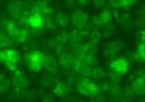
Nestin in Rat Cortical Stem Cells.
Nestin was detected in immersion fixed undifferentiated rat cortical stem cells using Mouse Anti-Rat Nestin Monoclonal Antibody (Catalog # MAB2736) at 10 µg/mL for 3 hours at room temperature. Cells were stained using the NorthernLights™ 557-conjugated Anti-Mouse IgG Secondary Antibody (red; Catalog # NL007) and counterstained with DAPI (blue). View our protocol for Fluorescent ICC Staining of Cells on Coverslips.
Detection of Mouse Nestin by Immunocytochemistry/Immunofluorescence
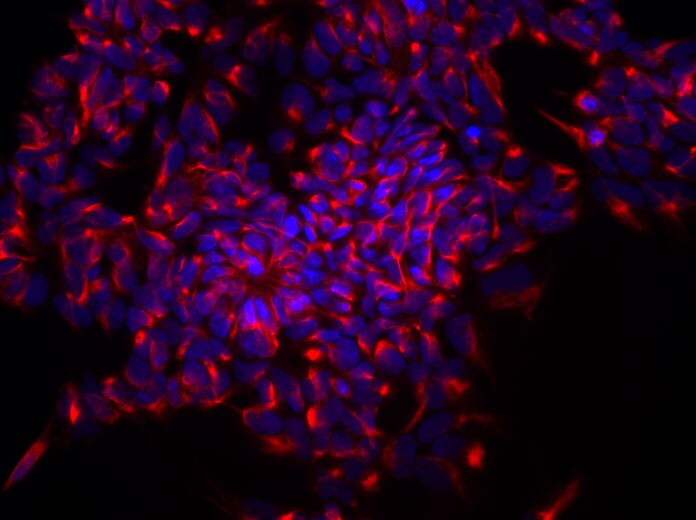
Zfp322a can enhance OSKM reprogramming and replace Sox2.(A) Zfp322a enhanced reprogramming efficiency and accelerated the onset of reprogramming process. OSKM serves as control experiment. (B) The iPSCs generated from OSKM plus Zfp322a presented alkaline phosphatase activity. There were more AP stained colonies generated from OKSM+Zfp322a compare to OKSM. (C) The iPSCs expressed endogenous Oct4, Nanog, Sox2, Rex1 and SSEA-1, indicating that they were ES-cell like. Immunostaining using anti-Oct4, anti-Nanog anti-Sox2, anti-Rex1 and anti-SSEA-1 antibodies were performed with GFP+ iPSCs generated from OKSM+Zfp322a. (D) GFP+ iPSCs generated by OKSM+Zfp322a were able to express ectoderm, mesoderm and endoderm lineage markers in the EB formation assay. iPSCs were stained with anti-Nestin, anti-Gata4 and anti-alpha smooth muscle actin (SMA) antibodies and pictures were taken at 60× magnification. DAPI (blue) served as nucleus marker. (E) Zfp322a was able to replace Sox2, but not Oct4 or Klf4 in OSKM reprogramming process. Results from three independent experiments were presented. (F) iPSCs generated from OKM plus Zfp322a were positive with AP staining and more AP positive colonies were observed in OKM+Zfp322a as compared to OKSM. (G) iPSCs generated by OKM plus Zfp322a expressed pluripotency markers Oct4, Nanog, Sox2, Rex1 and SSEA-1. (H) iPSCs derived from OKM+Zfp322a could differentiate into ectoderm, mesoderm and endoderm lineages, which were showed by anti-Nestin, anti-Gata4, anti-SMA staining respectively. Image collected and cropped by CiteAb from the following publication (https://dx.plos.org/10.1371/journal.pgen.1004038), licensed under a CC-BY license. Not internally tested by R&D Systems.
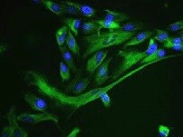
Nestin in C2C12 Mouse Cell Line.
Nestin was detected in immersion fixed C2C12 mouse myoblast cell line using Mouse Anti-Mouse/Rat Nestin Monoclonal Antibody (Catalog # MAB2736) at 25 µg/mL for 3 hours at room temperature. Cells were stained using the NorthernLights™ 557-conjugated Anti-Mouse IgG Secondary Antibody (red; NL007) and counterstained with DAPI (blue). Specific staining was localized to cytoplasm. Staining was performed using our protocol for Fluorescent ICC Staining of Non-adherent Cells.
Detection of Nestin by Immunocytochemistry/ Immunofluorescence
Zfp322a can enhance OSKM reprogramming and replace Sox2.(A) Zfp322a enhanced reprogramming efficiency and accelerated the onset of reprogramming process. OSKM serves as control experiment. (B) The iPSCs generated from OSKM plus Zfp322a presented alkaline phosphatase activity. There were more AP stained colonies generated from OKSM+Zfp322a compare to OKSM. (C) The iPSCs expressed endogenous Oct4, Nanog, Sox2, Rex1 and SSEA-1, indicating that they were ES-cell like. Immunostaining using anti-Oct4, anti-Nanog anti-Sox2, anti-Rex1 and anti-SSEA-1 antibodies were performed with GFP+ iPSCs generated from OKSM+Zfp322a. (D) GFP+ iPSCs generated by OKSM+Zfp322a were able to express ectoderm, mesoderm and endoderm lineage markers in the EB formation assay. iPSCs were stained with anti-Nestin, anti-Gata4 and anti-alpha smooth muscle actin (SMA) antibodies and pictures were taken at 60× magnification. DAPI (blue) served as nucleus marker. (E) Zfp322a was able to replace Sox2, but not Oct4 or Klf4 in OSKM reprogramming process. Results from three independent experiments were presented. (F) iPSCs generated from OKM plus Zfp322a were positive with AP staining and more AP positive colonies were observed in OKM+Zfp322a as compared to OKSM. (G) iPSCs generated by OKM plus Zfp322a expressed pluripotency markers Oct4, Nanog, Sox2, Rex1 and SSEA-1. (H) iPSCs derived from OKM+Zfp322a could differentiate into ectoderm, mesoderm and endoderm lineages, which were showed by anti-Nestin, anti-Gata4, anti-SMA staining respectively. Image collected and cropped by CiteAb from the following open publication (https://pubmed.ncbi.nlm.nih.gov/24550733), licensed under a CC-BY license. Not internally tested by R&D Systems.Applications for Nestin Antibody (307501)
Application
Recommended Usage
Immunocytochemistry
8-25 µg/mL
Sample: Immersion fixed undifferentiated rat cortical stem cells and C2C12 mouse myoblast cell line
Sample: Immersion fixed undifferentiated rat cortical stem cells and C2C12 mouse myoblast cell line
Western Blot
1 µg/mL
Sample: Recombinant Rat Nestin
Sample: Recombinant Rat Nestin
Reviewed Applications
Read 4 reviews rated 4.8 using MAB2736 in the following applications:
Formulation, Preparation, and Storage
Purification
Protein A or G purified from hybridoma culture supernatant
Reconstitution
Reconstitute at 0.5 mg/mL in sterile PBS. For liquid material, refer to CoA for concentration.
Loading...
Formulation
Lyophilized from a 0.2 μm filtered solution in PBS with Trehalose. *Small pack size (SP) is supplied either lyophilized or as a 0.2 µm filtered solution in PBS.
Shipping
Lyophilized product is shipped at ambient temperature. Liquid small pack size (-SP) is shipped with polar packs. Upon receipt, store immediately at the temperature recommended below.
Stability & Storage
Use a manual defrost freezer and avoid repeated freeze-thaw cycles.
- 12 months from date of receipt, -20 to -70 °C as supplied.
- 1 month, 2 to 8 °C under sterile conditions after reconstitution.
- 6 months, -20 to -70 °C under sterile conditions after reconstitution.
Calculators
Background: Nestin
References
- Hockfield, S. and R.D. McKay (1985) J. Neurosci. 5:3310.
- Lendahl, U. et al. (1990) Cell 60:585.
- Frederiksen, K. and R.D. McKay (1988) J. Neurosci. 8:1144.
- Tohyama, T. et al. (1992) Lab. Invest. 66:303.
- Uchida, N. et al. (2000) Proc. Natl. Acad. Sci. USA 97:14720.
- Frederiksen, K. et al. (1988) Neuron 1:439.
- Cattaneo, E. et al. (1990) Nature 347:762.
- Reynolds, B.A. and S. Weiss (1992) Science 255:1707.
- Rietze, R.L. et al. (2001) Nature 412:736.
- Carpenter, M.K. et al. (2001) Exp. Neurol. 172:383.
- Zulewski, H. et al. (2001) Diabetes 50:521.
- Lumelsky, N. et al. (2001) Science 292:1389.
- Lechner, A. et al. (2001) Biochem. Biophys. Res. Commun. 293:670.
- Shih, C.C. et al. (2001) Blood 98:2412.
Alternate Names
NES
Gene Symbol
NES
UniProt
Additional Nestin Products
Product Documents for Nestin Antibody (307501)
Certificate of Analysis
To download a Certificate of Analysis, please enter a lot or batch number in the search box below.
Note: Certificate of Analysis not available for kit components.
Product Specific Notices for Nestin Antibody (307501)
For research use only
Citations for Nestin Antibody (307501)
 Powered by Bioz
Customer Reviews for Nestin Antibody (307501) (4)
4.8 out of 5
4 Customer Ratings
Have you used Nestin Antibody (307501)?
Submit a review and receive an Amazon gift card!
$25/€18/£15/$25CAN/¥2500 Yen for a review with an image
$10/€7/£6/$10CAN/¥1110 Yen for a review without an image
Submit a review
Customer Images
Showing
1
-
4 of
4 reviews
Showing All
Filter By:
-
Application: Immunocytochemistry/ImmunofluorescenceSample Tested: iPS cellsSpecies: MouseVerified Customer | Posted 10/26/2021

-
Application: Immunocytochemistry/ImmunofluorescenceSample Tested: epithelial cellsSpecies: MouseVerified Customer | Posted 09/04/2021
-
Application: Immunocytochemistry/ImmunofluorescenceSample Tested: Neural stem cells derived from h9Species: HumanVerified Customer | Posted 08/19/2019Neural stem cells were fixed in 4%PFA for 15 minutes. The nestin antibody was used at 10ug/ml at 4 degree overnight. Secondary antibody was incubated at room temperature for 1 hour.
-
Application: ImmunocytochemistrySample Tested: See PMID 23798370Species: RatVerified Customer | Posted 02/19/2015
There are no reviews that match your criteria.
Protocols
Find general support by application which include: protocols, troubleshooting, illustrated assays, videos and webinars.
- Appropriate Fixation of IHC/ICC Samples
- Cellular Response to Hypoxia Protocols
- ClariTSA™ Fluorophore Kits
- Detection & Visualization of Antibody Binding
- ICC Cell Smear Protocol for Suspension Cells
- ICC Immunocytochemistry Protocol Videos
- ICC for Adherent Cells
- Immunocytochemistry (ICC) Protocol
- Immunocytochemistry Troubleshooting
- Immunofluorescence of Organoids Embedded in Cultrex Basement Membrane Extract
- Immunohistochemistry (IHC) and Immunocytochemistry (ICC) Protocols
- Preparing Samples for IHC/ICC Experiments
- Preventing Non-Specific Staining (Non-Specific Binding)
- Primary Antibody Selection & Optimization
- Protocol for VisUCyte™ HRP Polymer Detection Reagent
- Protocol for the Fluorescent ICC Staining of Cell Smears - Graphic
- Protocol for the Fluorescent ICC Staining of Cultured Cells on Coverslips - Graphic
- Protocol for the Preparation and Fluorescent ICC Staining of Cells on Coverslips
- Protocol for the Preparation and Fluorescent ICC Staining of Non-adherent Cells
- Protocol for the Preparation and Fluorescent ICC Staining of Stem Cells on Coverslips
- Protocol for the Preparation of a Cell Smear for Non-adherent Cell ICC - Graphic
- R&D Systems Quality Control Western Blot Protocol
- TUNEL and Active Caspase-3 Detection by IHC/ICC Protocol
- The Importance of IHC/ICC Controls
- Troubleshooting Guide: Western Blot Figures
- Western Blot Conditions
- Western Blot Protocol
- Western Blot Protocol for Cell Lysates
- Western Blot Troubleshooting
- Western Blot Troubleshooting Guide
- View all Protocols, Troubleshooting, Illustrated assays and Webinars

