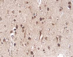
NGF Receptor in Human Brain.
Nerve Growth Factor Receptor (NGF R)/TNFRSF16 was detected in immersion fixed paraffin-embedded sections of human brain (cortex) using Human NGF R Monoclonal Antibody (Catalog # MAB367) at 25 µg/mL overnight at 4 °C. Tissue was stained using the Anti-Mouse HRP-DAB Cell & Tissue Staining Kit (brown; Catalog # CTS002) and counterstained with hematoxylin (blue). View our protocol for Chromogenic IHC Staining of Paraffin-embedded Tissue Sections.
This antibody specifically recognizes human NGF Receptor. In ELISAs, this antibody shows no cross-reactivity with 19 other tested family members.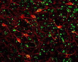
NGF Receptor in Mouse Brain.
Nerve Growth Factor Receptor (NGF R)/TNFRSF16 was detected in perfusion fixed frozen sections of mouse brain (cortex) using Mouse NGF R Antigen Affinity-purified Polyclonal Antibody (Catalog # AF1157) at 7 µg/mL overnight at 4 °C. Tissue was stained (red) and counterstained (green). View our protocol for Fluorescent IHC Staining of Frozen Tissue Sections.
This antibody specifically recognizes mouse NGF Receptor. In ELISA and Western blot experiments, this antibody shows approximately 5% cross-reactivity with recombinant human NGF R.
Secondary Antibodies
R&D Systems offers a wide range of biotinylated, HRP-conjugated, fluorochrome-labeled, and unlabeled species-specific secondary antibodies. Our NorthernLights™ fluorescent secondary antibodies are available with three distinct excitation and emission maxima, making them ideal for multi-color fluorescence microscopy.