VEGF Receptor 2 in Human Placenta.
Vascular Endothelial Cell Growth Factor Receptor 2 (VEGF R2)/KDR/Flk-1 was detected in immersion fixed paraffin-embedded sections of human placenta using Human VEGF R2 Antigen Affinity-purified Polyclonal Antibody (Catalog # AF357) at 15 µg/mL overnight at 4 °C. Tissue was stained using the Anti-Goat HRP-AEC Cell & Tissue Staining Kit (red; Catalog # CTS009) and counterstained with hematoxylin (blue). View our protocol for Chromogenic IHC Staining of Paraffin-embedded Tissue Sections.
This antibody specifically recognizes human VEGF R2. Reactivity with VEGF R2 from other species has not been determined.
VEGF Receptor 2 in Mouse Embryo.
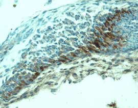
Vascular endothelial growth factor receptor 2 (VEGF R2)/KDR/Flk-1 was detected in immersion fixed frozen sections of mouse embryo (14 d.p.c.) using Mouse VEGF R2 Antigen Affinity-purified Polyclonal Antibody (Catalog # AF644) at 15 µg/mL overnight at 4 °C. Tissue was stained using the Anti-Goat HRP-DAB Cell & Tissue Staining Kit (brown; Catalog # CTS008) and counterstained with hematoxylin (blue). Specific labeling was localized to mesenchymal cells.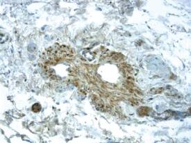
VEGF Receptor 2 in Human Placenta.
Vascular endothelial cell growth factor receptor 2 (VEGF R2)/KDR/Flk-1 was detected in immersion fixed paraffin-embedded sections of human placenta using Human VEGF R2 Monoclonal Antibody (Catalog # MAB3571) at 25 µg/mL overnight at 4 °C. Tissue was stained using the Anti-Mouse HRP-DAB Cell & Tissue Staining Kit (brown; Catalog # CTS002) and counterstained with hematoxylin (blue). View our protocol for Chromogenic IHC Staining of Paraffin-embedded Tissue Sections.
This antibody specifically recognizes human VEGF R2. In Western blot experiments, this antibody shows no cross-reactivity with recombinant human VEGF R1 or R3, or recombinant mouse VEGF R2.
VEGF Receptor 2 in bEnd.3 Mouse Cell Line.
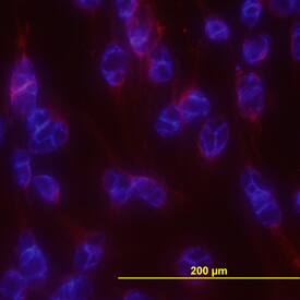
Vascular Endothelial Growth Factor Receptor 2 (VEGF R2)/KDR/Flk-1 was detected in immersion fixed bEnd.3 mouse endothelioma cell line using Mouse VEGF R2 Monoclonal Antibody (Catalog # MAB4432) at 10 µg/mL for 3 hours at room temperature. Cells were stained using the NorthernLights™ 557-conjugated Anti-Rat IgG Secondary Antibody (red; Catalog # NL013) and counterstained with DAPI (blue). View our protocol for Fluorescent ICC Staining of Cells on Coverslips.
This antibody specifically recognizes mouse VEGF R2. Reactivity with VEGF R2 from other species has not been determined.
Secondary Antibodies
R&D Systems offers a wide range of biotinylated, HRP-conjugated, fluorochrome-labeled, and unlabeled species-specific secondary antibodies. Our NorthernLights™ fluorescent secondary antibodies are available with three distinct excitation and emission maxima, making them ideal for multi-color fluorescence microscopy.
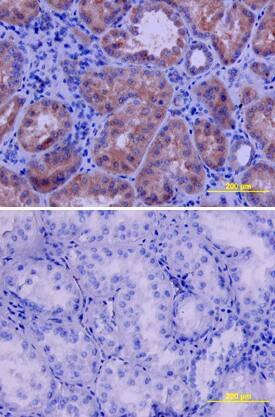
VEGF R2/KDR/Flk-1 in Human Kidney.
Vascular Endothelial Growth Factor Receptor 2 (VEGF R2/KDR/Flk-1) was detected in immersion fixed paraffin-embedded sections of human kidney using Human VEGF R2/KDR/Flk-1 Antigen Affinity-purified Polyclonal Antibody (Catalog # AF357) at 10 µg/mL overnight at 4 °C. Tissue was stained using the Anti-Goat HRP-DAB Cell & Tissue Staining Kit (brown; Catalog # CTS008) and counterstained with hematoxylin (blue). Lower panel shows a lack of labeling if primary antibodies are omitted and tissue is stained only with secondary antibody followed by incubation with detection reagents. View our protocol for Chromogenic IHC Staining of Paraffin-embedded Tissue Sections.
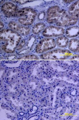
VEGF R2/KDR/Flk-1 in Human Kidney.
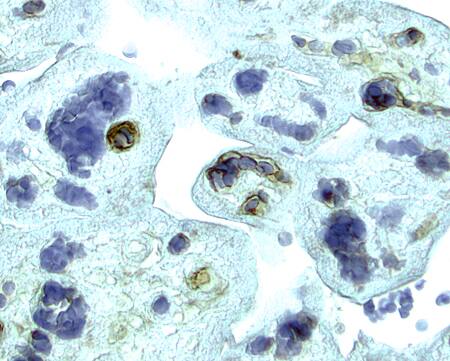
Vascular Endothelial Growth Factor Receptor 2 (VEGF R2/KDR/Flk-1) was detected in immersion fixed paraffin-embedded sections of human kidney array using Human VEGF R2/KDR/Flk-1 Monoclonal Antibody (Catalog # MAB3571) at 15 µg/mL overnight at 4 °C. Tissue was stained using the Anti-Mouse HRP-DAB Cell & Tissue Staining Kit (brown; Catalog # CTS002) and counterstained with hematoxylin (blue). Lower panel shows a lack of labeling if primary antibodies are omitted and tissue is stained only with secondary antibody followed by incubation with detection reagents. View our protocol for Chromogenic IHC Staining of Paraffin-embedded Tissue Sections.
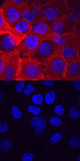
Phospho-VEGF R2/KDR/Flk-1 (Y1214) in A431 Human Cell Line.
Vascular Endothelial Growth Factor Receptor 2 (Phospho-VEGF R2/KDR/Flk-1 (Y1214)) was detected in immersion fixed A431 human epithelial carcinoma cell line untreated (lower panel) or treated (upper panel) with pervanadate using Rabbit Anti-Human Phospho-VEGF R2/KDR/Flk-1 (Y1214) Antigen Affinity-purified Polyclonal Antibody (Catalog # AF1766) at 10 µg/mL for 3 hours at room temperature. Cells were stained using the NorthernLights™ 557-conjugated Anti-Rabbit IgG Secondary Antibody (red; Catalog # NL004) and counterstained with DAPI (blue). View our protocol for Fluorescent ICC Staining of Cells on Coverslips.