Propidium iodide
Tocris Bioscience | Catalog # 5135

Key Product Details
Description
Alternative Names
Wavelength
Product Description
Key information: Propidium iodide (PI) is a widely used red-fluorescent DNA stain. It is membrane impermeant to live cells and so PI staining differentiates live and dead cells. PI can be used to stain nucleic acids in dead cells across different cell types, including mammalian cells, bacteria, and yeast.Application: flow cytometry, confocal microscopy, fluorescence microscopy.
Used for: apoptosis detection by PI staining, nuclear counterstaining in multicolor fluorescent assays, cell death measuring in a mixed live-cell population by PI staining and flow cytometry.
Properties and Photophysical Data: Propidium iodide fluorescence increases by approximately 20- to 30-fold upon binding to DNA or RNA (through intercalation). When unbound, excitation and emission maxima (λ) are 493 nm and 636 nm, respectively, when bound to DNA, excitation and emission maxima (λ) are 535 nm and 617 nm, respectively.
Optical Data

| Emission Color | Orange |
| λabs | 535 nm |
| λem | 617 nm |
| Application | Flow Cytometry, Fluorescence Microscopy, Confocal Microscopy |
Spectra Viewer
Plan Your Experiments
Use our spectra viewer to interactively plan your experiments, assessing multiplexing options. View the excitation and emission spectra for our fluorescent dye range and other commonly used dyes.
Spectra Viewer
Product Specifications for Propidium iodide
Molecular Weight
Formula
Storage
Purity
Chemical Name
CAS Number
PubChem ID
InChI Key
SMILES
The technical data provided above is for guidance only. For batch specific data refer to the Certificate of Analysis.
Solubility
| Solvent | Max Conc. mg/mL | Max Conc. mM | |
|---|---|---|---|
| Solubility | |||
| water | 3.34 | 5 with sonication |
Preparing Stock Solutions for Propidium iodide
The following data is based on the product molecular weight 668.39.
Batch specific molecular weights may vary from batch to batch due to the degree of hydration, which all affect the solvent volumes required to prepare stock solutions.
| Concentration / Solvent Volume / Mass | 1 mg | 5 mg | 10 mg |
|---|---|---|---|
| 0.05 mM | 29.92 mL | 149.61 mL | 299.23 mL |
| 0.25 mM | 5.98 mL | 29.92 mL | 59.85 mL |
| 0.5 mM | 2.99 mL | 14.96 mL | 29.92 mL |
| 2.5 mM | 0.60 mL | 2.99 mL | 5.98 mL |
Calculators
Background References
References are publications that support the biological activity of the product. See our Citations tab to view 18 publications citing the usage of this product.
- Sonnemann p53-dependent and p53-independent anticancer effects of different histone deacetylase inhibitors. Br.J.Cancer 2014 PMID: 24281001
- Yun Serine hydrolase inhibitors block necrotic cell death by preventing calcium overload of the mitochondria and permeability transition pore formation. J.Biol.Chem. 2013 PMID: 24297180
- Jeong SIRT4 protein suppresses tumor formation in genetic models of Myc-induced B cell lymphoma. J.Biol.Chem. 2013 PMID: 24368766
Product Documents for Propidium iodide
Certificate of Analysis
To download a Certificate of Analysis, please enter a lot or batch number in the search box below.
Product Specific Notices for Propidium iodide
For research use only
Citations for Propidium iodide
 Powered by Bioz
Customer Reviews for Propidium iodide (4)
Have you used Propidium iodide?
Submit a review and receive an Amazon gift card!
$25/€18/£15/$25CAN/¥2500 Yen for a review with an image
$10/€7/£6/$10CAN/¥1110 Yen for a review without an image
Submit a review
Customer Images
-
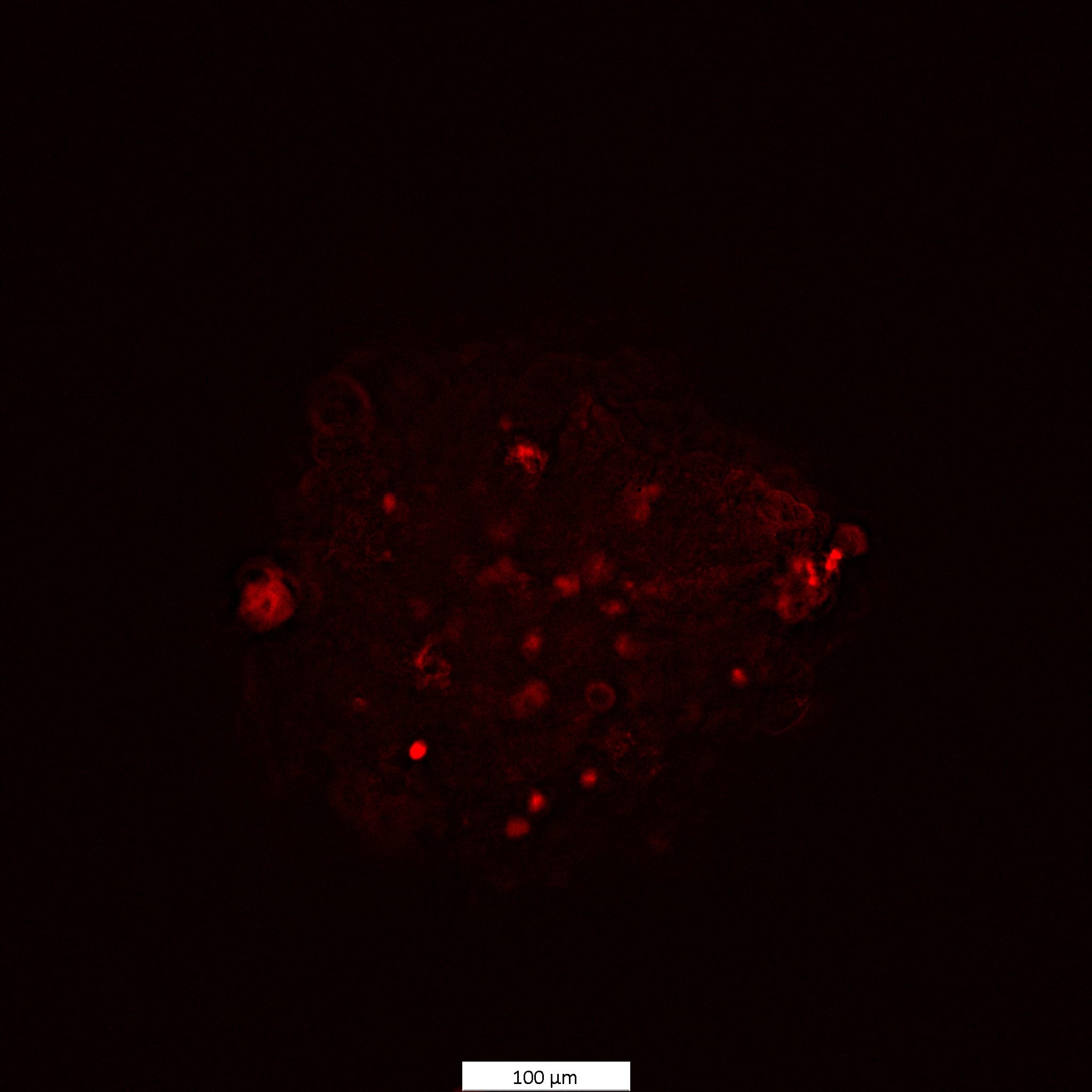
Species: HumanAssay Type: In VitroCell Line/Tissue: MCF7Verified Customer | Posted 11/26/2024MCF7 cells spheroids were stained with 3 uM Propidium Iodide for 1.5h

-
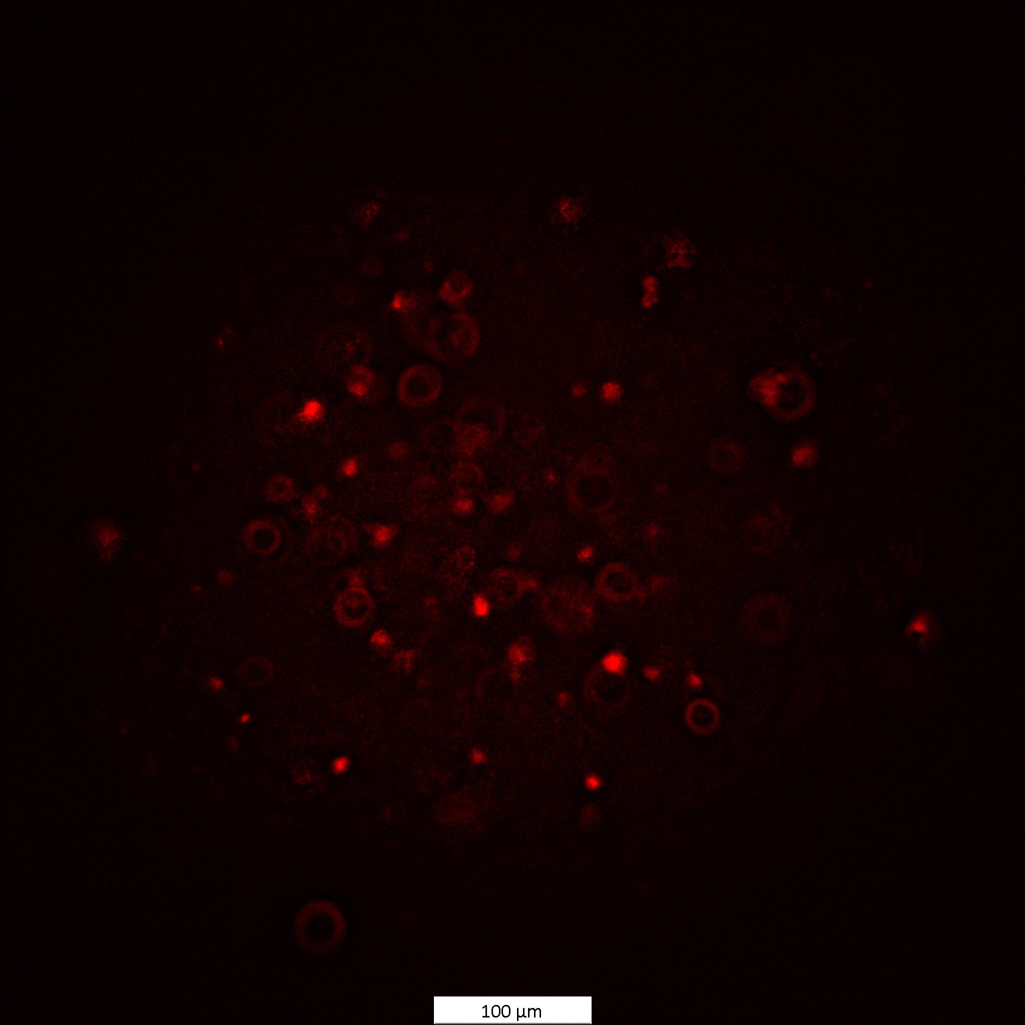
Species: HumanAssay Type: In VitroCell Line/Tissue: HCT116Verified Customer | Posted 11/26/2024Staining of HCT 116 spheroids using a 3 uM concentration
-
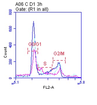
Species: HumanAssay Type: In VitroCell Line/Tissue: Jurkat CellsVerified Customer | Posted 08/17/2020PI was used in the cell cycle analysis of Jurkat cells. The cell pellet of the treated cells was resuspended in 200 μL propidium iodide (PI) solution containing 50 μg/mL PI, 0.1 mg/mL RNase A, and 0.05% Triton X-100 (v/v) (prepared in 1X PBS buffer). The cells were incubated for 30 min. in the dark at room temperature and by agitating every 10 mins. After incubation, the samples were analyzed using the flow cytometer.
-
Species: MouseAssay Type: Ex VivoVerified Customer | Posted 03/13/20185mM is great for visualization with an mcherry filter. It goes into water well.Propidium Iodide was used as an intracellular fluorescent dye. Picture is of the glass electrode with a 5mM solution. 250uM was also tried but had poor visibility.
There are no reviews that match your criteria.