Western Blot
蛋白免疫印迹是一种基础性分析技术,用于检测细胞或组织裂解液中的蛋白表达水平。联合免疫沉淀或亚细胞组分分离等技术使用时,还能探究蛋白亚细胞定位、翻译后修饰、蛋白加工及蛋白间相互作用。Bio-Techne 提供品类齐全的抗体、对照品、配套产品、成像系统及分析软件,完美适配传统与全自动蛋白免疫印迹实验需求。
传统蛋白免疫印迹
一抗
可靠且经印迹实验验证的抗体,是蛋白免疫印迹实验成功的核心。Bio-Techne 的蛋白免疫印迹抗体产品系列涵盖被广泛引用的单克隆抗体和重组一抗,全系产品 享有品质保障.。
申领 Bio-Techne 的蛋白免疫印迹电子手册
手册内含分步实操教程与问题排查方案,助您获得稳定且可重现的实验结果。

其他蛋白免疫印迹配套产品
除抗体和裂解液对照品外,Bio-Techne 还提供各类实验配套试剂耗材,包括 HRP 稳定剂、亚细胞组分分离试剂盒、蛋白分子量标准、印迹转印膜等。
蛋白免疫印迹配套产品
| HRP稳定剂 | 丽春红 S 染色液 | PBS 缓冲液片剂 | RunBlue三色预染蛋白分子量 Marker |
| 未染色重组蛋白分子量标准 | 重组蛋白免疫印迹标准品 | 亚细胞组分分离试剂盒 | 蛋白免疫印迹转印膜 |
| 多肽抑制剂 | 其他配套产品 |
Simple Western™全自动蛋白免疫印迹系统
传统蛋白免疫印迹实验操作繁琐、实验难度大。为解决传统蛋白免疫印迹技术痛点,Bio-Techne 旗下品牌 ProteinSimple 推出 Simple Western™全自动毛细管蛋白免疫印迹分析平台。传统印迹实验中的蛋白分离、固定、洗涤、检测等全部流程,均可在台式毛细管电泳仪内自动完成。
- Simple Western 提供高通量免疫印迹分析,可在短短 3 小时内完成多达 25 份样本的完全定量,通宵运行可检测 96 份样本。
- 搭载 Jess™ 平台 Stellar™检测模块,具备行业领先的近红外/红外荧光检测灵敏度。
- 平台配备 RePlex™ 技术,可洗脱首轮孵育抗体,重新进行抗体孵育或总蛋白归一化校正,省去传统实验繁琐的洗脱重新孵育步骤。
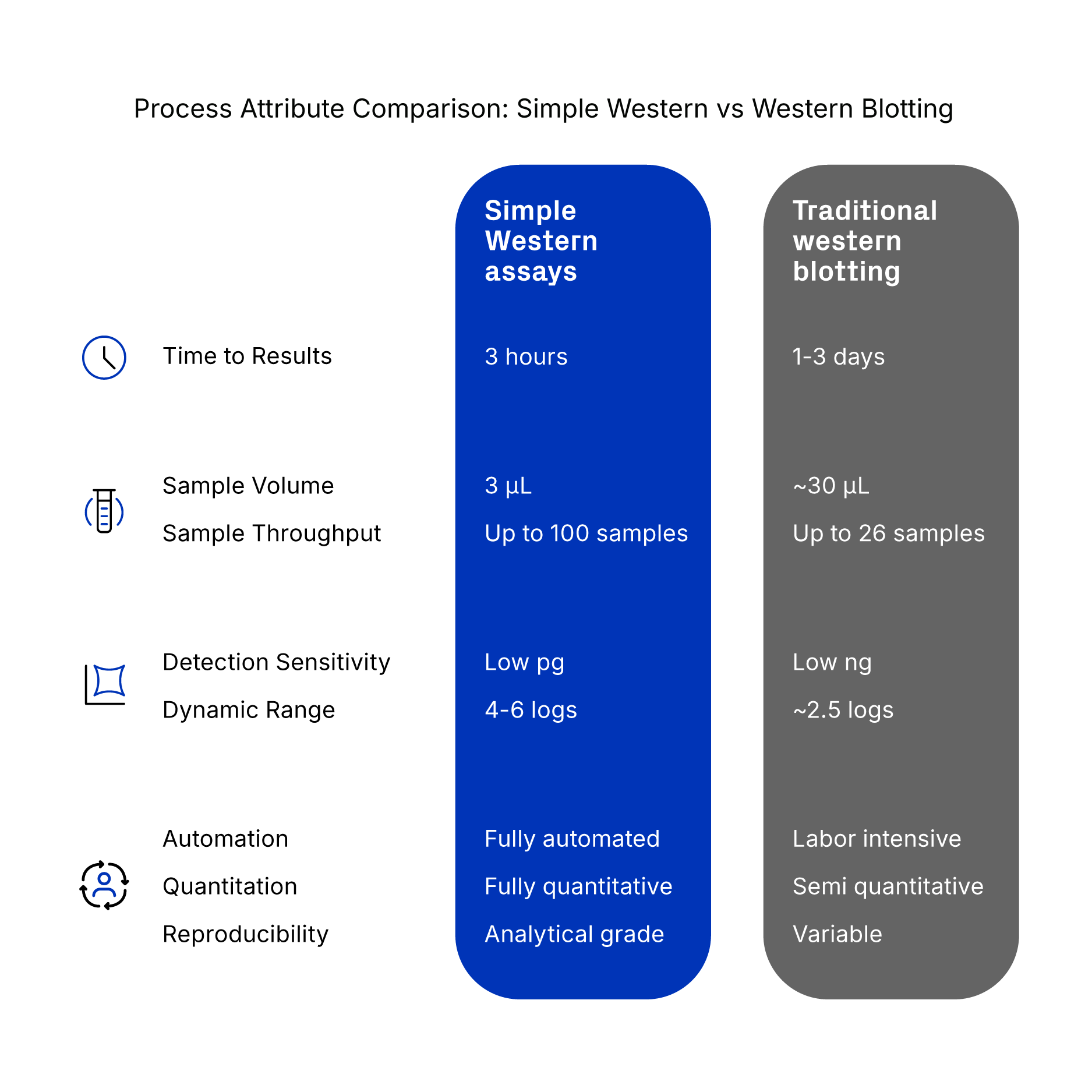
Jess 平台 Simple Western 系统与传统蛋白免疫印迹对比
Bio-Techne 提供经 Simple Western 验证的各类抗体
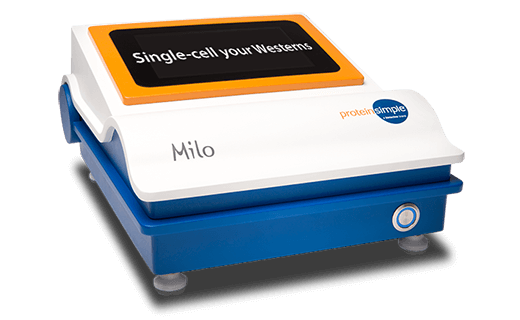
现在,您可以使用 Milo™ 在单个细胞中进行蛋白免疫印迹分析。
您是否涉足单细胞蛋白质组学研究?Milo™ 单细胞蛋白免疫印迹技术单次运行可检测约 1,000 个单细胞中的蛋白表达水平。可用于验证 RNAseq 数据、分析 FACS 分选细胞,作为全球首款单细胞蛋白免疫印迹技术,助力深度开展单细胞蛋白质组学研究。
定制抗体服务
若官网暂无所需抗体,可直接联系我们。定制抗体生产流程繁杂,可依托我们资深抗体研发团队的专业技术与丰富经验,完成各类定制抗体项目。

蛋白免疫印迹手册

分步式蛋白免疫印迹实操指南,适合入门学习,全面讲解实验原理、操作流程及问题排查方案。
蛋白免疫印迹问题排查资源
线上综合排查指南,涵盖无条带、杂带过多、背景过高等各类常见实验问题解决方案。
经验证单克隆抗体开发白皮书
本白皮书阐述了我们的单克隆抗体全套验证流程,包含抗原设计、可行性验证、特异性和灵敏度检测、质量控制和成品放行标准。
了解蛋白免疫印迹抗体染色与检测
关于蛋白免疫印迹
蛋白免疫印迹是借助抗体特异性识别细胞或组织裂解液中的靶标蛋白的经典实验技术。依托抗体高度特异性的结合特性,蛋白免疫印迹可在数千种不同蛋白质的混合物中精准实现单一靶标蛋白的定性与定量分析。在蛋白免疫印迹实验中,将样本加样至聚丙烯酰胺凝胶内,通过 SDS-PAGE 电泳根据其分子量大小进行蛋白分离。
- 样本制备:成功进行蛋白免疫印迹实验的第一步是样本制备,需要裂解细胞以分离蛋白,同时需抑制蛋白水解和去磷酸化,并进行蛋白变性处理。有关蛋白免疫印迹样本制备的详细信息,请查阅我们的《样本制备方案》。
- 分离: 样本加载到由浓缩胶与分离胶两层构成的聚丙烯酰胺凝胶上,蛋白基于其分子量通过 SDS-PAGE 电泳进行分离。浓缩胶中丙烯酰胺含量低,pH 值偏低,可使样本中的蛋白聚拢压缩,形成整齐清晰的条带。随后蛋白质进入碱性更强、胶浓度更高的分离胶中,依照分子量大小实现分离,分子量越小的蛋白电泳迁移速度越快。查看 SDS-PAGE凝胶电泳实验方案。
- 转膜:借助电场作用力,将凝胶内的蛋白转印至膜上。查看 蛋白转膜方案。
- 染色:在转印膜上加入靶向靶标蛋白的特异性一抗进行孵育。再加入偶联酶或荧光染料的二抗,以显色蛋白/抗体复合物。
- 显影成像:最终在分子量标准品(Marker)指示的其预期分子量附近,将显现特异性蛋白条带。关于抗体染色与成像的更多信息,请参阅我们的《蛋白免疫印迹实验方案》。
传统蛋白免疫印迹实验流程概述

(A)通过聚丙烯酰胺凝胶电泳(PAGE)完成蛋白分离;(B)将分离后的蛋白转印至膜(如硝酸纤维素膜或 PVDF 膜)上进行检测;(C)在转印膜上加入靶向靶标蛋白的一抗进行孵育,通常随后使用酶标二抗检测抗体-抗原复合物;该酶(如辣根过氧化物酶,HRP)作用于底物(如电化学发光,ECL),产生光信号;(D)该信号可通过放射自显影胶片或化学发光成像系统采集捕获。 了解更多蛋白免疫印迹实验优化相关信息。
完成蛋白免疫印迹后,可对印迹膜上进行洗脱处理,去除已结合的一抗和二抗,重新孵育其他抗体开展重复检测。 如需了解更多关于此过程的信息,请查阅我们的《膜洗脱与重新孵育方案》。
什么是 SDS-PAGE?
SDS-PAGE(十二烷基硫酸钠-聚丙烯酰胺凝胶电泳)是依据分子量大小分离蛋白的常用的电泳技术。SDS 是一种阴离子去污剂,带有负电荷,可结合蛋白并破坏维持其空间结构的非共价键,使蛋白发生变性;还能使蛋白带上与自身分子量相对应的均一净负电荷。变性蛋白电泳时仅依据其分子大小完成迁移分离。实验中一般先进行 SDS-PAGE 电泳分离蛋白,再开展蛋白免疫印迹:电泳实现蛋白分离,印迹则将蛋白从凝胶转印至膜上,利用抗体判定靶标蛋白的存在/缺失/表达水平。
什么是非变性 PAGE ?适用场景?
非变性凝胶体系不使用 SDS,上样缓冲液中也不添加 DTT、β-巯基乙醇等还原剂。蛋白质可维持天然空间构象和自身原有电荷,电泳迁移速率同时受分子量和电荷双重影响。多用于分析蛋白聚集状态、研究蛋白复合物、分离酶等实验。
蛋白免疫印迹实验上样量如何确定?
蛋白免疫印迹实验凝胶上样蛋白量受多种因素影响,主要取决于靶标蛋白表达丰度及待检测样本类型。经验参考标准:细胞裂解液、细胞核裂解液及膜蛋白样本,每孔总蛋白上样量 20-30 µg;检测纯化蛋白时,常规上样量为 10-100 ng。正式开展印迹实验前,需提前确定最佳上样量。
蛋白免疫印迹实验需要设置哪些对照?
规范设置实验对照,既能快速排查实验问题,也能有效验证实验结果可靠性,标准实验需配备以下几类对照:
1. 阳性对照裂解液——阳性对照裂解液取自已知表达靶标蛋白的细胞系或组织样本。此对照将在蛋白免疫印迹实验中呈现出一条阳性条带。该对照可验证整套实验流程无异常,以此判定样本无阳性信号是因本身不含靶标蛋白,而非实验操作失误。
2. 阴性对照裂解液——阴性对照裂解液取自已知不表达靶标蛋白的样本。此对照在蛋白免疫印迹实验中不会产生条带。该对照用于排查抗体是否存在非特异性结合现象。
3. 内源性阳性对照裂解液——内源性阳性对照裂解液取自已知表达靶标蛋白的样本。此对照应在检测重组蛋白(如标签蛋白)样本时使用。重组蛋白空间折叠结构可能与天然蛋白存在差异,折叠异常可能导致抗体无法结合抗原表位。借助该对照可区分阴性结果成因:究竟是重组蛋白表位被封闭,还是实验体系失效。
4. 内参对照——内参对照是指管家蛋白,这类蛋白几乎在所有组织和细胞中均稳定均一表达。该对照可排除因上样量不均、转膜异常造成的组间蛋白表达差异,也是实现样本间蛋白水平半定量分析的必要参照。
Simple Western 与传统蛋白免疫印迹有何区别?
Simple Western 是依托毛细管电泳技术搭配免疫检测搭建的全自动化的蛋白免疫印迹分析系统。该平台能自动完成电泳和免疫印迹技术涉及的所有步骤,包括蛋白上样、电泳分离、抗体孵育、洗膜、信号检测以及数据的定量分析。查看 Bio-Techne 的 Simple Western概览页面,了解更多 Simple Western 相关信息。
为什么蛋白免疫印迹实际条带分子量与理论预测值不符?
通常,蛋白在 PAGE 凝胶基质中主要依据分子量大小完成电泳迁移,分子量越小迁移速率越快。然而,迁移行为也可能受到其他因素的影响,最终导致实测条带大小与理论值存在偏差。常见影响因素包括翻译后修饰、翻译后剪切、剪接变异、相对电荷变异以及蛋白多聚体形成等。
在蛋白免疫印迹孵育一抗适用封闭液推荐?
我们蛋白免疫印迹 QC 验证中常用的封闭液为 3% BSA (货号 5217)或 5% NFDM(脱脂奶粉)。在某些情况下,会将 BSA 和 NFDM 混合使用。检测磷酸化翻译后修饰蛋白务必选用 BSA 作为封闭液,避免抗体与脱脂奶粉中的酪蛋白发生非特异性结合。
蛋白免疫印迹实验检测磷酸化表位时,应使用何种封闭液?
通用实验准则:检测磷酸化蛋白优先选用 5% w/v BSA (溶于 TBST),因为抗体会特异性识别并结合奶粉中的酪蛋白(本身属于磷酸化蛋白),导致实验背景信号偏高。
更多蛋白免疫印迹相关问题,
可查阅我们的 《蛋白免疫印迹问题排查指南》。