Immunocytochemistry (ICC)
免疫细胞化学(ICC)常被称为免疫荧光(IF),是一种通过光学显微镜检测培养细胞或原代细胞中抗原的免疫染色技术。多色 ICC 实验需使用荧光偶联抗体。
精选 ICC 产品
验证 iPSC 的干性
利用标志物抗体分析人类干细胞的多能性,可筛选并扩增状态优良、未分化的干细胞群。搭配多种标志物检测,大幅提升干细胞多能性判定结果的可信度。
聚焦信号转导研究
选用经 ICC 验证的抗体,直观观测细胞内信号转导过程。无论是观察突触形成及后续信号级联反应,还是监测线粒体内部生理活动,均有适配的信号转导研究专用抗体。
ClariTSA™ 荧光染料试剂盒
依托亮度出众的 ClariTSA™ 荧光染料提升靶标检测效果,尤其适用于低丰度靶标分析。该类染料可显著提高 IHC、ICC、ISH 等空间生物学实验的信噪比。ClariTSA™ 染料专为RNAscope™多重荧光 V2 检测优化定制,保障实验结果精准可靠。
多色 ICC 专用抗体
Bio-Techne 可全面满足各类多色 ICC 实验需求。我们拥有品类齐全的荧光偶联抗体,涵盖偶联光稳定性荧光染料(如 Alexa Fluor® 647、Alexa Fluor® 488)的一抗产品。
查看各类预偶联一抗:
| Alexa Fluor® 488 | Janelia Fluor® 549 | DyLightTM 550 |
| Alexa Fluor® 647 | Janelia Fluor® 646 | Biotin |
二抗与检测试剂
ICC 染色可采用直接染色法(使用偶联一抗),也可采用间接检测法(搭配偶联二抗)完成结果观测。在间接 IF 检测中,需使用荧光染料标记二抗来检测一抗-抗原复合物。若采用间接检测方法,搭配多种一抗务必保证其宿主来源物种互不相同。选择二抗时还需考虑一抗的亚类(如 IgG、IgM、IgA),以及二抗靶向位点——仅重链(Hc)、仅轻链(Lc)、轻重链兼顾(H+L)。
间接检测法可通过多个二抗结合同一个一抗实现信号放大,从而提升对低丰度靶标的检测能力。 了解直接检测法与间接检测法的更多区别。
按反应性分类的 ICC 二抗
| 抗山羊 | 抗小鼠 | 抗大鼠 |
| 抗人 | 抗兔 | 抗绵羊 |
检测试剂
除抗体染色外,荧光染料和探针还可用于进一步研究细胞结构、观察细胞形态。
| 细胞结构 | 试剂 | 适用范围 |
|---|---|---|
| 细胞核 | DAPI, DRAQ5TM | 活细胞或固定细胞;可透过细胞膜 |
| 细胞核 | DRAQ7TM | 活细胞或固定细胞;不会染色完整活细胞,可用于细胞活力检测 |
| 细胞质 | DRAQ9TM | 活细胞或固定细胞;可透过细胞膜 |
| 线粒体 | MitoBrilliantTM 646 | 活细胞或固定细胞 |
| 线粒体 | MitoBrilliantTM Live 646, MitoBrilliantTM Live 549 | 活细胞;其活性依赖于膜电位(Δψm) |
ICC 对照品
规范设置实验对照,是确保 ICC 实验结果解读准确的关键。ICC 对照包括背景染色对照和抗体特异性对照。
抗体特异性对照
Bio-Techne 对所有经 ICC 实验验证的抗体均执行严格的质控检测,海量成像实验数据充分印证产品品质。 常用抗体特异性验证方式:基因敲除(KO)细胞系、阳性和阴性细胞系、蛋白刺激实验、RNA-蛋白共检测。Bio-Techne 还提供封闭肽等配套对照试剂。
背景染色对照
背景杂色会严重干扰 ICC 成像结果判读。采用间接检测法时,必须设置不加一抗的空白对照:样本不进行一抗孵育,其余二抗、显色等实验步骤保持一致。
同型对照用作阴性对照,可有效区分非特异性背景信号与特异性抗体信号。同型对照抗体与实验所用一抗亚型、类别完全一致,但不靶向实验目的抗原。
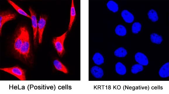
基因敲除 ICC 对照。检测野生型 HeLa 细胞(阳性)、敲除 KRT18 基因 HeLa 细胞(阴性)中 KRT18 的表达情况。敲除细胞无特异性染色信号,这表明一抗具备良好特异性。实验细胞均使用DAPI(NBP2-31156)进行复染。
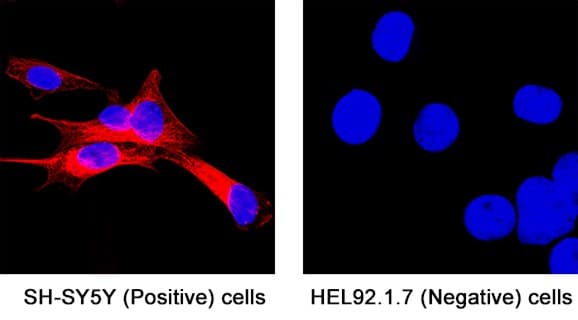
阳性与阴性细胞系对照。在已知表达神经元特异性 β-Ⅲ 微管蛋白的 SH-SY5Y 细胞中检出靶标蛋白;不表达该蛋白的 HEL92.1.7 细胞无染色信号。实验细胞均使用DAPI(NBP2-31156)进行复染。
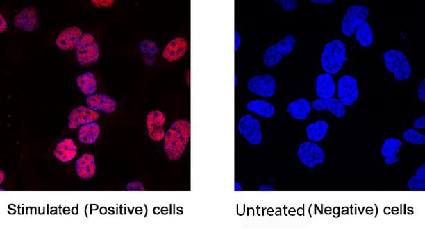
蛋白诱导表达对照经缺氧处理后,HeLa 细胞核内 HIF-1α 蛋白表达上调(左侧),而未经处理对照组细胞中未检出对应信号。实验细胞均使用DAPI(NBP2-31156)进行复染。
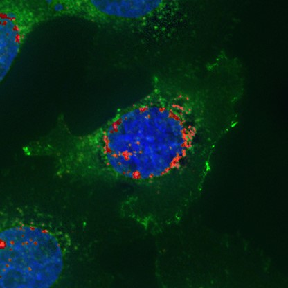
RNA-蛋白共检测对照。同一细胞内表皮生长因子受体(EGFR)mRNA(红色,采用 RNAscopeTM 方案)与 EGFR 蛋白(绿色,通过 ICC 法)的共同检测结果。mRNA 及其编码蛋白在同一细胞内的共定位,可进一步验证抗体特异性。
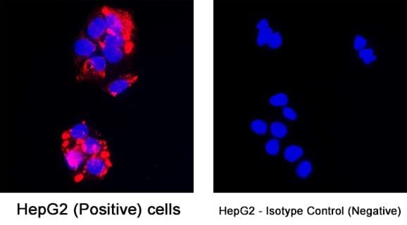
将 HepG2 细胞分别与小鼠抗人 AFP 抗体(MAB1368)IgG1 亚型与非免疫性 IgG1 抗体(MAB002)共同孵育,两者工作稀释浓度均为 25 μg/mL。MAB1368 抗体在细胞质中显示出强烈的特异性细胞标记,而与 IgG1 同型对照孵育的细胞则无染色信号。
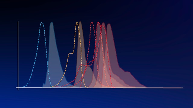
Spectra Viewer
借助Spectra Viewer灵活规划实验,搭配多重荧光组合方案。可查询我们全系荧光染料及其他常用染料的激发和发射光谱。
荧光染料与探针产品册

手册详述荧光染料、探针及染色试剂应用原理,收录全系列产品信息,助力挑选适配试剂,优化实验效果。
免疫细胞化学(ICC)手册

本免疫细胞化学(ICC)手册讲解 ICC 技术,侧重 多色 ICC/IF 实验,包含详细实验方案及问题排查建议。既适合新手入门学习,也可供资深实验人员复盘精进实验技巧。
ICC相关博客文章
ICC 是一种广泛应用的免疫染色技术,利用标记抗体原位特异性结合细胞内靶标蛋白,从而实现细胞中特定蛋白的定位与可视化检测。借助荧光显微镜即可完成,以此判定细胞是否表达特定蛋白,以及该蛋白在细胞内的具体分布位置。ICC 技术所用样本为离体细胞,如培养细胞;而免疫组化(IHC)则采用组织切片作为实验样本。了解ICC 与 IHC 的更多区别。
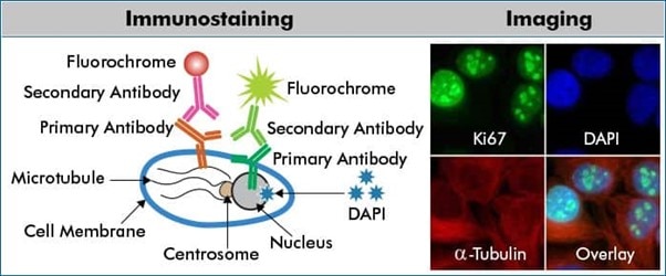
上图为多色 ICC/IF 间接法蛋白检测实验示意图。实验采用抗 Ki67 (核蛋白;货号 NBP2-54791)和抗 α-微管蛋白(微管蛋白;货号 NB100-690)孵育 HeLa 细胞,随后分别使用 DyLight 488(绿色)和 DyLight 550(红色)偶联的二抗进行检测。固定后细胞用细胞核染料DAPI(蓝色;货号 NBP2-31156)进行复染。
荧光免疫检测中,抗体偶联荧光基团,经特定波长光源激发后便可发出荧光信号。常规检测可选用标记一抗或二抗;也可先使用生物素化抗体,再搭配荧光标记链霉亲和素,进一步提升实验检测灵敏度。
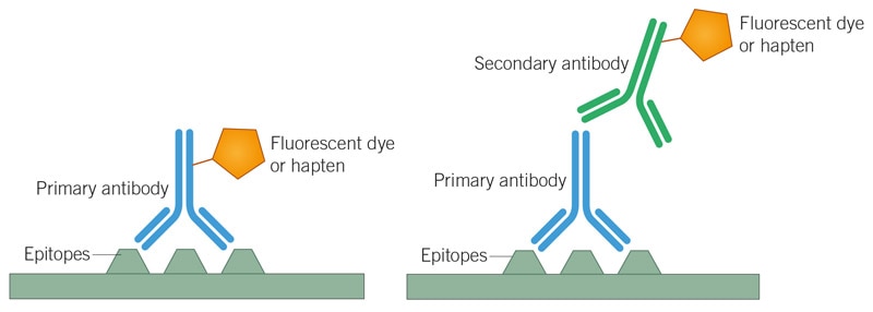
直接和间接检测
直接法:多色 ICC/IF 实验直接使用偶联荧光基团的一抗开展实验。间接法:先采用无荧光标记的一抗结合靶标分子,再使用荧光标记二抗,特异性识别一抗完成信号放大。
直接法与间接法如何选择?
不存在绝对最优方案,需根据实验需求灵活选定。下表列举的考量因素,有助于确定哪种方法最符合您的实验需求。
| 直接检测法 | 间接检测法 | |
|---|---|---|
| 检测灵敏度 | 直接检测法灵敏度偏低,无法借助二抗实现信号放大。 | 单个一抗分子可结合多个二抗分子,信号放大效果强,检测灵敏度高。 |
| 实验耗时 | 省去二抗孵育步骤,实验流程更简短。 | 需额外进行二抗孵育与多次洗涤,整体实验周期更长。 |
| 多重检测 | 更易实现多重检测,可混用同种属来源的多种一抗。 | 易出现二抗交叉反应,多重检测操作难度更高。 |
| 背景信号强弱 | 无二抗引发的交叉反应,非特异性背景信号较低。 | 受二抗非特异性结合影响,整体背景染色通常较高。 |
| 实验灵活度 | 可用偶联一抗品类有限,实验灵活性较差。 | 可按需搭配不同荧光偶联二抗,实验灵活度更强。 |
DRAQ5™ 是 BioStatus Limited 的注册商标。