
Key Product Details
Species Reactivity
Validated:
Mouse
Cited:
Mouse, Transgenic Mouse
Applications
Validated:
Western Blot, Flow Cytometry, CyTOF-ready
Cited:
Immunohistochemistry, Western Blot, Neutralization, Flow Cytometry, Immunocytochemistry, Immunoprecipitation, Electrophysiology
Label
Unconjugated
Antibody Source
Polyclonal Goat IgG
Loading...
Product Specifications
Immunogen
S. frugiperda insect ovarian cell line Sf 21-derived recombinant mouse IL‑17 RA/IL‑17 R
Extracellular domain
Extracellular domain
Specificity
Detects mouse IL‑17 RA/IL‑17 R in direct ELISAs and Western blots. In Western blots, approximately 5% cross-reactivity with recombinant human IL‑17 RA/IL‑17 R is observed and less than 1% cross-reactivity with recombinant mouse (rm) IL-17 RC and rmIL-17 RD is observed.
Clonality
Polyclonal
Host
Goat
Isotype
IgG
Scientific Data Images for Mouse IL-17RA/IL-17R Antibody
Detection of Mouse IL‑17 RA/IL‑17 R by Western Blot.
Western blot shows lysates of RAW 264.7 mouse monocyte/macrophage cell line. PVDF membrane was probed with 1 µg/mL of Goat Anti-Mouse IL-17 RA/IL-17 R Antigen Affinity-purified Polyclonal Antibody (Catalog # AF448) followed by HRP-conjugated Anti-Goat IgG Secondary Antibody (Catalog # HAF017). A specific band was detected for IL-17 RA/IL-17 R at approximately 150 kDa (as indicated). This experiment was conducted under reducing conditions and using Immunoblot Buffer Group 1.
Detection of IL‑17 RA/IL‑17 R in RAW 264.7 Mouse Cell Line by Flow Cytometry.
RAW 264.7 mouse monocyte/macro-phage cell line was stained with Goat Anti-Mouse IL-17 RA/IL-17 R Antigen Affinity-purified Polyclonal Antibody (Catalog # AF448, filled histogram) or control antibody (Catalog # AB-108-C, open histogram), followed by Phycoerythrin-conjugated Anti-Goat IgG Secondary Antibody (Catalog # F0107).Applications for Mouse IL-17RA/IL-17R Antibody
Application
Recommended Usage
CyTOF-ready
Ready to be labeled using established conjugation methods. No BSA or other carrier proteins that could interfere with conjugation.
Flow Cytometry
2.5 µg/106 cells
Sample: RAW 264.7 mouse monocyte/macrophage cell line
Sample: RAW 264.7 mouse monocyte/macrophage cell line
Western Blot
1 µg/mL
Sample: RAW 264.7 mouse monocyte/macrophage cell line
Sample: RAW 264.7 mouse monocyte/macrophage cell line
Reviewed Applications
Read 1 review rated 4 using AF448 in the following applications:
Flow Cytometry Panel Builder
Bio-Techne Knows Flow Cytometry
Save time and reduce costly mistakes by quickly finding compatible reagents using the Panel Builder Tool.
Advanced Features
- Spectra Viewer - Custom analysis of spectra from multiple fluorochromes
- Spillover Popups - Visualize the spectra of individual fluorochromes
- Antigen Density Selector - Match fluorochrome brightness with antigen density

Formulation, Preparation, and Storage
Purification
Antigen Affinity-purified
Reconstitution
Reconstitute at 0.2 mg/mL in sterile PBS. For liquid material, refer to CoA for concentration.
Loading...
Formulation
Lyophilized from a 0.2 μm filtered solution in PBS with Trehalose. *Small pack size (SP) is supplied either lyophilized or as a 0.2 µm filtered solution in PBS.
Shipping
Lyophilized product is shipped at ambient temperature. Liquid small pack size (-SP) is shipped with polar packs. Upon receipt, store immediately at the temperature recommended below.
Stability & Storage
Use a manual defrost freezer and avoid repeated freeze-thaw cycles.
- 12 months from date of receipt, -20 to -70 °C as supplied.
- 1 month, 2 to 8 °C under sterile conditions after reconstitution.
- 6 months, -20 to -70 °C under sterile conditions after reconstitution.
Calculators
Background: IL-17RA/IL-17R
IL‑17 R consists of a 291 amino acid (aa) extracellular domain, a 21 aa transmembrane segment, and a 521 aa cytoplasmic domain (4). The cytoplasmic domain contains a region homologous to the TIR domain of the TLR/IL-1 R family (5). Mouse IL-17 R shares 84% and 72% aa sequence identity with rat and human IL-17 R, respectively. Within the extracellular domain, it shares 18-25% sequence identity with mouse IL-17 RB, C, D, and E. While the expression of IL-17 is restricted to activated T cells, IL-17 R exhibits a broad tissue distribution (4). Even in the absence of ligand, IL-17 R exists on the cell surface as a multimer (6). IL-17 R can bind IL-17 but must associate with IL-17 RC to transduce signals (7, 8). Interestingly, human IL-17 R does not appear to form productive complexes with mouse IL-17 RC (8). The IL-17 R can also signal in response to IL-17F (9). IL-17 R ligation promotes T cell activation and the production of IL-6, G-CSF, SCF, and multiple pro‑inflammatory chemokines (4, 7, 9, 10). IL-17A and IL-17F synergize with TNF-alpha in the induction of CXCL1, G-CSF, and IL-6 (9, 11). This effect requires the presence of both TNF RI and TNF RII (9). IL-17 interactions with IL-17 R also inhibit the TNF-alpha induced upregulation of fibroblast CCL5 and VCAM-1 (11). CCL5 and VCAM-1 induced effects are differentially sensitive to blockade with IL-17 R specific antibodies, suggesting that IL-17 R triggers divergent intracellular signals (11). In vivo, IL‑17 R activity is important for increased generation of neutrophils and their recruitment to sites of inflammation (10, 12, 13). IL-17 R is required for host defense against microbial infection and for the progression of arthritis from inflammation to destructive joint erosion (10, 13).
References
- Iwakura, Y. and H. Ishigame (2006) J. Clin. Invest. 116:1218.
- Moseley, T.A. et al. (2003) Cytokine Growth Factor Rev. 14:155.
- Kawaguchi, M. et al. (2004) J. Allergy Clin. Immunol. 114:1265.
- Yao, Z. et al. (1995) Immunity 3:811.
- Novatchkova, M. et al. (2003) Trends Biochem. Sci. 28:226.
- Kramer, J.M. et al. (2006) J. Immunol. 176:711.
- Hymowitz, S.G. et al. (2001) EMBO J. 20:5332.
- Toy, D. et al. (2006) J. Immunol. 177:36.
- McAllister, F. et al. (2005) J. Immunol. 175:404.
- Ye, P. et al. (2001) J. Exp. Med. 194:519.
- Schnyder, B. et al. (2005) Cytokine 31:191.
- Tan, W. et al. (2006) J. Immunol. 176:6186.
- Lubberts, E. et al. (2005) J. Immunol. 175:3360.
Long Name
Interleukin 17 Receptor
Alternate Names
CD217, Cdw217, IL-17 R, IL-17 RA, IL17RA
Entrez Gene IDs
Gene Symbol
IL17RA
Additional IL-17RA/IL-17R Products
Product Documents for Mouse IL-17RA/IL-17R Antibody
Certificate of Analysis
To download a Certificate of Analysis, please enter a lot or batch number in the search box below.
Note: Certificate of Analysis not available for kit components.
Product Specific Notices for Mouse IL-17RA/IL-17R Antibody
For research use only
Related Research Areas
Citations for Mouse IL-17RA/IL-17R Antibody
 Powered by Bioz
Customer Reviews for Mouse IL-17RA/IL-17R Antibody (1)
4 out of 5
1 Customer Rating
Have you used Mouse IL-17RA/IL-17R Antibody?
Submit a review and receive an Amazon gift card!
$25/€18/£15/$25CAN/¥2500 Yen for a review with an image
$10/€7/£6/$10CAN/¥1110 Yen for a review without an image
Submit a review
Customer Images
Showing
1
-
1 的
1 review
Showing All
Filter By:
-
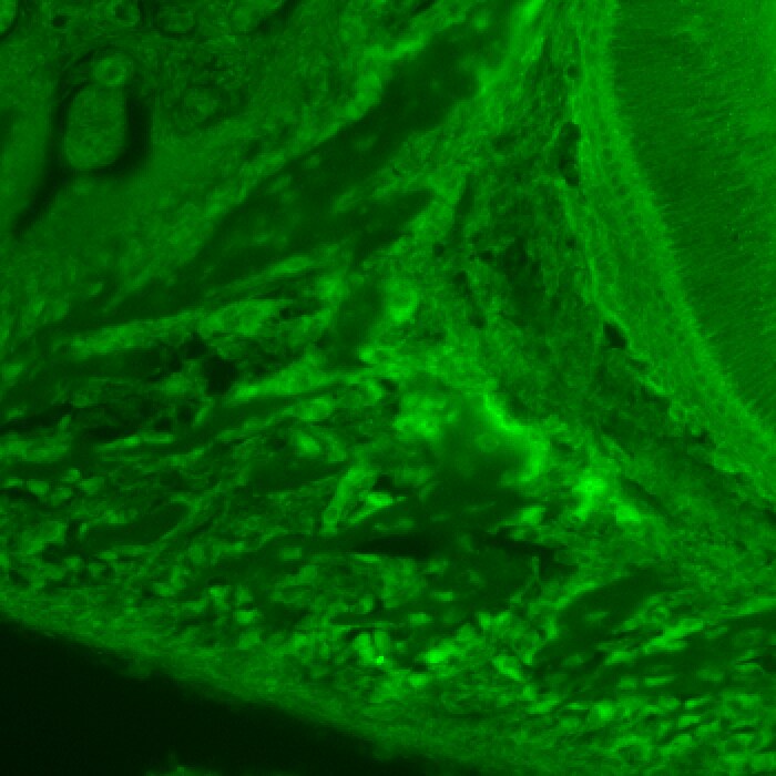
Application: Immunocytochemistry/ImmunofluorescenceVerified Customer | Posted 01/24/2024

There are no reviews that match your criteria.
Protocols
Find general support by application which include: protocols, troubleshooting, illustrated assays, videos and webinars.
- 7-Amino Actinomycin D (7-AAD) Cell Viability Flow Cytometry Protocol
- Cellular Response to Hypoxia Protocols
- Extracellular Membrane Flow Cytometry Protocol
- Flow Cytometry Protocol for Cell Surface Markers
- Flow Cytometry Protocol for Staining Membrane Associated Proteins
- Flow Cytometry Staining Protocols
- Flow Cytometry Troubleshooting Guide
- Intracellular Flow Cytometry Protocol Using Alcohol (Methanol)
- Intracellular Flow Cytometry Protocol Using Detergents
- Intracellular Nuclear Staining Flow Cytometry Protocol Using Detergents
- Intracellular Staining Flow Cytometry Protocol Using Alcohol Permeabilization
- Intracellular Staining Flow Cytometry Protocol Using Detergents to Permeabilize Cells
- Propidium Iodide Cell Viability Flow Cytometry Protocol
- Protocol for Liperfluo
- Protocol for the Characterization of Human Th22 Cells
- Protocol for the Characterization of Human Th9 Cells
- Protocol: Annexin V and PI Staining by Flow Cytometry
- Protocol: Annexin V and PI Staining for Apoptosis by Flow Cytometry
- R&D Systems Quality Control Western Blot Protocol
- Troubleshooting Guide: Fluorokine Flow Cytometry Kits
- Troubleshooting Guide: Western Blot Figures
- Western Blot Conditions
- Western Blot Protocol
- Western Blot Protocol for Cell Lysates
- Western Blot Troubleshooting
- Western Blot Troubleshooting Guide
- View all Protocols, Troubleshooting, Illustrated assays and Webinars


