
Key Product Details
Validated by
Biological Validation
Species Reactivity
Validated:
Mouse
Cited:
Mouse, Bovine, Transgenic Mouse
Applications
Validated:
Western Blot, Simple Western
Cited:
Immunohistochemistry, Western Blot, Immunoprecipitation, In vivo assay
Label
Unconjugated
Antibody Source
Polyclonal Goat IgG
Loading...
Product Specifications
Immunogen
Mouse myeloma cell line NS0-derived recombinant mouse MFG-E8 (R&D Systems, Catalog # 2805-MF)
Ala23-Cys463
Accession # P21956
Ala23-Cys463
Accession # P21956
Specificity
Detects mouse MFG-E8 in direct ELISAs and Western blots. In these formats, less than 2% cross‑reactivity with recombinant human MFG-E8 is observed.
Clonality
Polyclonal
Host
Goat
Isotype
IgG
Scientific Data Images for Mouse MFG-E8 Antibody
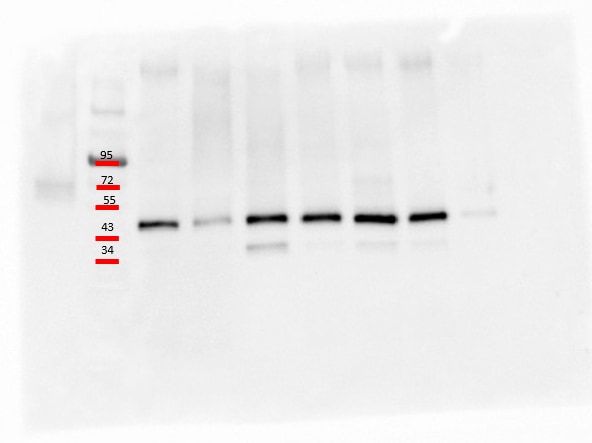
Detection of Mouse MFG‑E8 by Western Blot.
Western blot shows lysates of mouse mammary gland tissue. PVDF membrane was probed with 0.25 µg/mL of Goat Anti-Mouse MFG-E8 Antigen Affinity-purified Polyclonal Antibody (Catalog # AF2805) followed by HRP-conjugated Anti-Goat IgG Secondary Antibody (Catalog # HAF019). A specific band was detected for MFG-E8 at approximately 60-70 kDa (as indicated). This experiment was conducted under reducing conditions and using Immunoblot Buffer Group 1.
Detection of Mouse MFG‑E8 by Simple WesternTM.
Simple Western lane view shows lysates of recombinant mouse (rm) MFG-E8, loaded at 50 ng/mL. A specific band was detected for MFG-E8 at approximately 87 kDa (as indicated) using 50 µg/mL of Goat Anti-Mouse MFG-E8 Antigen Affinity-purified Polyclonal Antibody (Catalog # AF2805) followed by 1:50 dilution of HRP-conjugated Anti-Goat IgG Secondary Antibody (Catalog # HAF019). This experiment was conducted under reducing conditions and using the 12-230 kDa separation system.
Detection of Human MFG-E8 by Western Blot
MFGE8 and NO Are Produced and Secreted by Co-cultured BV2 Microglia Only When Co-cultured in Contact with pFTAA+Neurons(A and B) Representative blot (A) ofMFGE8 in BV2 cells lysates cultured alone or co-cultured in direct contact, or via atranswell, with DRG neurons from 5-month-old P301S-tau, 5-month-old C57, 5-month-old Alz17 mice, or 2-month-old P301S mice.Asterisk: either another isoform ofMFGE8 or a breakdown product. (B) Densitometry of MFGE8 expression normalized to beta -actin; significantly more MFGE8 is produced only in contact co-cultures containing pFTAA+ neurons. Mean± SD, n = 3 independent experiments (*p < 0.05), twoway ANOVA, Dunnett’s correction.(C and D) Elevated MFGE8 (C) or NO (D) in medium conditioned by BV2 cells co-cultured in contact with DRG neurons from5-month-old P301S mice compared with 5-month-old Alz17, 5-month- old C57, or 2-month-old P301S mice (MFGE8: ****p < 0.0001versus all; NO: ****p < 0.0001 versus 5-month-old C57 or 2-month-old P301S mice; ***p < 0.001 versus 5-month-oldAlz17 mice). N.D., none detected. Mean ± SD, n = 3 independent experiments, two-way ANOVA, Bonferroni corrected.(E and F) Elevated MFGE8 (E) or NO (F) in medium conditioned by primary microglia from C57 mice co-cultured in contactwith DRG neurons from 5-month-old P301S mice; microglia from MFGE8 KO mice are negative controls. Mean ± SD, n = 3independent microglial preparations, one-way ANOVA, **p < 0.01 (MFGE8), *p < 0.05 (NO).(G) Increased MFGE8 immunostaining intensity in frontal motor cortex of 5-month-old P301S mice compared with 5-month-oldC57 mice; MFGE8 KO mouse is negative control; 25 μm section at inter- aural 5.12 mm, bregma 1.32 mm; brown, DAB; blue,cresyl violet. Scale bar, 130 mm. Inset, 65 μm.(H and I) Elevated MFGE8 (H) in 5-month-old P301S-tau brains. Lysatesfrom cortex(Ctx), brain stem (BS), and cerebellum(Cb) of 5-month-old C57, 5-month-old P301S-tau, and 5-month-old Alz17 mice probed with anti-MFGE8; MFGE8 KO brain lysate isnegative control. rec, re
Detection of Human MFG-E8 by Western Blot
MFG-E8 deficiency impairs wound closure and wound angiogenesis.a The serum levels of MF-E8 in HCs (n = 30), diabetes (n = 33), and DFU patients (n = 25) were detected with ELISA. b The serum levels of MFG-E8 in normal or diabetic mice (n = 6) post-wounding for 0, 3, 7, 14 days were determined by ELISA. c Western blot analysis of MFG-E8 expression in skin tissues of normal or diabetic mice after wound for 3 days, GAPDH as a loading control. d The serum fed glucose levels of WT (n = 24) or Mfge8−/− mice (n = 28) when treated with vehicle or STZ for 28 days were measured. e Representative digital imaging of wounds from age-matched diabetic WT or Mfge8−/− mice postwounding up to day 14. f Wound closures kinetics of diabetic WT and Mfge8−/− mice (n = 6). g Representative images of H&E-stained skin tissues from WT or Mfge8−/− mice injected with vehicle or STZ after wound for 3 days. h The amount of CD31+ epithelial cells (ECs) and alpha -smooth muscle actin ( alpha -SMA+) pericytes in wound skins of WT or Mfge8−/− mice treated with vehicle or STZ at day 14 after wound. i Quantification of alpha -SMA+ vessels in five random microscopic fields in n = 6 mice per group was performed. For all experiments, data are presented as mean ± SEM, *P < 0.05, **P < 0.01, ***P < 0.001. Image collected and cropped by CiteAb from the following publication (https://pubmed.ncbi.nlm.nih.gov/32963812), licensed under a CC-BY license. Not internally tested by R&D Systems.
Detection of Human MFG-E8 by Western Blot
MFGE8 and NO Are Produced and Secreted by Co-cultured BV2 Microglia Only When Co-cultured in Contact with pFTAA+Neurons(A and B) Representative blot (A) ofMFGE8 in BV2 cells lysates cultured alone or co-cultured in direct contact, or via atranswell, with DRG neurons from 5-month-old P301S-tau, 5-month-old C57, 5-month-old Alz17 mice, or 2-month-old P301S mice.Asterisk: either another isoform ofMFGE8 or a breakdown product. (B) Densitometry of MFGE8 expression normalized to beta -actin; significantly more MFGE8 is produced only in contact co-cultures containing pFTAA+ neurons. Mean± SD, n = 3 independent experiments (*p < 0.05), twoway ANOVA, Dunnett’s correction.(C and D) Elevated MFGE8 (C) or NO (D) in medium conditioned by BV2 cells co-cultured in contact with DRG neurons from5-month-old P301S mice compared with 5-month-old Alz17, 5-month- old C57, or 2-month-old P301S mice (MFGE8: ****p < 0.0001versus all; NO: ****p < 0.0001 versus 5-month-old C57 or 2-month-old P301S mice; ***p < 0.001 versus 5-month-oldAlz17 mice). N.D., none detected. Mean ± SD, n = 3 independent experiments, two-way ANOVA, Bonferroni corrected.(E and F) Elevated MFGE8 (E) or NO (F) in medium conditioned by primary microglia from C57 mice co-cultured in contactwith DRG neurons from 5-month-old P301S mice; microglia from MFGE8 KO mice are negative controls. Mean ± SD, n = 3independent microglial preparations, one-way ANOVA, **p < 0.01 (MFGE8), *p < 0.05 (NO).(G) Increased MFGE8 immunostaining intensity in frontal motor cortex of 5-month-old P301S mice compared with 5-month-oldC57 mice; MFGE8 KO mouse is negative control; 25 μm section at inter- aural 5.12 mm, bregma 1.32 mm; brown, DAB; blue,cresyl violet. Scale bar, 130 mm. Inset, 65 μm.(H and I) Elevated MFGE8 (H) in 5-month-old P301S-tau brains. Lysatesfrom cortex(Ctx), brain stem (BS), and cerebellum(Cb) of 5-month-old C57, 5-month-old P301S-tau, and 5-month-old Alz17 mice probed with anti-MFGE8; MFGE8 KO brain lysate isnegative control. rec, re
Detection of Human MFG-E8 by Western Blot
The activation of NLRP3 inflammasome was aggravated in MFG-E8-deficient mice.a The infiltrated active caspase-1+ and IL-1 beta + macrophages in dermis of wound skins from WT or Mfge8−/− mice (n = 6) treated with vehicle or STZ, post-wounding at day 3, were determined with immunofluorescence. b Quantification of the expression of active caspase-1, caspase-1, and MFG-E8 in wound skin tissue from WT or Mfge8−/− mice (n = 6) treated with vehicle or STZ at day 3 post-wounding, beta -actin as loading control. c Calculation of the ratio of active caspase-1/caspase-1 in wound skin tissues from each group mice. d Infiltration of death cells into wound skin tissues of WT or Mfge8−/− mice (n = 6) post-wounding at day 3 was identified by TUNEL. Representative images were shown, original magnification ×200. e The average TUNEL-positive cells were counted at more than five random microscopic fields. For all experiments, data are presented as mean ± SEM, *P < 0.05, **P < 0.01, ***P < 0.001. Image collected and cropped by CiteAb from the following publication (https://pubmed.ncbi.nlm.nih.gov/32963812), licensed under a CC-BY license. Not internally tested by R&D Systems.
Detection of Human MFG-E8 by Western Blot
MFGE8 and NO Are Produced and Secreted by Co-cultured BV2 Microglia Only When Co-cultured in Contact with pFTAA+Neurons(A and B) Representative blot (A) ofMFGE8 in BV2 cells lysates cultured alone or co-cultured in direct contact, or via atranswell, with DRG neurons from 5-month-old P301S-tau, 5-month-old C57, 5-month-old Alz17 mice, or 2-month-old P301S mice.Asterisk: either another isoform ofMFGE8 or a breakdown product. (B) Densitometry of MFGE8 expression normalized to beta -actin; significantly more MFGE8 is produced only in contact co-cultures containing pFTAA+ neurons. Mean± SD, n = 3 independent experiments (*p < 0.05), twoway ANOVA, Dunnett’s correction.(C and D) Elevated MFGE8 (C) or NO (D) in medium conditioned by BV2 cells co-cultured in contact with DRG neurons from5-month-old P301S mice compared with 5-month-old Alz17, 5-month- old C57, or 2-month-old P301S mice (MFGE8: ****p < 0.0001versus all; NO: ****p < 0.0001 versus 5-month-old C57 or 2-month-old P301S mice; ***p < 0.001 versus 5-month-oldAlz17 mice). N.D., none detected. Mean ± SD, n = 3 independent experiments, two-way ANOVA, Bonferroni corrected.(E and F) Elevated MFGE8 (E) or NO (F) in medium conditioned by primary microglia from C57 mice co-cultured in contactwith DRG neurons from 5-month-old P301S mice; microglia from MFGE8 KO mice are negative controls. Mean ± SD, n = 3independent microglial preparations, one-way ANOVA, **p < 0.01 (MFGE8), *p < 0.05 (NO).(G) Increased MFGE8 immunostaining intensity in frontal motor cortex of 5-month-old P301S mice compared with 5-month-oldC57 mice; MFGE8 KO mouse is negative control; 25 μm section at inter- aural 5.12 mm, bregma 1.32 mm; brown, DAB; blue,cresyl violet. Scale bar, 130 mm. Inset, 65 μm.(H and I) Elevated MFGE8 (H) in 5-month-old P301S-tau brains. Lysatesfrom cortex(Ctx), brain stem (BS), and cerebellum(Cb) of 5-month-old C57, 5-month-old P301S-tau, and 5-month-old Alz17 mice probed with anti-MFGE8; MFGE8 KO brain lysate isnegative control. rec, re
Detection of Mouse MFG-E8 by Western Blot
MFGE8 regulates TG hydrolase activity through CES1D(A and B) (A) Representative western blot showing CES1D protein level in WT and Mfge8 KO primary enterocytes from three independent experiments. GAPDH was used as loading control. N = 8 mice per group. (B) Densitometric analysis of the western blots (including A).(C) Representative western blot showing CES1D protein level in WT and beta 5 KO primary enterocytes from two independent experiments. HSP90 was used as loading control. N = 5 WT and N = 4 beta 5 KO mice in total.(D) Densitometric analysis of the western blots of CES1D protein (including C).(E) TG hydrolase activity in WT, Ces1d KO, and Mfge8 KO primary enterocytes 1 h after incubation with rMFGE8 or RGE. N = 5 independent experiments.(F) Western blot of CES1D and MFGE8 protein levels in proximal small intestinal enterocytes of Mfge8 KO mice with transgenic inducible expression of MFGE8 in enterocytes (MFGE8 re-expressed, Vil rtTA+ TetO Mfge8+) and single transgenic (control), WT, and Mfge8 KO enterocyte controls. GAPDH was used as loading control.(G) Densitometric analysis of the blot presented in (F).(H) TG hydrolase activity in primary enterocytes isolated from the same groups of mice in (E) and (F). N = 3 mice in each group.(I and J) 3H signal in (I) the proximal jejunum and (J) serum 2 h after oral administration of [3H]oleic acid to WT, Mfge8 KO, Ces1d KO, and Mfge8/Ces1d double-KO mice. N = 3–4 mice in each group. All data are expressed as mean ± SEM. *p < 0.05, **p < 0.01, ***p < 0.001. Data in (B), (D), and (G) were analyzed by unpaired t test. Data in (E) and (H) to (J) were analyzed by one-way ANOVA followed by Bonferroni’s post test. Image collected and cropped by CiteAb from the following open publication (https://pubmed.ncbi.nlm.nih.gov/36924494), licensed under a CC-BY license. Not internally tested by R&D Systems.
Detection of Mouse MFG-E8 by Western Blot
Characterization of the surface functionalized mExo with FA. (A) Schematic illustration showing a process of surface engineering and Dox loading. The box with a blue dashed line demonstrates the modified chemical structure of 16:0 PE-FA obtained from Avanti Polar Lipids. (B) Size distribution of mExo species (mExo, mExo-FA, Dox@mExo, and Dox@mExo-FA). (C) TEM images of mExo, mExo-FA, Dox@mExo, and Dox@mExo-FA. Scale bar: 100 nm. (D) Western blotting analysis of TSG101, CD9, MFG-E8, calnexin, and casein. TSG101 and CD9, representative positive exosome markers; MFG-E8, milk-exosomal proteins; calnexin, negative marker of exosome; casein, representative contaminants from milk Image collected and cropped by CiteAb from the following open publication (https://pubmed.ncbi.nlm.nih.gov/38031117), licensed under a CC-BY license. Not internally tested by R&D Systems.
Mouse MFG-E8 ELISA Standard Curve
Recombinant Mouse MFG‑E8 (Catalog # 2805-MF) was serially diluted and captured by Rat Anti-Mouse MFG‑E8 Monoclonal Antibody (Catalog # MAB2805) coated on a Clear Polystyrene Microplate (Catalog # DY990). Goat Anti-Mouse MFG‑E8 Antigen Affinity-purified Polyclonal Antibody (Catalog # AF2805) was biotinylated and incubated with the protein captured on the plate. Detection of the standard curve was achieved by incubating Streptavidin-HRP (Catalog # DY998)Applications for Mouse MFG-E8 Antibody
Application
Recommended Usage
Simple Western
50 µg/mL
Sample: Recombinant Mouse (rm) MFG-E8
Sample: Recombinant Mouse (rm) MFG-E8
Western Blot
0.25 µg/mL
Sample: Mouse mammary gland tissue
Sample: Mouse mammary gland tissue
Reviewed Applications
Read 3 reviews rated 5 using AF2805 in the following applications:
Formulation, Preparation, and Storage
Purification
Antigen Affinity-purified
Reconstitution
Reconstitute at 0.2 mg/mL in sterile PBS. For liquid material, refer to CoA for concentration.
Loading...
Formulation
Lyophilized from a 0.2 μm filtered solution in PBS with Trehalose. *Small pack size (SP) is supplied either lyophilized or as a 0.2 µm filtered solution in PBS.
Shipping
Lyophilized product is shipped at ambient temperature. Liquid small pack size (-SP) is shipped with polar packs. Upon receipt, store immediately at the temperature recommended below.
Stability & Storage
Use a manual defrost freezer and avoid repeated freeze-thaw cycles.
- 12 months from date of receipt, -20 to -70 °C as supplied.
- 1 month, 2 to 8 °C under sterile conditions after reconstitution.
- 6 months, -20 to -70 °C under sterile conditions after reconstitution.
Calculators
Background: MFG-E8
References
- Raymond, A. et al. (2009) J. Cell. Biochem. 106:957.
- Stubbs, J.D. et al. (1990) Proc. Natl. Acad. Sci. USA 87:8417.
- Hoffhines, A.J. et al. (2008) J. Biol. Chem. 284:3096.
- Silvestre, J.-S. et al. (2005) Nat. Med. 11:499.
- Borges, E. et al. (2000) J. Biol. Chem. 275:39867.
- Hanayama, R. et al. (2002) Nature 417:182.
- Ait-Oufella, H. et al. (2007) Circulation 115:2168.
- Kranich, J. et al. (2010) J. Exp. Med. 207:2271.
- Hanayama, R. et al. (2004) Science 304:1147.
- Kranich, J. et al. (2010) J. Exp. Med. 205:1293.
- Atabai, K. et al. (2009) J. Clin. Invest. 119:3713.
- Bu, H.-F. et al. (2007) J. Clin. Invest. 117:3673.
- Jinushi, M. et al. (2009) J. Exp. Med. 206:1317.
- Kvistgaard, A.S. et al. (2004) J. Dairy Sci. 87:4088.
Long Name
Milk Fat Globule EGF Factor 8
Alternate Names
BA46, Breast epithelial antigen BA46, Lactahedrin, Medin, MFGE8, SED1
Gene Symbol
MFGE8
UniProt
Additional MFG-E8 Products
Product Documents for Mouse MFG-E8 Antibody
Certificate of Analysis
To download a Certificate of Analysis, please enter a lot or batch number in the search box below.
Note: Certificate of Analysis not available for kit components.
Product Specific Notices for Mouse MFG-E8 Antibody
For research use only
Citations for Mouse MFG-E8 Antibody
 Powered by Bioz
Customer Reviews for Mouse MFG-E8 Antibody (3)
5 out of 5
3 Customer Ratings
Have you used Mouse MFG-E8 Antibody?
Submit a review and receive an Amazon gift card!
$25/€18/£15/$25CAN/¥2500 Yen for a review with an image
$10/€7/£6/$10CAN/¥1110 Yen for a review without an image
Submit a review
Customer Images
Showing
1
-
3 的
3 reviews
Showing All
Filter By:
-
Application: Western BlotSample Tested: Colon tissueSpecies: MouseVerified Customer | Posted 10/01/2024

-
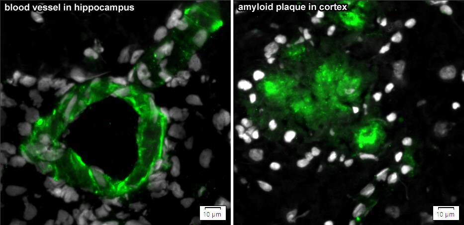
Application: ImmunohistochemistrySample Tested: Alzheimer's disease brainSpecies: MouseVerified Customer | Posted 06/30/2023Immunohistochemistry/fluorescence (Frozen sections). 10 µm cryostat sections from fresh frozen brain sample. Post-fixation with 4% formaldehyde for 15min. Blocking: PBS + 1% BSA +1% ovalbumin +10% normal donkey serum +0.3% LDAO +0.3M Glycine. Incubation buffer: PBS + 1% BSA +1% ovalbumin +10% normal donkey serum. Primary AB: goat anti-mouse MFG-E8 (#AF2805) 2 µg/ml, 2h at room temperature. DRAQ5 5µM (nuclei marker) together with primary AB. Secondary AB: Donkey anti-goat/AlexaFluor488 4 µg/ml, 1h at RT. Autofluorescence suppression: 4mM CuSO4 in 50mM ammoniumacetate buffer, pH 5, 30min at RT. Imaging: SlideView VS200 slide scanner with X-Cite NOVEM illumination.
-
Application: Western BlotSample Tested: SpermSpecies: PorcineVerified Customer | Posted 09/07/2017Dilution: 1:1000 Secondary used: Goat IgG Horseradish Peroxidase-conjugated Antibody from R&D Systems.
There are no reviews that match your criteria.
Protocols
Find general support by application which include: protocols, troubleshooting, illustrated assays, videos and webinars.
- Cellular Response to Hypoxia Protocols
- R&D Systems Quality Control Western Blot Protocol
- Troubleshooting Guide: Western Blot Figures
- Western Blot Conditions
- Western Blot Protocol
- Western Blot Protocol for Cell Lysates
- Western Blot Troubleshooting
- Western Blot Troubleshooting Guide
- View all Protocols, Troubleshooting, Illustrated assays and Webinars
Loading...