Amine Reactive Comp-Bead 2 Population Kit
Novus Biologicals | Catalog # NBP3-00496
Loading...
Key Product Details
Applications
Flow Cytometry
Kit Type
Kit
Product Summary for Amine Reactive Comp-Bead 2 Population Kit
The Amine Reactive Compensation Beads are designed for labeling amine-reactive dyes (live/dead fixable viability stains) when setting compensation in flow cytometry.
Particle size: 7.0 -7.9 micron
Particle material: polystyrene
- Ensure consistent and accurate compensation in any channel
- Evaluate cell viability of animal cells, bacteria, yeast, and fungi
- Contains both amine reactive and nonreactive beads for generating positive and negative populations
Particle size: 7.0 -7.9 micron
Particle material: polystyrene
Loading...
Product Specifications
Application Notes
Dye and bead concentrations may be further optimized for best results. Centrifugation force and time may need to be increased if needed. Protect the beads from light after exposure to the dye. Use immediately after staining.
Scientific Data Images for Amine Reactive Comp-Bead 2 Population Kit
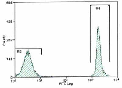
Amine Reactive Comp-Bead 2 Population [NBP3-00496] - Histogram. Amine Reactive Comp-Beads stained with Biolegend Zombie Green(Tm).

Flow Cytometry: Amine Reactive Comp-Bead 2 Population Kit [NBP3-00496]
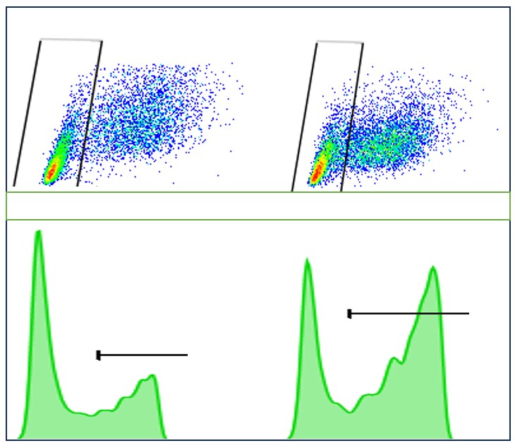
Flow Cytometry: Amine Reactive Comp-Bead 2 Population Kit [NBP3-00496] - Beads used for flow cytometry compensation calculations. Image from a verified customer review.Kit Contents for Amine Reactive Comp-Bead 2 Population Kit
- High Binding Beads - 5 mL
- Negative Binding Beads - 5 mL
Formulation, Preparation, and Storage
Formulation
0.016 M PBS (pH 7.4)
Preservative
0.02% Sodium Azide
Concentration
Concentration is not relevant for this product. Please see the protocols for proper use of this product.
Shipping
The product is shipped with polar packs. Upon receipt, store it immediately at the temperature recommended below.
Storage
Store at 4C in the dark. Do not freeze.
Background: Amine Reactive Comp-Bead 2 Population
Compensation beads are a highly desirable alternative to traditional cell-based single-color compensation. Positive beads can come pre-loaded with anti-IgG for conjugated antibody capture of mouse, rat, or hamster origin. Uncoated (negative) beads ensure standardization of the autofluorescence in each channel. Beads are preferable to cell-based compensation for several reasons:
1) No precious sample is wasted, and more sample can be used for experimental acquisition.
2) Since 100% of positive beads are able to capture the antibody, the exact experimental fluorochrome can be used regardless of a marker's cellular expression. Dimly expressed markers may not be bright enough for compensation using cells.
3) Reduced autofluorescence of beads allows for more precise calculation of spectral overlap.
Alternate Names
Comp Beads, Compensation Beads, Flow Cytometry Compensation, Live Dead Compensation Beads, Live/Dead Compensation Beads
Additional Amine Reactive Comp-Bead 2 Population Products
Product Documents for Amine Reactive Comp-Bead 2 Population Kit
Certificate of Analysis
To download a Certificate of Analysis, please enter a lot or batch number in the search box below.
Product Specific Notices for Amine Reactive Comp-Bead 2 Population Kit
This product is for research use only and is not approved for use in humans or in clinical diagnosis. Kits are guaranteed for 6 months from date of receipt.
Customer Reviews for Amine Reactive Comp-Bead 2 Population Kit (1)
4 out of 5
1 Customer Rating
Have you used Amine Reactive Comp-Bead 2 Population Kit?
Submit a review and receive an Amazon gift card!
$25/€18/£15/$25CAN/¥2500 Yen for a review with an image
$10/€7/£6/$10CAN/¥1110 Yen for a review without an image
Submit a review
Customer Images
Showing
1
-
1 of
1 review
Showing All
Filter By:
-
Verified Customer | Posted 09/16/2024We used beads for flow cytometry compensation calculations, and in the image, confidential information has been removed. The different populations are clearly distinguishable following two population comp-bead based compensation.

There are no reviews that match your criteria.
Protocols
View specific protocols for Amine Reactive Comp-Bead 2 Population Kit (NBP3-00496):
1. Allow beads to come to room temperature, then vortex briefly.
2. Add 1 drop (about 50 uL) of the High Binding beads to a 1.5 mL microcentrifuge tube.
3. Wash the beads by adding 0.5 mL of PBS (free of surfactant and blocker) to the microcentrifuge tube, centrifuge at 300 x G for 5 minutes, decant and repeat.
4. Decant and resuspend in 50 uL PBS.
5. Prepare the amine reactive dye according to the manufacturer's instructions.
6. Add 1 - 4 uL of the amine reactive dye to the bead suspension and vortex briefly.
7. Incubate for 30 minutes. Protect tube from light.
8. Add 1 mL of PBS to the same tube and vortex briefly.
9. Centrifuge at 300 x G for 5 minutes, decant and repeat.
10. Resuspend the beads in PBS containing 0.05% BSA with brief vortex.
11. Add a drop (about 50 uL) fo the Negative Binding beads to the labeled High Binding beads.
12. Analyze on the flow cytometer using a live gate around the singlet population in the FSC/SSC dot plot.
13. Create a fluorescent histogram for the appropriate detectors and perform compensation to achieve the desired results.
Find general support by application which include: protocols, troubleshooting, illustrated assays, videos and webinars.
- 7-Amino Actinomycin D (7-AAD) Cell Viability Flow Cytometry Protocol
- Extracellular Membrane Flow Cytometry Protocol
- Flow Cytometry Protocol for Cell Surface Markers
- Flow Cytometry Protocol for Staining Membrane Associated Proteins
- Flow Cytometry Staining Protocols
- Flow Cytometry Troubleshooting Guide
- Intracellular Flow Cytometry Protocol Using Alcohol (Methanol)
- Intracellular Flow Cytometry Protocol Using Detergents
- Intracellular Nuclear Staining Flow Cytometry Protocol Using Detergents
- Intracellular Staining Flow Cytometry Protocol Using Alcohol Permeabilization
- Intracellular Staining Flow Cytometry Protocol Using Detergents to Permeabilize Cells
- Propidium Iodide Cell Viability Flow Cytometry Protocol
- Protocol for Liperfluo
- Protocol for the Characterization of Human Th22 Cells
- Protocol for the Characterization of Human Th9 Cells
- Protocol: Annexin V and PI Staining by Flow Cytometry
- Protocol: Annexin V and PI Staining for Apoptosis by Flow Cytometry
- Troubleshooting Guide: Fluorokine Flow Cytometry Kits
- View all Protocols, Troubleshooting, Illustrated assays and Webinars
Loading...